
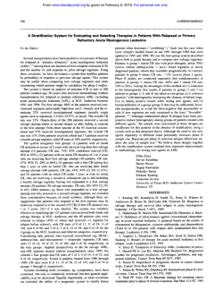
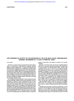
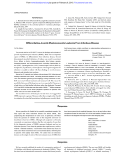
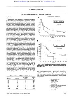
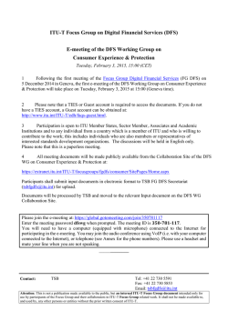
Immunophenotype of Adult Acute Lymphoblastic Leukemia
From www.bloodjournal.org by guest on February 6, 2015. For personal use only.
Immunophenotype of Adult Acute Lymphoblastic Leukemia,
Clinical Parameters, and Outcome: An Analysis of a Prospective Trial
Including 562 Tested Patients (LALA87)
By Claude Boucheix, Bernard David, Catherine Sebban, Evelyne Racadot, Marie-Christine Bene, Alain Bernard,
Lydia Campos, Helene Jouault, FranGois Sigaux, Eric Lepage, Patrick Herve, and Denis Fiere for the French Group
on Therapy for Adult Acute Lymphoblastic Leukemia
The aim of the multicentric trial LALA87 was t o test the
efficacy of different postremission therapies in adults (15 t o
60 year olds) with acute lymphoblastic leukemia (ALL). An
immunologic subclassification based on surface marker expression was proposed. Among the 562 tested patients, 511
were assigned either to the B lineage (361 cases, 6 3 % ) or
to theT lineage (150 cases, 26%). T-ALL were significantly
associated with male sex, age less than 35 years, mediastinal
mass, central nervous system involvement, highwhite
blood cell count, and low anemia. In a univariate and multivariate analysis, T-cell leukemia had a more favorable
outcome than Btell leukemia with respective median disease-free survivals (DFSs) of 28 and 14 months (P < .005).
However, the type of postremission therapy modifies the
value of the immunophenotype prognosticfactor. Inthe che-
motherapy arm, T-ALL patients (26 patients) had a more
favorable outcome than B-ALL patients (57 patients) ( P <
.003). In the autologous bone marrow transplantation
(ABMT) arm, the apparent better outcome of T-ALL patients
(35 T/50 B) did notreach statistical significance ( P = .2) and
there was no difference in the allogeneic bone marrow transplantation (alloBMT) arm (37 T/71 B: P = .g). In the B-celllineage leukemias, subclassification by stages and myeloid
antigen coexpression (109'0) were not associated with different prognosis. CD10+ T-ALL (31 patients) were associated
with a better DFS compared with the CD10- T-ALL (73 patients) with respective median DFS, not reached and 18.5
months ( P = .04).
0 1994 by The American Societyof Hematology.
T
bone marrow transplantation ( ~ ~ ~ O B Mand
T autologous
)~~.~~
bone marrow transplantation (ABMT).28-30
Only multicentric
studies with random assignment and careful analysis might
help to define adapted therapies in relation to the diversity
of the disease.
Patients included in a large French multicentric trial
LALA8731were submitted to an initial evaluation including
immunological phenotyping. This report describes immunological phenotypic data and analyzes their relationships with
clinical characteristics and outcomes. The main objective
was to assess the relevance of a reliable immunologic classification with three objectives: (1) to identify a clinical and
biologic profile according to phenotypic subgroups; (2) to
assess the effect of phenotype on outcome; and (3) to evaluate the optimal postremission therapy in each group.
HE RELATIONSHIP between lymphoid ontogeny and
human acute lymphoblastic leukemia (ALL) has been
more precisely unravelled by the description of the differentiation pathways of the T-and B-lymphoid lineages."8 The
sequence of events leading to the development of mature
lymphoid cells is associated with cell surface modifications
that can be easily analyzed by the use of monoclonal antibodies (MoAbs). The importance of the immunophenotypic
analysis of ALL has been documented by numerous studies
in adults and children. However, the great number of phenotypic patterns has introduced a further level of complexity in
the classification of ALL. If certain leukocyte differentiation
antigens used for these analyses show some level of lineage
specificity (for instance the CD3 T-cell lineage), other surface molecules display a wider tissue distribution. For instance, the CDlOKALLA antigen initially described in nonTleukemia' and now identified as neutral endopeptidase,
has a large tissue distribution, but its transient expression
during B-cell differentiation helps to define a particular
stage; it is also expressed on a fraction of T-ALL (for review,
see Lebien and McCormack"). On the other hand, the myeloid markers CD13 and CD33 have notbeen found on
normal lymphoid cells and their expression in some ALL
has been referred to as myeloid antigen coexpression. The
clinical significance of the presence of myeloid markers has
been widely debated,""'but
because of the small size of
some of the patient groups, the mixture of adult and pediatric
cases and the differences in the treatment protocols, no final
conclusions can be drawn from studies completed thus far.
Beside immunophenotyping aspects, the heterogeneity of
the disease is observed at both clinical (age of onset, sex,
symptoms, and prognosis) and biologic levels (cytology and
cytogenetic aspects). Despite some recent progress, the prognosis of adult ALL remains poor. If complete remission (CR)
can be achieved in 70% to 80% of patients, relapses occur
frequently and long-term disease-free survival (DFS) does
not exceed 25% to 30%.20-25Three kinds of postremission
therapy can be proposed: chemotherapy (CT), allogeneic
Blood, Vol 84, No 5 (September I),
1994: pp 1603-1612
MATERIALS AND METHODS
Patients
From November 1, 1986 to July 31, 1991, 634 patients treated in
43 hematologic centers entered the LALA87 trial. Eligibility criteria
From HGpital Paul-Brousse, Institut National de la Sante' et de la
Recherche Me'dicale, Unite' 268, Villejuif; HGpital Edouard-Herriot,
Lyon: Centre Regional de Transfusion Sanguine, Besancon: Faculte'
de Me'decine, Laboratoire d'lmmunologie, Nancy: H6pital de 1'Archet, Nice: Hepita1 Henri-Mondor, Cre'teil: H6pital Saint-Louis,
Paris.
Submitted December 30, I993; accepted April 28, 1994.
Supported by an INSERM Research Contract No. 89 69001, RCseau de Recherche Clinique INSERM 488009, and Association pour
la Recherche contre le Cancer Contract No. 6763.
Address reprint requests to Claude Boucheix, MD, H6pital PaulBrowse, Institut National de la Sante' et de la Recherche Me'dicale,
Unite' 268, 94800 Villejuif; France.
The publication costs of this article were defrayed in part by page
charge payment. This article must therefore be hereby marked
"advertisement" in accordance with 18 U.S.C. section I734 solely to
indicate this fact.
0 1994 by The American Sociery of Hematology.
0006-4971/94/8405-0.00/0
1603
From www.bloodjournal.org by guest on February 6, 2015. For personal use only.
BOUCHEIX ET AL
1604
LALA 87
i
INDUCTION
l
I
$1
l
V
I
CONSOLIDATION
POST-REMISSION THERAPY
Allogeneic
116
1
I
BMT
(+6 > 40 years)
+I
> 50 years
CT
INCLUDED
634
CRl
ANALYSED
572
__.+ 436
(76%)
(15 60 years)
~
JI
Relapses
21
BM harvest
and purge
\
RANDOMIZATION
+
were morph~logically~~
and cytochemically confirmed diagnosis of
ALL (L3 FAB subtype excluded), absence of prior malignancy,
severe illness or psychiatric disease, and age from l 5 to 60 years.
After exclusions, 572 patients were analyzed. Median age ofthe
population was 33 years.
The design of the protocol and the main clinical results have been
reported el~ewhere.~'
The general design withthe flow chart of
the patients is summarized in Fig 1. Briefly, eligible patients were
submitted to an induction CT regimen including cyclophosphamide,
vincristine, prednisone, and a random allocation between two anthracyclins (daunombicin or zorubicin). If CR was not obtained at day
28, a salvage therapy with amsacrine and cytosine arabinoside was
administered. A central nervous system (CNS) prophylaxis was associated. Patients from 15 to 40 achieving CR, with an available HLAcompatible sibling donor were scheduled for alloBMT. Patients over
50 years received consolidation and maintenance CT. Patients from
40 to 50 years and patients up to 40 without HLA sibling donor
were randomized after a first course of consolidation between consolidation and maintenance CT or consolidation and ABMT. They received inboth arms three courses of consolidation including the
anthracycline used for induction, cytosine arabinoside and L-Asparaginase (modified 3A protocol). In the CT a r m , the maintenance
In the
CT consisted of eight cycles of the L10 modified protoc01.~~
ABMT arm, the protocol assumed MoAb depletion of the BM by
the MoAbs D66/CD2, A50/CD5, and I21/CD7 (provided by A. Bernard) for T-ALL and the MoAbs ALBZKDlO (Immunotecb, Marseille) and SB4/CD19 (Sanofi, Montpellier, France) for B-ALL.
Asta-2 (Asta Werke, Frankfurt, Germany) depletion was proposed
for patients with less than two of these markers on their leukemic
cells.
Stopping date was set for September 1992. Median follow-up was
33 months. Four hundred and thirty six patients (78%) achieved CR
after induction or induction and salvage therapy. One hundred and
sixteen patients were scheduled to receive alloBMT with an estimated 3-year DFS rate of 43% ? 5%. The ABMT ann (95 patients)
and CT arm (96 patients) produced comparable 3-year DFS rates
(39% v 32%). Patients over 50 years had a 3-year DFS rate of 24%.
Immunophenotype Analysis
To classify the disease according to the differentiation pathways
and to choose appropriate antibodies in case of BM depleted autograft, heparinized BM or blood samples were used for immunophe-
ABMT
95
1
Seattle
Grafted 63
Fig 1. General design
and
flow chart of the LALA87 trial.
notyping using a panel of MoAbs. Immunophenotyping was usually
performed on BM cells by indirect immunofluorescence and flow
cytometry, to analyze a pure (or nearly pure) blast cell population.
The immunologic classification used in this trial was based on the
expression of the CDl, CD2, CD3, CD5, CD7, CD4, CD8, HLADR, CDlO, 0 1 9 , CD20 markers and the absence of surface Igs.
However, it had been suggested that the panel of antibodies tested
be extended. Therefore numerous patients were tested also for CD9,
CD13, CD24, CD33, and HLA class I1 antigens.
Because of the large number of centers involved in the study, the
origin of the MoAbs varied from one center to another, butthe
different antibodies belonged to the same cluster according to the
CD n ~ m e n c l a t u r e .Results
~ ~ . ~ ~ were reviewed, interpreted, and registered in an independent database (with the same entry number as
the main database) by an immunologic committee that issued regular
information to the centers involved in the trial. After reinterpretation
and correction for the blast cell percentage and negative controls,
the arbitrary threshold of 20% labeled blast cells was considered the
limit for positivity of a given marker (20% blast cells expressing
the antigen). If necessary, the immunologic committee requested
new testing of the cells with additional MoAbs on cryopreserved
cells. The fusion between the complete immunologic database with
the main database was performed in January 1993.
We proposed an immunologic classification B1, B2, B3A, B3B,
TlA, TIB, T 2 , T3, as defined in Fig 2. To reduce the number of
subgroups, the lineage assignment was determined by the presence
of the pan-lineage markers, CD19 for the B-cell lineage, CDUCDSI
CD7 for the T-cell lineage. The level of differentiation was evaluated
by the presence of the CDlO and CD20 antigens for the B-cell
lineage and the CD1 and CD3 antigens for the T-cell lineage. For
the latter, in the absence of CD3 or CD1 expression or in the absence
of testing, the presence of the CD4 and/or CD8 markers was considered as reflecting a higher level of differentiation and assigned these
patients to a T2T3 subgroup. Classification in intermediate stages
or only within the T-cell lineage was decided in the absence of testing
of differentiation stage markers. Finally, in cases of coexpression, the
most differentiated marker was considered for the classification; eg,
coexpression of CD1 and CD3 assigned the leukemia to the T3
stage.
Statistical Methods
The endpoints were CR rate, overall survival, and DF'S. Survival
duration was calculated from the date of randomization until death
From www.bloodjournal.org by guest on February 6, 2015. For personal use only.
lALA87.IMMUNOPHENOTYPEANDOUTCOME
1605
of differentiation and lineage involvement is shown on Table
1. Terminologies such as B1B2 or T1T2 indicate that one or
several markers were not tested precluding a more accurate
classification.
B-lymphoidlineage ALL
B1
B3
I
B2
B
I
I
HLA-DR
b
I
CD19
CD 10
CD20
T-lymphoidlineage ALL
I
I
m
I
I
I
T1
T
2
I
T
3
CD7
CD5
CD2
CD 1
CD4
CD8
CD3
UD = undifferentiated ALL
UC = unclassified ALL
Fig 2. Classification of adult B- and T-ALL according to immunophenotypes in the LALA87 trial.
or date last known alive. DFS was calculated from date of CR until
relapseorlastknownsurvivaldateinCR.Deathsin
CRwere
counted as adverse events. Survival curves were estimated by the
method of Kaplan and M e i e Univariate
~ ~ ~
analyses were performed
using the chi-squaretestandthe
generalized Wilcoxon test of
Gehan.38Test statistics for comparison of endpoints were regarded
as significant if the P value was < .05. All survival analyses were
made on an intention to treat basis. Multivariate analyses of prognostic factors were performed according to Cox's proportional hazard
model for covariate analysis of censored survival data.39
45
RESULTS
General Characteristics of Immunophenotypic Data
B2
36
As shown in Table 1, 572 patients (15 to 60 years old)
were evaluable for clinical analysis. Immunophenotyping
was not performed in 10 patients. According to our criteria,
51 1 patients over 562 (90.9%) could be assigned to the B
lineage (361 cases, 63%) or to the T lineage (150 cases,
26%). Fifteen patients (2.7%) were negative for all markers
of the B or T cell lineages, and thus were considered as
undifferentiated. Because of insufficient data, 36 patients
(6.2%) remained unclassified (UC); in 9 of these patients, the
inability to perform a significant immunophenotype could be
related to BM fibrosis. The distribution related to the stage
Comparative Clinical Analysis of the Major
Immunophenotype Groups
Main pretherapeuticcharacteristics.
Clinical andbiologic characteristics, related to each type and subtype, are
reported in Table 2 for all patients between 15 and 60 years
old. T-ALL and B-ALL significantly differed in their presentation. Patients with T-ALL were statistically significantly
younger (77% of them are younger than 35 years v 50% for
B-ALL: P < .00l). Patients with T-ALL were more often
male (75% v 55% in B-ALL, P < .001). Mediastinal mass
was present in 49% of T-ALL versus 2% in B-ALL (P <
.001) and white blood cell ( W C )count was greater than
30 X 109/L in 55% of T-ALL versus 30.5% in B-ALL (P
< .001). Anemia was less frequent in T-ALL (65% v 85%
in B-ALL, P < .OOl). CNS involvement was observed in
10.5% of T-ALL whereas it was present in only 5% of BALL (P < .04).
Relationships between
phenotype
and
outcome.
As
shown in Table 2 for all patients from 15 to 60 years, global
CR rates were 81% for T-ALL and 74% for B-ALL. This
difference was not statistically significant (P = .08). For TALL, 106 patients over 150 (71%) achieved CR at day 28
after the start of the induction course and of the 38 patients
who received the scheduled salvage therapy with amsacrine
and cytosine arabinoside, 16/38 (42%) patients achieved CR.
For B-ALL, 248 patients over the 361 achieved CR at day 28
(69%) and among the 68 patients who received the salvage
therapy, 19 (28%) of them achieved CR.
Patients with T-ALL had a significantly better DFS and
better overall survival when compared with patients with BALL. The respective median DFS for T- and B-ALL were
Table 1. Frequency of Immunologic Subtypes
in the LALA87 Trial
BLineage
Subgroups
CD13
T-
CD13
Lineage
andlor
No.
B1
B1
1
B2
142
B283
B3
6
B3A
117
838
14
Totals (63%)
361
CD33'
andlor
No.
Subgroups
T
4/36
T1
0
T1A
15/120
T1B
1/18
T1T2
012
2/30 T2
5/97
T2T3
019
T3
251282
14
4
11
27
2
40
9
43
150 (26%)
CD33'
OB
214
311 1
3/25
012
014
2/32
121117
Of 572 patients, 15 were undifferentiated, 36 were unclassified, and
10 were not tested. The B283 group represents patients not tested
for the CD20 antigen; the T group represents mainly patients tested
for CD2, CD5, and CD7 but not for the other T-cell differentiation
markers. The T2T3 group contained patients not tested for CD3 and
positive for either CD1, CD4, and/or CD8 markers. The number of
patients expressing CD13 and/or CD33 among those tested for these
antigens is also indicated.
From www.bloodjournal.org by guest on February 6, 2015. For personal use only.
1606
BOUCHEIX ET AL
I
I
S
*LD
S
N
N
1
I
%
m
r
I
d
N
~
N
S
S
I
m
0
m
3
f
m ;
$- 2
c
-.g '6
C
w
-
S E N 0
-
<
"
2
I
s
r
m
$i
-
gm-
$
*
(D
-
m
"Qz$,E
"
B
p'
N
d
e
sh
S
m
I
m
-
$
o
m
4 "
N
W
O
~
I
"
m
-
I
-
m
S
g
m
F
m2 s:+; z 2
m
:?m
zqall,g
z
~
-
2 s Q k - c ? m p
9.-
0
m
"
~ m %
o +
~ -mm &m mk N
m o
(D
m
-d
0
:-c?
r
(Dl.-
s
t rn g
5
g
-? m G
h ~ k r
d
r
a2
.-c
D
..-.
"
-m
m
m"
(
S ru?Sl.%g
=--~mR~~$dzwm~~~~~~
m
,
~
I
p
4
t
"
N
I
N
a s
gE
m
S
u?tu?P
J - O h m
-r ;
$ mg
-
S
z -m
z g~z zZZ?azeENa ~z
7
-P
5
h U
N O W % W O d r
N
N
E
N
a
g
h
b
z
k d N h g r
+
-
u
*
N
-
'C
v
m
N
S
:,
*
"
2 ~
o mN $ C~ -~z oQ z ~o gI
a
c
~m
"
,
I
.-c5
d
-
m
o?"s
re n~r .b i S
mrm
4 (eD m
~ og d=l .~mmmra~N ; Z G
p
N
-
N
~
N
-
N
"u?;z;
S
"
$N.,
S %Nz L z gz , poN z' nmm" " ; ~ " - 2 m
m
c
r
P
~
zZ z z
N
P
z
"
-E
r
.-cE
.-a
g
c
r
:
.-um u a,
o a
=.- G
m .S
-
$:
4
r
r
"
d
$
2
N
N
cE2g
L
si
f
=.
8
9
6
9
0
C
V
I
S
S
I
a
z..
e
-
~ a mg- $ Lo~ 2c9"~- Sg . ,g* $ g az z5o!zR F
z .Br
"! .P
m
c
r
r
.e
m
U A
e a
Z
'
-
.-
S
i
z
v v v
-
t-
v)
9 9 9
s
gg;s
r
0 0 0
?.L ( D
.-$ 22
a2
02E
U
g ; "2
\ i v ' v
6 6
cri
g
.S
.g
-5
z
:k
-
8
0
m
9 2&
m
3
6
C
-
vi
? ,g=m%
6m
. ;"a*?hg&g PP N SXmN*?
4
-P
m
mL Z
C
z
r
3:
s
N
C
h
-
N
S
2m ,SE
zz:"
g
m
)
z..
?
U
B
!
i
i
4
c
-
.-
0
c
C ?
m - €
0
5 d - S -
"
Q
.-
Er$+G-
"l"5d
-s
2
E"
-l&zE0 5 %
E
U
-
3
Q
gci
3
E m 1 3 g c i ..
9 S E : .pt
._
2 .5
v)
-
.S2
2
x, g+ <, .. m*&?,
E8 ;:~ gS8g-gzxosSB
!&? ? - s .c2E +$? C$k;Oz
o m z v m B m a
~ ~ v I ~ ) z Y I Z G P V I S$s Emz E
ss
0
~
z
~
~
From www.bloodjournal.org by guest on February 6, 2015. For personal use only.
LALA87, IMMUNOPHENOTYPE AND OUTCOME
1607
100
DFS 3 ycnrs
%
montha
0
**
"b.0..
*tt
4.*4*.**
40
52.4 43
T-ALL (N-103)
33.5 15
B-ALL (N-172)
pes
4
8:
89(52)
DFS
0
. ..........
8.0
b ..................
.
p
"***4..*
b
Allogmneic BMT ann S 40 p m
9:
RCI.
N(%)
37(36)
4..*..
4.
-
T-ALL (N-37)
B-ALL (N-71)
months
22
23
3ycan &.l.
%
N(%)
44 14(38)
45
26(37)
p - 0.9
...
...b . . . . . . ......................
......................
0.01
...... ...............
4
Auto&gvw BMT ann S 40 years
%
DFS
bbb :
8;. .b
0
:6.6.
*.
T-ALL (N-29)
B-ALL (N-38)
months
n.r.
25
58
. ...................................
.........
p
b.........,
i
3ycam
%
20(53)
43
Rd.
N(%)
ll(38)
0.27
I
'...... .......................
2
3
4
5 years
p
b
0..
i
i
3
4
- o.ooo7
5 years
Fig 3. T- versus B-ALL DFS curves for patients5 40 years in the various armsof the ALL87 trial. A curve of a11 patients c 40 years is shown
for comparison. Patients S 40 years in CR with an HLA-compatible sibling were selected immediately for alloBMT; the DFS stam with the
obtention of CR. For the CT and A B M arms, patients in CR were randomized after one cyde of consolidation therapy andthe DFS stam at
the date of CR obtention. All statistical analyses were performed on an intention to treat basis h r . , not reached; N. number; Rel, relapses)
28 months and 14 months (P = .005). Respective overall
survivals for T- and B-ALL were 25 months and 16 months
(P = .001).Ina multivariate analysis, Tphenotypewas
found as an independent favorable prognostic factor (P =
.W) as well as initial WBC count less than 30 X 109/L
(P < .001) for overall survival. For DFS, T phenotype was
also found as an independent prognostic factor with age less
than 35, initial WBC count less than 3O.lO9/L and platelets
greater than 100 X 10'' /L (P < .001).
Impact of the phenotype group on the outcome of the
drfferent postremission therapies. When stratifying according to the different post-remission therapy regimens(CT
arm, allo-BMT arm and ABMT a
rm)a statistically significant difference between the outcome of T- and B-ALL was
only observed in the CT arm (Fig 3). The relapse rate for
T-ALL was identical in each arm, but varied considerably
for B-ALL.
Among the 96 patients randomized in the CT arm, 26 of
them (27%) were T-ALL, whereas 57 (59%) were B-ALL.
There was a better DFS for patients with T-ALL, with a
median DFS not reached compared with 16 months for patients with B-ALL, and a 3-year DFS probability rate of
59% for T-ALL versus 20% for B-ALL (< .003).
Among the 95 patients randomized in the ABMT arm, 35
of them (37%) were T-ALL, whereas 50 (53%) wereBALL. The observed difference favoring T-ALL was not statistically significant (P= .2). Median DFS was not reached
for T-ALL, but was 12 months for B-ALL. The respective
3-year DFS probability rates were 51% and 35%.
Amongthe 116 patientsscheduled for alloBMT,35 of
them (32%) hadT-ALL,whereas
71 (61%) hadB-ALL.
The outcome for T- and B-ALL wassimilar with respective
medianDFSof
22 and 23months and respective 3-year
survival probability rates of 44% and 45% (P = .9).
To illustrate the influence of postremission therapy on the
outcome according to the immunologic phenotype,Fig3
displays the DFS curves for all patients = 40 years according
to the three therapies.
Immunologic Subclassification and Clinical Outcome
B-lymphoid lineage ALL. Itwas possible to makean
accurate assignment, using theclassificationproposed above,
for 318/361 of the B-lineage ALL (88%) with alarge majority of B2 (40%) and B3A subtypes (32%). For these 318
patients accurately assigned to a given subgroup of the Blymphoid lineage (Bl,B2,B3), no difference between these
different immunologic subgroups was observed in terms of
CR rate, DFS, and overall survival (Fig 4).
The presence or absence of CD10 antigen did not change
survival parameters.Myeloidantigen coexpression CD13
and/or CD33 was examined in 282 of these patients; leukemic cells of 25 patients (8.9%) were positive for at least one
of the two markers. CD13 was observed in 16/258 patients,
CD33 was observed in 15/249 patients. Among the 20 positive patients tested for both markers, 6 expressed simultaneously CD13 and CD33. No significant relationship with the
differentiation stage was detected and no effect on survival
could be observed by statistical analysis. A number (91.4%,
From www.bloodjournal.org by guest on February 6, 2015. For personal use only.
1608
BOUCHEIX ET AL
100
LALA87 T-ALL subgroups
.. ...
80
n
6.
8
I
'
..............x . . . . . . . . . . . . . . . . . .
...*:........
W
CI
T1A-ALL (N-6)
TlB-ALL (N-20)
T2-ALL (N-35)
T3-ALL
(N-39)
O
U.a.
&!A.
P.......
X
L
*............
h: :A.........
......
. . . . . . . . . . . . . . . . . . . p. . . .0.53
........
=
40
20
0
X
B1-ALL (N-32)
BZ-ALL (N-100)
B3-ALL (N-105)
Fig 4. DFScurvesaccording
to the wbdauificationof B- and
I
1
2
3
221/258) of B-ALL tested for the CD9 antigen4' expressed
this marker, but without any prognostic impact.
The level of differentiation was not related to the other
main parameters such as age or WBC count.
T-lymphoidlineage ALL. For the T-lymphoid lineage,
an accurate differentiation stage assignment was possible
for 81% of the 150 cases. T-cell ALL were almost equally
distributed among the three levels of differentiation with T1
(30, 6%), T2 (26, 6%), and T3 (28, 7%). The evolution
according to the level of differentiation (TlA, TlB,T2, T3)
did not show significant differences between the subgroups.
Comparisons on the basis of individual markers showed no
significant differences in term of DFS according to the presence of either CD1 or CD3. However, the 3-year survival
was significantly better for patients whose cells expressed
the CD1 antigen (63% for CDI+ patients, 42% for CDl+
patients, P = .03).
Among the 130 patients with T-ALL tested for CDIO, 38
of them (29%) expressed CD10, whereas 92 (71%) did not
express CDIO. Best overall survival was for CD10+ T-ALL
patients, but the trend was not significant ( P = .M). However, there was a favorable outcome for DFS of the T-ALL
4
5 Yeus
T-ALL. The median DFS and 3year DFS are indicated in the table.
patients with CD10 antigen on leukemic cells (Fig 5). The
frequency of CD10 expression increased with the level of
differentiation; as to the age, 26 of 99 (26%) below 35 years
and 12 of 31 (39%) above 35 years were CD10'. In contrast,
HLA class I1 (121131 tested, 9%) and CD9 antigen expression (20/101 tested, 19.8%) werenot correlated withthe
prognosis.
Myeloid antigen coexpression (CD13 and/or CD33) was
examined in 117 patients; leukemic cells of12 patients
(10.2%) were positive for at least one of the two markers,
more frequently CD33 than CD13. Indeed, CD13 was observed in 4 of 107 patients, whereas CD33 was observed in
IO of 95 patients. Among the 11 positive patients tested for
the two markers, 2 of them expressed both. A significant
relation with the differentiation stage was detected because
4 of 11 T1A were positive with 9 of 44 for the whole TI
stage, whereas only 2 of 30 and 2 of 32 were positive for
the T2 and T3 stages, respectively. Because the criteria of
inclusion were morphologic and cytochemical, we can't exclude that some of the TIA patients had MO AML.
Undifferentiated ALL. The number of patients with undifferentiated ALL (15 cases) was too small for evaluating
From www.bloodjournal.org by guest on February 6, 2015. For personal use only.
LALA87.
1609IMMUNOPHENOTYPE AND OUTCOME
'Q...
*g
b
1
LALA87 CD20 in T-ALL
4
i..
8.
**
6.
* - g . * -.;
...b . . .
4*,*
0
DFS 3years
months
%
n.r.
64
18.5
40
CDlO POS. (N-31)
CDlO ntg. (N-73)
b..................
**. .
b.....
*
*...
4
.............
p = 0.04
4.*
W......................
Fig 5. DFScurves of CD10'
versusCD10-T-ALL.(n.r.,
not
reachdl
i
2
a prognosis impact. On the whole, the outcome of these
patients was similar to that of B-lineage-related ALL.
Twelve patients were tested for myeloid markers and only
one of them was positive (CD13); 10 of the 11 patients tested
for HLA class I1 were positive.
Prognostic Factors According to Immunophenotype
Groups: Univariate and Multivariate Analysis
Initial WBC count and age are the most commonly described prognostic factors in adult ALL. They were validated
in the LALA87 trial.3' When looking at the influence of the
phenotype on these parameters in a univariate analysis, it
appears that the WBC count was the most important prognostic factor in the B-lineage ALL, whereas age was the most
important criterion for T-ALL. The main outcomes according to age and WBC count for B- and T-ALL are detailed in Table 3.
A multivariate analysis was performed in T-ALL with
age, WBC count, platelets, and presence or absence of CDlO
antigen. Only WBC count ( P = .05)was found for overall
survival, whereas for DFS, age less than 35 (P = .004),
5 years
4
3
WBC count less than 30 X 109L (P < .OOl), and presence
of CDlO ( P = .001) were three independent favorable prognostic factors.
Similarly, in B-ALL, WBC count was the only prognostic
factor found for overall survival (P = .02) whereas WBC
count less than 30 X 109L (P< .001) and platelets greater
than 100 X 10"L (P< .OO1) were found as the two independent favorable prognostic factors for DFS.
DISCUSSION
A considerable number of markers is presently available
for the immunophenotyping of ALL. In the LALA87 trial,
we tested a set of markers commonly used in 1987 to evaluate the prognostic impact of an immunologic classification
on the outcome of patients included in this protocol and to
search for an adapted therapeutic strategy. All patients were
submitted to a similar induction protocol and postremission
was randomly allocated according to age and availability of
a HLA sibling donor. Furthermore, most of the included
patients benefited from an accurate immunologic analysis
allowing clinicobiologic correlations. The previously re-
Table 3. Relevance of the Usual Prognosis Criteria of Adult ALL According to the B or T lmmunophenotype for All Patients Analyzed
Median
Survival
No.
B-ALL
36 1
WBC < 30 X 1 0 ~ ~ 251
WBC > 30 X 1091~
110
Age 1 3 5
77
183
Age >35
70.8
178
T-ALL
150
WBC < 30 X 1091~
84.665
WBC > 30 X 1 0 7 ~ 77.6 85
Age <35
(941 80.9 115
Age >35
34
Abbreviation: NR, not reached.
% CR (no.)
% 3-yr
Survival
(mod
76.4 (192)
31.9
19.5
68 (75)
16.7 11 15.2
(141)
21
(126)
14
28.6
(55)
(66)
82.8 (29)
28
56.7NR
42.4 16.6
NR
13
8.5
2 3.4
8
t4
37.9 2 3.9
1426.4 2 3.7
2 6.3
t 5.5
54.9 t 4.7
t 8.2
PValue
.0001
.03
.04
,002
DFS
(mod
% 3-yr DFS
(?SE)
15
36 2 3.8
2 4.5
32.8 t 4.3
2 4.4
15
NR
14
NR
55.2
41.4
54.1
27.2
2 7.0
2 6.4
t 5.4
2 9.4
PValue
,0002
.4
.03
,004
From www.bloodjournal.org by guest on February 6, 2015. For personal use only.
BOUCHEIX ET AL
1610
ported difference in the clinical presentation and the prognosis between T- and B-lineage leukemia^'.^' appears to be
very significant in this trial. We confirmed the generally
better outcome of T-ALL and our results are consistent with
the fact that T-ALL and B-ALL are two distinct clinical
entities. The favorable impact of T phenotype was found in
this trial in a large multivariate analysis onboth overall
survival and DFS. A further level of stratification introduced
by the investigation of cytogenetic and molecular rearrangements would be important, at least for B-ALL, because patients with bcr-ab1 rearrangement have a poor prognosis. In the LALA87 trial, karyotype was performed in 274
patients and Ph chromosome was found in 58; because CR
was obtained in only 57% of these patients and only one of
them is still alive, these patients contribute to the poor prognosis of B-ALL in this trial.
Interestingly, the outcomes with the available postremission therapy regimens strongly differ especially in the alloBMT arm where the difference between the B- and T-cell
leukemias disappeared. No statistical difference was observed in the ABMT arm. However, the lack of statistical
significance could be explained by the small sample size of
each patient group. Indeed, there is no obvious reason for this
difference of outcome according to the type of postremission
therapy. Nevertheless, it has to be noticed that patients in
the alloBMT arm did not receive cytosine arabinoside, which
is considered to be a very effective drug in T-ALL4'.
The immunophenotypic subclassification of B cell leukemias did not show significant differences according to the
level of differentiation. The usual comparison of CD10+
versus CD10- non T-ALL (usually referred as null-ALL) is
not very informative. It has to be noticed that the CD10non T-ALL, frequently grouped in the literature are a mixture
of undifferentiated, B1, and B3B ALL in the classification
usedin this trial. In our opinion, there isnot longer any
biologic relevance to classifying ALL in this way. cytoplasmic p chains, not included in this study for technical
reasons, are an additional potentially useful marker; they are
usually associated with late differentiation stages of B-ALL,
but they do not fit strictly within a given stage of the classification proposed in this study.' The incidence of myeloid
antigen coexpression as judged by the presence of the CD13
or CD33 antigens was limited to less than 10%of the patients
and no clinical correlation could be observed. The frequency
and clinical importance of myeloid markers has often been
reported in the litterat~re""~;
however, no satisfactory interpretation can be drawn from these studies because results
are contradictory. In the large series of 633 children from
Ludwig et al," the 43 B-ALL patients with myeloid markers
had a high remission rate and the LFS was identical to the
corresponding myeloid marker-negative patients; also, Bradstock et all3did not observe adverse effects of myeloid markers in a mixed population of adult and children with B-ALL.
On the other hand, Sobol et all' observed myeloid antigen
coexpression in 17 of 55 adult B-ALL and they reported a
lower rate of CR and a shorter survival. Large numbers of
uniformly treated patients are needed to assess the prognosis
of these markers within a given protocol.
Subclassification of T-ALL showed no significant differ-
ences. However, the T1A group (1 1 patients with isolated
CD7 positivity), seemed to have a slightly worse prognosis
in terms of survival. An interesting point in the group of TALL is the impact of the CDlO expression; the CDlO+-ALL
(30% of T-ALL) had a better prognosis than the CD10-,
even when using a multivariate analysis. A similar conclusionwas drawn from a large study in childhood ALL.42
In this trial, CDlO expression seems to be an independent
prognostic factor that needs to be confirmed byother studies.
On the other hand, no effect on prognosis of the HLA class
I1 (8% of the T-ALL) marker was observed. Regarding the
13 cases of myeloid antigen coexpression (9%) in T-ALL,
most of them were found in early differentiation stages. The
biologic significance of these leukemias and related normal
cells has been addressed in several paper^^'.^^; in some cases,
it has been shown that the leukemic cells gave rise to both
T-cell and myeloid colonies in ~ i t r o . ~ '
In conclusion, this study, within the limits of the treatment
regimens, supports some previous observations on the link
between immunologic phenotype and prognosis for the major T- and B-cell-lineage-related adult ALL. The design of
the trial outlines some particular findings such as the loss of
prognostic significance of the immunophenotype for patients
receiving alloBMT in first remission and the different values
of classical prognostic parameters between T- and B-cell
leukemia. These statements need to be confirmed by other
studies to be incorporated in the design of future therapeutic
trials.
ACKNOWLEDGMENT
Participating centers were HBpital Saint Louis, Paris, France (Drs
C. Gisselbrecht, J.M. Miclea,
S . Castaigne), HBpital Edouard Herriot,
Lyon, France (Drs D. Fibre, C. Sebban, J. Troncy), Centre HospiJ. Reiffers,B.David),
talier,Bordeaux,France(DrsA.Broustet,
Centre Hospitalier, Toulouse, France (Drs J. Pris, M. Attal, F. Huguet), Centre Hospitalier,
Nancy, France (Dr F. Witz), HBpital Henri
Mondor,CrCteil,France (Drs J.P. Vernant, Cordonnier, Rochant),
Centre Hospitalier, Besangon, France (Drs J.Y. Cahn, M. Flesh, P.
HervC), HBpital PitiC SalpCtribre, Paris, France (Drs V. Leblond, L.
Sutton), HBpital Cochin, Paris, France (Drs F. Dreyfus, B. Varet),
Centre Hospitalier, Nice, France (Dr N. Gratecos), Centre associCs
deBelgique(DrsJ.L.Michaux,A.Delannoy,A.Bosly),Centre
Hospitalier, Grenoble, France(Dr M. Michallet), Centre Hospitalier,
Lille,France (Dr J.P. Jouet), InstitutGustaveRoussy,Villejuif,
France (Drs M. Hayat, J.L. Pico), Centre Hospitalier, Dijon, France
(Drs D. Caillot, H. Guy), HBpital Saint Antoine, Paris, France (Dr
A.Najman),CentreAnticancCreuxdeMarseille,France(DrsD.
Maraninchi, J.A. Gastaut), Centre Hospitalier, Caen, France
(Dm X.
Troussard, M. Leporrier), Centre Hospitalier, Reims, France(Dr B.
Pignon), Centre Hospitalier, Strasbourg, France (Dr Dufour), Centre
Hospitalier, Saint Etienne, France (Drs J. Jaubert, D. Guyotat), Centre Hospitalier, Lyon Sud, France (Dr D. Espinousse), HBpital Val
de Grice, Paris,France (Drs G. Nedellec, R.Martoia, G. Auzanneau), Centre Hospitalier, Colmar, France
(Drs B. Audhuy, R. Mors),
CentreHospitalier,Limoges,France(Dr D. Bordessoule),Centre
Hospitalier, Brest, France (Dr J. Bribre), Centre Hospitalier, Avignon, France (Dr G. Lepeu), Centre Hospitalier,Annecy, France (Dr
C. Martin), Centre Hospitalier, Clermont Ferrant, France(Dr P. Travade),HBpitalAntoine BCclbre, Clamart, France (Dr G. Tertian),
Centre AnticancCreux de Rennes, France (Dr C. Gandhour), Centre
Hospitalier, Sud, Rennes, France (Dr R. Leblay), HBpital St Anne
Toulon,France (Dr D. Jaubert). HBpitalBeaujon, Clichy, France
From www.bloodjournal.org by guest on February 6, 2015. For personal use only.
"87,
IMMUNOPHENOTYPE AND OUTCOME
(Dr J.L. Bernard), Centre Hospitalier, St Pierre de la RCunion, France
(Dr C. Gamier), Centre Antoine Lacassagne, Nice, France (Dr A.
Thyss), Centre Hospitalier Leclerc, Dijon, France (Dr J.M. Nahboltz), Centre Hospitalier, Meaux, France (Dr C. Allard), Centre
Hospitalier, Poitiers, France (Dr F. Guilhot), Centre Hospitalier, Colombes, France (Dr F. Teillet), HBpital RenC Huguenin, Saint Cloud,
France (Dr M. Janvier).
REFERENCES
1. Brouet JC, Seligman M: The immunological classification of
acute lymphoblastic leukemias. Cancer 42:817, 1978
2. Reinhen EL, Kung PC, Goldstein G , Levey RH, Schlossman
S F Discrete stages of human intrathymic differentiation: Analysis
of normal thymocytes and leukemic lymphoblasts of T cell lineage.
Proc Natl Acad Sci USA 77:1588, 1980
3. Nadler LM, Korsmeyer SJ, Anderson KC, Boyd AW, Slaughenhoupt B, Park E, Jensen J, Coral F, Mayer RJ, Sallan SE, Ritz J,
Schlossman S F B-cell origin of non T-cell acute lymblastic leukemia : A model for discrete stages of neoplastic and normal pre-B
cell differentiation. J Clin Invest 74:322, 1984
4. Foon KA, Todd RF: Immunologic classification of leukemia
and lymphoma. Blood 68: 131, 1986
5. Freedman AS, Nadler LM: Cell surface markers in hematologic
malignancies. Semin Oncol 14:193, 1987
6. Loken MR, Shah VO, Dattlio KL, Civin CI: Flow cytometric
analysis of human bone marrow: II. Normal B lymphocyte development. Blood 70:1316, 1987
7. Garand R, Bene MC, andthe GEIL: Incidence, clinical and
laboratory features, and prognostic significance of immunophenotypic subgroups in acute lymphoblastic leukemia: The GEIL experience, in Ludwig WD, Thiel E (eds): Recent Advances in Cell Biology of Acute Leukemia. Impact on Clinical Diagnosis and Therapy.
Recent Results in Cancer Research, v01 131. Berlin, Germany,
Springer-Verlag, 1993, p 283
8. Traweek ST: Immunophenotypic analysis of acute leukemia.
Am J Clin Pathol 99:504, 1993
9. Greaves MF, Brown C, Rapson NT, Lister A: Antisera to acute
lymphoblastic leukemia cells. Clin Immunol Immunopathol 4:67,
1975
10. LeBien TW, McCormack RT: The common acute lymphoblastic leukemia antigen (CD10)-emancipation from a functional
enigma. Blood 73:625, 1989
1l . Soboi RE, Mick R, Royston I, Davey FR, Ellison RR, Newman R, Cuttner J, Griffin JD, Collins H, Nelson DA, Bloomfield
CD: Clinical importance of myeloid antigen expression in adult acute
lymphoblastic leukemia. N Engl J Med 316: 1111, 1987
12. Crist W, Pullen J, Boyett J, Falleta J, van Eys J, Borowitz,
Jackson J, Dowel1 B,Russell C, Quddus F, Ragab A, Vietti T: Acute
lymphoid leukemia in adolescents: Clinical and biologic features
predict a poor prognosis. A Pediatric Oncology Group study. J Clin
Oncol 6:34, 1988
13. Bradstock KF, Kirk J, Grimsley PG, Kabral A, Hughes WG:
Unusual immunophenotypes in acute leukaemias: incidence and clinical correlations. Br J Haematol 72512, 1989
14. Childs CC, Hirsch-Ginsberg C, Walters RS, Anderson BS,
Reuben J, Trujillo JM, Cork A, Stass SA, Freireich ET, Zipf TF:
Myeloid surface antigen-positive acute lymphoblastic leukemia
(My+ ALL): Immunophenotypic, ultrastructural, cytogenetic and
molecular characteristics. Leukemia I1:777, 1989
15. Ludwig WD, Bartram CR, Harbott J, Koller U, Haas OA,
Hansen-Hagge T, Heil G, Seibt-Jung H, Teichmann JV, Ritter J,
Knapp W, Gadner H, Thiel E, Riehm H: Phenotypic and genotypic
heterogeneity in infant acute leukemia I. acute lymphoblastic leukemia. Leukemia 6:431, 1989
16. Guyotat D, Campos L, Shi ZH, Charrin C, Treille D, Magaud
1611
JP, Fiere D: Myeloid surface antigen expression in adult acute
lymphoblastic leukemia. Leukemia 9:664, 1990
17. Pui CH, Behm FG, Singh B, Rivera GK, Schell MJ, Roberts
WM, Crist WM, Mirro J Jr: Myeloid-associated antigen expression
lacks prognostic value in childhood acute lymphoblastic leukemia
treated with intensive multiagent chemotherapy. Blood 75: 198, 1990
18. Wiersma SR. Ortega J, Sobel E, Weinberg KI: Clinical importance of myeloid-antigen expression in acute lymphoblastic leukemia
of childhood. N Engl J Med 324:800, 1991
19. Pui CH, Behm FG, Crist WM: Clinical and biologic relevance
of immunologic marker studies in childhood acute lymphoblastic
leukemia. Blood 82:343, 1993
20. Hoelzer D, Thiel E, Loffler H, Buchner T, Ganser A, Heil G ,
Koch P, Freund M, Diedrich H, Ruhl H, Maschmeyer G, Lipp T,
Nowrousian MR, Burkert M, Gerecke D, Ralle H,Miiller U,
Lunscken C, Fiille H, Ho AD, Kiichler R, Busch F W , Schneider W,
Gorg C, Emmerich B, Braumann D, Vaupel HA, von Paleske A,
Bartels H, Neiss A, Messerer D: Prognostic factors in a multicenter
study for treatment of acute lymphoblastic leukemia in adults. Blood
71:123, 1988
21. Hoelzer D: High-dose chemotherapy in adult acute lymphoblastic leukemia. Semin Hematol 28:84, 1991
22. Berman E, Weiss M, Kempin S, Gee T, Bertino J, Clarkson
B: Intensive therapy for adult acute lymphoblastic leukemia. Semin
Hematol 28:72, 1991
23. Linker CA, Levitt LJ, O'Donnell M, Forman SJ, Ries CA:
Treatment of adult acute lymphoblastic leukemia with intensive cyclical chemotherapy: A follow-up report. Blood 11:2814, 1991
24. Blume KG, Forman SJ, Snyder DS, Nademanee AP, O'Donne1 MR, Fahey JL, Krance RA, Sniecinski LT, Stock AD, Findley
DO, Lipsett JA, Schmidt GM, Nathwani MB, Hill LR, Metter GE:
Allogeneic bone marrow transplantation for acute lymphoblastic leukemia during first complete remission. Transplantation 43:389, 1987
25. Weisdorf DJ, McGlave PB, Ramsay NK, Miller WJ, Nesbit
ME, Woods WG, Goldman AL,Kim TH, Kersey JH: Allogeneic
bone marrow transplantation for acute leukemia: Comparative outcomes for adults and children. Br J Haematol 69:351, 1988
26. Vemant JP, Marit G, Maraninchi D, Guyotat D, Kuentz M,
Cordonnier C, Reiffers J: Allogeneic bone marrow transplantation
in adults with acute lymphoblastic leukemia in first complete remission. J Clin Oncol 6:227, 1988
27. Horowitz MM, Messerer D, Hoelzer D, Gale RP, Neiss A,
Atkinson K, Barrett AJ, Biichner T, Freund M, Heil G , Hiddemann
W, Kolb HJ, L6ffler H, Marmont AM, Maschmeyer G , Rimm A A ,
Rozman C, Sobocinski KA, Speek B, Thiel E, Weisdorf DJ, Zwaan
FE, Bortin MM: Chemotherapy compared with bone marrow transplantation for adults with acute lymphoblastic leukemia in first remission. Ann Int Med 115:13, 1991
28. Gorin NC, Herve P, Aegerter P, Goldstone A, Linch D, Maraninchi D, Bumett A, Helbig W, Meloni G , Verdonck LF, De Witte
T, Rizzoli V, Carella A, Parlier Y, Auvert B, Goldman J: Autologous
bone marrow transplantation for acute leukemia in remission. Br J
Haematol 64:385, 1986
29. Kersey JH, Weisdorf D, Nesbit ME, LeBien TW, Woods WG,
McGlave PB, Kim T, Vallera DA, Goldman AI, Bostrom B, Hurd
D, Ramsay NKC: Comparison of autologous and allogeneic bone
marrow transplantation for treatment of high-risk refractory acute
lymphoblastic leukemia. N Eng J Med 317:461, 1987
30.Kersey JH: The role ofmarrow transplantation in acute
lymphoblastic leukemia. J Clin Oncol 7: 1589, 1989
31. Fiere D, Lepage E, Sebban C, Boucheix C, Gisselbrecht C,
Vemant JP, Varet B, Broustet A, Cahn JY, Rigal-Huguet F, Witz
F, Michaux JL, Michallet M, Reiffers J: Adult acute lymphoblastic
leukemia: A multicentric randomized trial testing Bone Marrow
From www.bloodjournal.org by guest on February 6, 2015. For personal use only.
1612
Transplantation as postremission therapy (LALA87 protocol). J Clin
Oncol 10:1190 1993
32. Bennett JM, Catovsky D, Daniel MT, Flandrin G, Galton
DAG, Gralnick HR, Sultan C: Proposals for the classification of the
acute leukaemias. Br J Haematol 33:451, 1976
33. Gaynor J, Chapman D, Little C, McKenzie S , Miller W,
Andreeff M, Arlin Z, Berman E, Kernpin S, Gee T, Clarkson B: A
cause-specific hazard rate analysis of prognostic factors among 199
adults with acute lymphoblastic leukemia. J Clin Oncol 6:1014, 1988
34. Bernard A, Boumsell L, Dausset J, Milstein C, Schlossman
SF: Leukocyte Typing. Berlin, Germany, Springer-Verlag, 1984
35. Reinherz EL, Haynes BF, Nadler LM, Berstein ID: Leukocyte
Typing 11. Berlin, Germany, Springer-Verlag, 1986
36. McMichael AJ: Leukocyte Typing 111:White Cell Differentiation Antigens. Oxford, UK, Oxford University, 1987
37. Kaplan EL, Meier P: Non parametric estimation from incomplete observations. J Am Stat Assoc 53:457, 1958
38. Pet0 R, Pike ML, Armitage P: Design andanalysis of randomized clinicals requiring prolonged observation of each patient. Br J
Cancer 35: I , 1997
39. Cox DR: Regression models and life tables. J R Stat SOCB
34: 187, 1972
40. Kersey JH, Lebien T, Abramson CS, Newman R, Sutherland
R, Greaves MF: p24, a human leukemia associated and lymphohem-
BOUCHEIX ET AL
opoietic progenitor cell surface structure identified with monoclonal
antibody Ba2. J Exp Med 153:726, 1981
41. Hoelzer D: Prognostic factors in acute lymphoblastic leukemia. Leukemia 6:49, 1992
42. Pui CH, Rivera GK, Hancock ML, Raimondi SC, Sandlund
JT, Mahmoud HH, Ribeiro RC,Furman WL, Hunvitz CA, Crist
WM. Behm FG: Clinical significance of CD10 expression in childhood acute lymphoblastic leukemia. Leukemia 7:35, 1993
43. Haynes BF, Denning SM, Singer KH, Kurtzberg J: Ontogeny
of T-cells precursors: A model for the initial stages of human T-cell
development. Immunol Today 3:87, 1989
44. Kurtzberg J, Waldmann AW, Davey MP, Bigner SH, Moore
JO, Hershfield MS, Haynes BF: CD7'. CD4-, CD8- acute leukemia:
A syndrome of malignant pluripotent lymphohematopoietic cells.
Blood 2:381, 1989
45. Thiel E, Kranz BR, Raghavachar A, Bartram CR. Loffler H,
Messerer D, Ganser A, Ludwig WD, Buchner T, Hoelzer D: Prethymic phenotype and genotype of pre-T (CD7'/ER-)-cell leukemia
and its clinical significance within adult acute lymphoblastic leukemia. Blood 5:1247, 1989
46. Garand R, Voisin S, Papin S, Praloran V, Lenormand B,
Favre M, Philip P, Bemier M, Vanhaecke D, Falkenrodt A, Bene
MC, Faure G andthe GEIL: Characteristics of Prom-ALL subgroups: Comparison with late T-ALL. Leukemia 7: 161, 1993
From www.bloodjournal.org by guest on February 6, 2015. For personal use only.
1994 84: 1603-1612
Immunophenotype of adult acute lymphoblastic leukemia, clinical
parameters, and outcome: an analysis of a prospective trial including
562 tested patients (LALA87). French Group on Therapy for Adult
Acute Lymphoblastic Leukemia
C Boucheix, B David, C Sebban, E Racadot, MC Bene, A Bernard, L Campos, H Jouault, F
Sigaux and E Lepage
Updated information and services can be found at:
http://www.bloodjournal.org/content/84/5/1603.full.html
Articles on similar topics can be found in the following Blood collections
Information about reproducing this article in parts or in its entirety may be found online at:
http://www.bloodjournal.org/site/misc/rights.xhtml#repub_requests
Information about ordering reprints may be found online at:
http://www.bloodjournal.org/site/misc/rights.xhtml#reprints
Information about subscriptions and ASH membership may be found online at:
http://www.bloodjournal.org/site/subscriptions/index.xhtml
Blood (print ISSN 0006-4971, online ISSN 1528-0020), is published weekly by the American
Society of Hematology, 2021 L St, NW, Suite 900, Washington DC 20036.
Copyright 2011 by The American Society of Hematology; all rights reserved.


© Copyright 2026