
Differentiating Juvenile Myelomonocytic Leukemia From
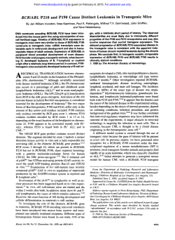
From www.bloodjournal.org by guest on February 6, 2015. For personal use only.
CORRESPONDENCE
365
REFERENCES
1. Bernardi F, Faioni EM, Castoldi E, Lunghi B, Castaman G, Sacchi
E, Mannucci PM: A factor V genetic component differing from Factor V
R506Q contributes to the activated protein C resistance phenotype.
Blood 90:1552, 1997
2. Lunghi B, Iacoviello L, Gemmati D, di Iasio MG, Castoldi E,
Pinotti M, Castaman G, Redaelli R, Mariani G, Marchetti G, Bernardi F:
Detection of new polymorphic markers in the factor V gene: Association with factor V levels in plasma. Thromb Haemost 75:45, 1996
3. Jenny RJ, Pittman DD, Toole JJ, Kriz RW, Aldape RA, Hewick
RM, Kaufman RJ, Mann KG: Complete cDNA and derived amino
acid sequence of human factor V. Proc Natl Acad Sci USA 84:4846,
1987
4. Tishkoff SA, Dietzsch E, Speed W, Pakstis AJ, Kidd JR, Cheung
K, Bonne´-Tamir B, Santachiara-Benericetti AS, Moral P, Krings M,
Paabo S, Watson E, Risch N, Jenkins T, Kidd KK: Global patterns of
linkage disequilibrium at the CD4 locus and modern human origins.
Science 271:1380, 1996
Differentiating Juvenile Myelomonocytic Leukemia From Infectious Disease
To The Editor:
fied herpesviruses, might contribute to understanding pathogenesis as
well as to diagnosis and management.
Two recent articles in BLOOD1,2 review the findings and outcome of
juvenile myelomonocytic leukemia (JMML). Both omit an important
aspect of JMLL: its differentiation from infectious disease. Several
disseminated microbial infections of infancy can result in persistent
fever, failure to thrive, hepatosplenomegaly, skin lesions, anemia,
thrombocytopenia, and myelomonocytosis, including Epstein-Barr virus (EBV), cytomegalovirus (CMV), human herpes virus-6 (HHV-6),
histoplasma, mycobacteria, and toxoplasma. Thorough investigation for
infection is needed in infants with these findings to avoid erroneous
diagnosis and mistaken interventions.
Herrod et al3 reported two infants with persistent EBV infection and
findings consistent with JMML, including increased numbers of F and i
cells and abnormal granulocyte-macrophage colony formation in vitro.
Both recovered without treatment and remained well. This raises the
possibility that some of the long-term survivors reported by Niemeyer et
al and Arico et al had similar infections rather than leukemia. Neonatal
CMV and HHV-6 infections can also mimic JMML.4,5 Might erroneous
diagnosis account for the better prognosis reported for patients with
JMML who are less than 6 months old?2
The excellent reviews of Niemeyer et al and Arico et al suggest that
JMML represents a group of diseases rather than a single entity. Careful
investigation for microbial associations, including more recently identi-
Donald Pinkel
Driscoll Children’s Hospital
Corpus Christi, TX
REFERENCES
1. Niemeyer CM, Arico M, Basso G, Biondi A, Cantu´ Rajnoldi A,
Creutzig U, Haas O, Harbott J, Hasle H, Kerndrup G, Locatelli F, Mann
G, Stollmann-Gibbels B, Van’t Veer-Korthof, van Wering E, Zimmerman M, and members of the European Working Group on Myelodysplastic Syndromes in Childhood: Chronic myelomonocytic leukemia in
childhood: A retrospective analysis of 110 cases. Blood 89:3534, 1997
2. Arico M, Biondi A, Pui C: Juvenile myelomonocytic leukemia.
Blood 90:479, 1997
3. Herrod H, Dow L, Sullivan J: Persistent Epstein-Barr virus
infection mimicking juvenile chronic myelogenous leukemia: Immunologic and hematologic studies. Blood 61:1098, 1983
4. Kirby M, Weitzman S, Freedman M: Juvenile chronic myelogenous leukemia: Differentiation from infantile cytomegalovirus infection. Am J Pediatr Hematol Oncol 12:292, 1990
5. Lorenzana A, Lyons H, Sawaf H, Higgins M, Carrigan D,
Emmanuel P: Human herpes virus-6 (HHV-6) infection in an infant
mimicking juvenile chronic myelogenous leukemia (JCML). J Pediatr
Hematol Oncol 19:370, 1997
Response
We are grateful to Dr Pinkel for his carefully considered remarks. We
fully agree that some infectious disease can mimic JMML, thus
jeopardizing the interpretation of some cases. In particular, Dr Pinkel
raises the question of whether some of the long-term survivors we
described, in fact, have JMML. Although such suspicion is obviously
warranted, our experience indicates that the vast majority of cases that
fit the diagnostic picture of JMML represent leukemia and not an
infectious process. Even when the patients show rapid recovery with or
without ‘‘minimal treatment,’’ reactivation of the disease may occur. In
some cases these recurrences have an accelerated phase, mirroring that
in patients with rapidly fatal JMML. We cannot rule out infectious
diseases as the origin of some of the more unusual cases of JMML that
have been reported in the medical literature, but we do not believe these
exceptions account for more than 10% of the total number. Perhaps current
advances in the diagnosis will help to resolve this intriguing issue.
Maurizio Arico`
IRCCS Policlinico S. Matteo
Pavia, Italy
Andrea Biondi
Ospedale S. Gerardo
Monza, Italy
Ching-Hon Pui
St Jude Children’s Research Hospital
Memphis, TN
Response
We have recently published the results of a retrospective analysis of
110 children with chronic myelomonocytic leukemia (CMML).1 There
has since been an international consensus to rename the disease juvenile
myelomonocytic leukemia (JMML). The new term JMML will include
all leukemias of childhood previously classed CMML,1,2 juvenile
chronic myelogenous leukemia (jCML),3,4 or infantile monosomy 7
From www.bloodjournal.org by guest on February 6, 2015. For personal use only.
366
CORRESPONDENCE
Table 1. Criteria for the Diagnosis of JMML
Category
Suggestive clinical features
Minimal laboratory criteria
(all 3 have to be fulfilled)
Item
1.
2.
3.
4.
5.
1.
2.
Criteria requested for
definite diagnosis
(at least 2)
3.
1.
2.
3.
4.
5.
Hepatosplenomegaly (97%)
Lymphadenopathy (76%)
Pallor (64%)
Fever (54%)
Skin rash (36%)
No Ph chromosome, no bcr-abl
rearrangement
Peripheral blood monocyte count
Ͼ1 ϫ 109/L
Bone marrow blasts Ͻ20%
Hemoglobin F increased for age
Myeloid precursors on peripheral
blood smear
White blood count Ͼ10 ϫ 109/L
Clonal abnormality (including
monosomy 7)
GM-CSF hypersensitivity of
myeloid progenitors in vitro
Numbers in parenthesis refer to the percentage of patients with the
particular clinical feature.1
Abbreviation: Ph, Philadelphia.
syndrome,3 because their clinical and biological similarities suggest that
they are spectrums of the same disease. We believe that the broad
agreement on nomenclature will facilitate cooperative treatment trials
and hasten research on the pathogenesis of JMML.
As addressed by Dr Pinkel, the clinical and morphological picture of
JMML can be mimicked by a variety of infectious organisms. In
addition, granulocyte-macrophage colony-stimulating factor hypersensitivity of myeloid progenitor cells, thought to play a central role in the
pathogenesis of JMML,5 has been noted in vitro in children with viral
infections.6 A basic tenet for the definition of myeloid leukemias is the
demonstration of the clonal origin from a malignant hematopoietic
progenitor cell.7 The clonal nature is often inferred by evidence of a
chromosomal abnormality or an activating mutation of a protooncogene. In this respect about half of the children with JMML have
evidence of a clonal disorder; 35% are known to have a chromosomal
abnormality1 and 15% to have a point mutation of the Nras or Kras
oncogene in their hematopoietic cells.8 More recently, the study of
X-chromosome inactivation patterns showed evidence for monoclonal
origin of mononuclear cells in all female JMML patients analyzed.9 In
the absence of a marker of clonality, the establishment of the diagnosis
JMML and firm exclusion of an infectious origin can be difficult.
To address this issue in our retrospective study1 we collected data on the
serology for cytomegalovirus (CMV; n ϭ 56), herpes virus type I (HSV;
n ϭ 27), and Epstein-Barr virus (EBV; n ϭ 51) from the time of diagnosis.
Thirty-eight percent of children were positive for CMV, 44% for HSV, and
47% for EBV. The prevalences of antibodies to these viruses were similar to
those observed in normal infant populations in Western Europe.10-13 There
were no significant differences in age at diagnosis or length of survival
between JMML patients with or without previous or recent CMV, HSV, or
EBV infection. Dr Pinkel raises the concern that some of our long-term
survivors might have had infections rather than leukemia. Of the seven
patients with a survival of more than 5 years without bone marrow
transplantation, four have succumbed to their disease (Table 7 in Niemeyer et
al1). Of the three remaining patients, one girl currently alive with disease 6.5
years after diagnosis is known to have an Nras mutation (A. Biondi, personal
communication). Another patient had monosomy 7 in his bone marrow cells
documented twice within 6 months after diagnosis, whereas a normal
karyotype was found 4 and 9 years later. He had no evidence of disease when
seen last 9.6 years after diagnosis.14 The patient with the longest survival,
currently 13 years after diagnosis, had no marker of clonality. His smears and
clinical data were thoroughly reviewed, but it cannot be excluded that he
suffered from an infection rather than from leukemia. Viral studies from
the time of the diagnosis were not available.
We agree with Dr Pinkel that a careful investigation for an infectious
cause is mandatory in all children suspected as suffering from JMML.
Until the chromosomal and molecular abnormalities of the majority of
JMML patients with so-called ‘‘normal karyotype’’ have been unraveled, the diagnosis of JMML will have to be based on a number of clinical
and laboratory features (Table 1). The suggestive clinical features, the
minimal laboratory criteria, and the criteria requested for definite diagnosis
may prove to be a guideline in establishing the diagnosis of JMML.
For the European Working Group of MDS in Childhood (EWOG-MDS):
C.M. Niemeyer, MD
Universita¨ts-Kinderklinik
Freiburg, Germany
S. Fenu, MD
Cattedra di Ematologia
Universita´ degli Studi di Roma
Roma, Italy
H. Hasle, MD
Aarhus Kommunehospital
Aarhus, Denmark
G. Mann, MD
St Anna Kinderspital
Vienna, Austria
J. Stary, MD
2nd Medical Faculty, Charles University
2nd Clinic of Pediatrics
Pragues, Czech Republic
E. van Wering, MD
Dutch Childhood Leukemia Study Group
The Hague, The Netherlands
REFERENCES
1. Niemeyer CM, Arico´ M, Basso G, Biondi A, Cantu´ Rajnoldi A,
Creutzig U, Haas O, Harbott J, Hasle H, Kerndrup G, Locatelli F, Mann
G, Stollmann-Gibbels B, van’t Veer-Korthof, van Wering E, Zimmermann M, and members of the European Working Group on Myelodysplastic Syndromes in Childhood: Chronic myelomonocytic leukemia in
childhood: A retrospective analysis of 110 cases. Blood 89:3534, 1997
2. Castro-Malaspina H, Schaison G, Passe S, Pasquier M, Berger R,
Bayle-Weisgerber C, Miller D, Seligmann M, Bernard J: Subacute and
chronic myelomonocytic leukemia in children (juvenile CML). Cancer
54:675, 1984
3. Freeman MH, Estrov Z, Chan HSL: Juvenile chronic myelogenous leukemia. Am J Pediatr Hematol Oncol 10:261, 1988
4. Passmore SJ, Hann IM, Stiller CA, Ramani P, Swansbury GJ,
Gibbons B, Reeves BR, Chessels JM: Pediatric myelodysplasia: A study of
68 children and a new prognostic scoring system. Blood 85:1742, 1995
5. Emanuel PD, Bates LJ, Castleberry RP, Gualtieri RJ, Zuckerman
KS: Selective hypersensitivity to granulocyte-macrophage colonystimulating factor by juvenile chronic myeloid leukemia hematopoietic
progenitors. Blood 77:925, 1991
6. Lorenzana A, Lyons H, Sawaf H, Higgins M, Carrigan D,
Emmanuel P: Human herpes virus-6 (HHV-6) infection in an infant
mimicking juvenile chronic myelogenous leukemia (JCML). J Pediatr
Hematol Oncol 19:370, 1997
7. Fialkow PJ, Gartler SM, Yoshida A: Clonal origin of chronic
myelocytic leukemia in man. Proc Natl Acad Sci USA 58:1468, 1967
8. Kalra R, Paderanga DC, Olson K, Shannon KM: Genetic analysis
is consistent with the hypothesis that NF 1 limits myeloid cell growth
through p21ras. Blood 84:3435, 1994
9. Busque L, Gilliland DG, Prchal JT, Sieff CA, Weinstein HJ, Sokol
JM, Belickova M, Wayne AS, Zuckerman KS, Sokol L, Castleberry RP,
Emanuel PD: Clonality in juvenile chronic myelogeous leukemia.
Blood 85:21, 1995
From www.bloodjournal.org by guest on February 6, 2015. For personal use only.
CORRESPONDENCE
367
10. Leinikki P, Granstro¨m M-L, Santavuori P, Pettay O: Epidemiology of cytomegalovirus infections during pregnancy and infancy. Scand
J Infect Dis 10:165, 1987
11. Ahlfors K, Ivarsson S-A, Johnsson T, Svensson I: Congenital and
acquired cytomegalovirus infections. Acta Paediatr Scand 67:321, 1978
12. Stuart-Harris C: The epidemiology and clinical presentation of
herpes virus infections. J Antimicrob Chemother 12:1, 1983
13. Lamy ME, Favart AM, Cornue C, Mendez M, Segas M,
Bortonboy G: Study of Epstein-Barr virus (EBV) antibodies. Acta Clin
Belg 37:281, 1982
14. Stollmann B, Fonatsch C, Havers W: Persistent Ebstein-Barr
virus infection associated with monosomy 7 or chromosome 3 abnormality in childhood myeloproliferative disorders. Br J Haematol 60:183,
1985
Hereditary Hyperferritinemia-Cataract Syndrome: Two Novel Mutations
in the L-Ferritin Iron-Responsive Element
To the Editor:
Cazzola et al1 recently reported two kindreds with hereditary
hyperferritinemia cataract syndrome (HHCS) associated with novel
point mutations within a regulatory stem-loop motif in the L-ferritin
mRNA termed the iron-responsive element (IRE). Affected individuals
showed a characteristic clinical phenotype of elevated serum ferritin
concentration and cataract developing early in life. The proposed
pathogenesis of this disorder is that nucleotide substitutions within the
IRE disrupt its specific interaction with the cytoplasmic iron regulatory
protein (IRP). Failure of optimal IRP-IRE binding in turn leads to
failure of suppression of L-ferritin translation.
There are now increasing numbers of reports that describe the
genotype-phenotype relationship in kindreds with naturally occurring IRE
mutations, and as Cazzola et al1 report, the phenotype varies with the position
of the mutation in the IRE. These descriptions now provide clinical data that
support the structural model of the IRE-IRP interaction deduced from in
vitro binding studies using artificially created IRE mutants.2-4
We have identified two further kindreds with HHCS and novel
mutations in the L-ferritin IRE that further support this model.
Kindred I. The 51-year-old male proband of English origin developed visual symptoms in his mid-thirties from cataracts, but was
otherwise asymptomatic. Investigations revealed a serum ferritin of
1,389 µg/L but normal transferrin saturation. Similar abnormalities were
noted in the proband’s sister, and liver biopsy specimens from both
these individuals showed no iron overload. Sequencing of genomic
DNA from the proband showed a heterozygous point mutation that
corresponded to a ϩ39 C = U substitution in the L-ferritin mRNA.
Kindred 2. The 42-year-old female proband of English origin was
investigated for anemia detected at one of her regular blood transfusion
sessions. Although her red cell indices and transferrin saturation were
consistent with mild iron deficiency, her serum ferritin was elevated at 1,020
µg/L. The proband herself had had previous surgical extraction of cataracts,
and there were premature cataracts in 8 other family members. The son of the
proband required cataract extraction at 5 years old. Hyperferritinemia was
confirmed only in family members with cataract. Analysis of genomic DNA
also showed a heterozygous point mutation, this time corresponding to a ϩ36
C = A substitution in the L-ferritin mRNA. This substitution created an Mse
I restriction site within the amplified sequence, and restriction digests from
additional family members confirmed that the substitution segregated with
the hyperferritinemia-cataract phenotype.
The nucleotide substitutions detected in kindreds 1 and 2 lie in the
apical loop and upper stem of the IRE, respectively (Fig 1). We note that
in both kindreds individuals display a severe phenotype, and this is
consistent with the observations of Cazzola et al that mutations near the
apex of the IRE result in higher serum ferritin concentrations and denser
cataracts. These results also comply with data from in vitro binding
studies; nucleotide substitutions in the apical loop of the IRE dramatically reduce IRP affinity, consistent with its putative role as the IRP
binding site.2,3 Individuals from kindred 1 with a naturally occurring
mutation at this site are therefore expected to have a severe defect in
L-ferritin regulation. In the case of kindred 2, artificially created
Fig 1. Schematic representation of the L-ferritin IRE adapted from
Cazzola et al showing the updated distribution of genotypic abnormalities in HHCS. Substitutions ؉39 C = U in kindred 1 and ؉36 C = A lie
within the apical loop and upper stem, respectively. (Adapted and
reprinted with permission.1)
nucleotide substitutions in the IRE upper stem exert a profound effect
on IRP binding in vitro, but only if complementary base pairing in the
stem is disrupted.4 Pairing of nucleotides may facilitate IRE-IRP
binding by maintaining an optimum secondary structure of the IRE. The
From www.bloodjournal.org by guest on February 6, 2015. For personal use only.
1998 91: 365-367
Differentiating Juvenile Myelomonocytic Leukemia From Infectious Disease
Donald Pinkel
Updated information and services can be found at:
http://www.bloodjournal.org/content/91/1/365.full.html
Articles on similar topics can be found in the following Blood collections
Information about reproducing this article in parts or in its entirety may be found online at:
http://www.bloodjournal.org/site/misc/rights.xhtml#repub_requests
Information about ordering reprints may be found online at:
http://www.bloodjournal.org/site/misc/rights.xhtml#reprints
Information about subscriptions and ASH membership may be found online at:
http://www.bloodjournal.org/site/subscriptions/index.xhtml
Blood (print ISSN 0006-4971, online ISSN 1528-0020), is published weekly by the American Society of
Hematology, 2021 L St, NW, Suite 900, Washington DC 20036.
Copyright 2011 by The American Society of Hematology; all rights reserved.


© Copyright 2026