Es
docs
Explorar
Iniciar sesión
Crear una nueva cuenta
Download
Report
Healthcare
Provisional PDF - BioMed Central
A Conjugate Vaccine Attenuates Morphine
MAXWELL ACADEMY PVT. LTD. - Professional Training in Chennai
1-s2.0-S1569905614603699-main
Download [ PDF ] - journal of evolution of medical and dental sciences
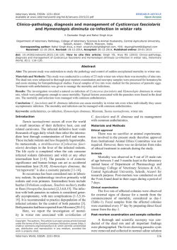
Clinico-pathology, diagnosis and management of
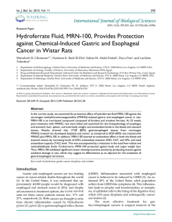
Hydroferrate Fluid, MRN-100, Provides Protection against Chemical
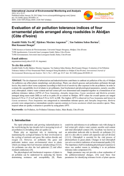
Evaluation of air pollution tolerance indices of four ornamental
arquivo de notícias - UNESP: Câmpus de Botucatu
2015 February Newsletter - Sun City Home Owners Association
Comparison with a Natural Reward
© Copyright 2026
Acerca de EsDocs
DMCA / GDPR
Alertar
![Download [ PDF ] - journal of evolution of medical and dental sciences](http://s2.esdocs.com/store/data/000458860_1-b5abb1dbf29b0a0496894dba1a110341-250x500.png)