
arquivo de notícias - UNESP: Câmpus de Botucatu
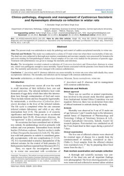
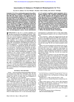
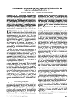
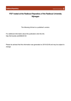
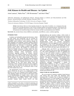
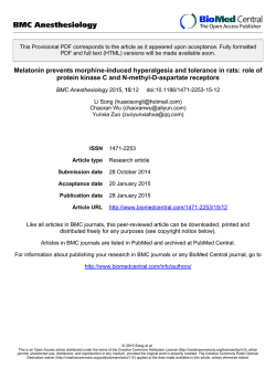
Toxicology and Applied Pharmacology 194 (2004) 132 – 140
www.elsevier.com/locate/ytaap
Chemically induced immunotoxicity in a medium-term multiorgan
bioassay for carcinogenesis with Wistar rats
Ana Lu´cia Tozzi Spinardi-Barbisan, a Ramon Kaneno, b Luı´s Fernando Barbisan, c
Joa˜o Lauro Viana de Camargo, a and Maria Aparecida Marchesan Rodrigues a,*
b
a
Departamento de Patologia, TOXICAN, Faculdade de Medicina, UNESP, 18618-000 Botucatu, SP, Brazil
Departamento de Microbiologia e Imunologia, Instituto de Biocieˆncias, UNESP, 18618-000 Botucatu, SP, Brazil
c
Departamento de Morfologia, Instituto de Biocieˆncias, UNESP, 18618-000 Botucatu, SP, Brazil
Received 27 May 2003; accepted 19 September 2003
Abstract
A variety of chemicals can adversely affect the immune system and influence tumor development. The modifying potential of chemical
carcinogens on the lymphoid organs and cytokine production of rats submitted to a medium-term initiation-promotion bioassay for
carcinogenesis was investigated. Male Wistar rats were sequentially initiated with N-nitrosodiethylamine (DEN), N-methyl-N-nitrosourea
(MNU), N-butyl-N-(4hydroxybutyl)nitrosamine (BBN), dihydroxy-di-n-propylnitrosamine (DHPN), and 1,2-dimethylhydrazine (DMH)
during 4 weeks. Two initiated groups received phenobarbital (PB) or 2-acetylaminofluorene (2-AAF) for 25 weeks and two noninitiated
groups received only PB or 2-AAF. A nontreated group was used as control. Lymphohematopoietic organs, liver, kidneys, lung, intestines,
and Zymbal’s gland were removed for histological analysis. Interleukin (IL)-2, IL-12, interferon gamma (IFN-g), tumor necrosis factor alpha
(TNF-a), IL-10, and transforming growth factor beta1 (TGF-h1) levels were determined by ELISA in spleen cell culture supernatants. At the
fourth week, exposure to the initiating carcinogens resulted in cell depletion of the thymus, spleen and bone marrow, and impairment of IL-2,
IL-12, and IFN-g production. However, at the 30th week, no important alterations were observed both in lymphoid organs and cytokine
production in the different groups. The results indicate that the initiating carcinogens used in the present protocol exert toxic effects on the
lymphoid organs and affect the production of cytokines at the initiation step of carcinogenesis. This early and reversible depression of the
immune surveillance may contribute to the survival of initiated cells facilitating the development of future neoplasia.
D 2003 Elsevier Inc. All rights reserved.
Keywords: Initiation-promotion model; Chemical carcinogens; Cytokines; Immunotoxicity; Lymphoid organs
Introduction
A variety of environmental chemical contaminants may
trigger deleterious effects on the immune system increasing the susceptibility to infections, autoimmune diseases,
and cancer development. Among them, chemical carcinogens like polychlorinated biphenyls, chlorinated dibenzo-pdioxins, pesticides, and heavy metals have shown immunotoxic activity (Descotes et al., 2000; De Wall et al.,
1992; Kimber and Dearman, 2002; Luster and Rosenthal,
1993; Luster et al., 1990; Vandebriel et al., 1998a; Voccia
et al., 1999).
* Corresponding author. Fax: +55-14-3815-2348.
E-mail address: [email protected] (M.A.M. Rodrigues).
0041-008X/$ - see front matter D 2003 Elsevier Inc. All rights reserved.
doi:10.1016/j.taap.2003.09.012
Experimental studies have demonstrated that chemicals
such as cyclosporin A, 2,3,7,8,-tetrachlorodibenzo-p-dioxin
(TCDD), tributyltin oxide (TBTO), and azathioprine, among
other substances, modulate both the cytokine-receptor
mRNA expression and cytokine protein levels (Vandebriel
et al., 1998a, 1998b). Besides, alterations of cytokine levels
have been reported in several types of cancer (Merendino et
al., 1999; Merogi et al., 1997; O’Hara et al., 1998). Since
chemical carcinogens influence both the immune system
and cancer development, the evaluation of cytokine production may be a valuable tool to investigate the participation of
the immune system on tumor development (Cohen et al.,
1999; Foster, 2001; Vandebriel et al., 1998b).
Cytokines are important in promoting the proliferation
and differentiation of hematopoietic cells and in regulating
A.L.T. Spinardi-Barbisan et al. / Toxicology and Applied Pharmacology 194 (2004) 132–140
the nature of immune response. Interleukin-12 (IL-12) has
the ability to skew the immune response in favor of Th1,
that is, induce the differentiation of naive T-helper cells
(Th0) to the Th1-profile inhibiting Th2 cell development.
Because Th1 cells produce interferon gamma (IFN-g), IL2, IL-3, and tumor necrosis factor alpha and beta (TNF-a
and TNF-h), they are more suited for the enhancement of
cellular immunity, although Th2 cells (which produce IL-3,
IL-4, IL-5, IL-6, IL-9, IL-10, IL-13, and TNF-a) are better
qualified in helping B cells to differentiate into antibodyproducing cells (Mossman and Sad, 1996). Cytokines such
as IL-2, IL-12, and IFN-g may activate natural killer (NK)
cells and cytotoxic T cells enhancing the host immune
response against malignant cells (Ikeda et al., 2002;
Oppenheim and Fujiwara, 1996; Trinchieri, 1995). Tumor
necrosis factor (TNF-a) is a major mediator of inflammation with action directed towards both tissue destruction
and recovery from damage (Balkwill, 2002). Besides,
TNF-a may act as an endogenous tumor promoter, contributing to the tissue remodeling and stromal development
necessary for tumor growth and spread (Balkwill, 2002;
Schreiber and Rowley, 1999; Seung et al., 1999). Other
cytokines, such as transforming growth factor beta (TGFh) and IL-10, may act as immune suppressive factors and
promote the growth of some tumors (O’Hara et al., 1998;
Seung et al., 1999).
A medium-term multiorgan system, based on the initiation-promotion concept of chemical carcinogenesis, has
been proposed as an alternative and complementary approach to the conventional long-term bioassay for detection
of chemical carcinogens (Ito et al., 1992, 1998; Takahashi
et al., 1992). It consists of a multiorgan initiation of
carcinogenesis with five genotoxic carcinogens followed
by the exposure of the animals to a test substance to
evaluate its promoting potential (Ito et al., 1992, 1998;
133
Takahashi et al., 1992). A task group gathered at the
International Agency for Research on Cancer (IARC) in
1999 recognized that this bioassay could be considered
appropriate for identifying carcinogens in rodents (IARC,
1999). This alternative model allows to investigate the
immune alterations that can occur at the initiation step of
the carcinogenic process, after DNA damage induced by the
initiating chemicals, or at the promotion step, when proliferation of altered cell clones had given rise to preneoplastic
and neoplastic lesions (Spinardi et al., 1999; SpinardiBarbisan et al., 2000).
The aim of the present study was to investigate the
influence of the chemical carcinogens used in a mediumterm multiorgan bioassay in male Wistar rats on lymphoid
organs and cytokine production at different stages of chemical carcinogenesis.
Material and methods
Animals. Male Wistar rats, 4-week-old, were purchased
from CEMIB—Centro Multidisciplinar de Investigacßa˜o
Biolo´gica (UNICAMP, Campinas, SP, Brazil) and housed
in polypropylene cages (four per cage) in a environmentcontrolled room maintained at 22 F 2 jC, 55 F 10%
relative humidity and a 12:12 h light/dark cycle. Animals
were supplied with filtered water and fed NUVILAB-CR1
(NUVITAL, Curitiba, PR, Brazil) ad libitum. After a 4-week
acclimation period, animals weighing 225– 250 g were used
in this study.
Chemical agents. N-nitrosodiethylamine (DEN), N-methylN-nitrosourea (MNU), 1,2-dimethylhydrazine (DMH), and
2-acetylaminofluorene (2-AAF) were purchased from Sigma Chemical Co. (St. Louis, MO, USA); N-butyl-N-
Fig. 1. Experimental design (for details, see Materials and methods).
134
A.L.T. Spinardi-Barbisan et al. / Toxicology and Applied Pharmacology 194 (2004) 132–140
Table 1
Body weight gain and organ relative weights (mean F SD) of animals submitted to a medium-term multiorgan carcinogenesis model (4th and 30th weeks)
Groups
Number of
animals
Body weight gain (g)
4th week
Control
DMBDDb
20
20
30th week
Control
PBc
2-AAFd
DMBDD
DMBDD/PB
DMBDD/2-AAF
15
19
20
20
20
17
Relative weight (%)
Liver
Kidneya
Spleen
Thymus
106.60 F 11.29
3.10 F 28.99*
4.05 F 0.25
3.47 F 0.43*
0.31 F 0.02
0.38 F 0.06*
0.38 F 0.03
0.57 F 0.11*
0.16 F 0.02
0.09 F 0.03*
234.93
245.63
166.60
176.40
193.05
137.35
3.15
3.74
5.10
3.11
3.79
6.29
0.24
0.25
0.28
0.28
0.28
0.33
0.31
0.30
0.41
0.38
0.40
0.44
0.04
0.07
0.05
0.05
0.05
0.06
F
F
F
F
F
F
52.38
43.83
26.43*
35.29*
46.29*,***
45.96*
F
F
F
F
F
F
0.33
0.35
0.62*
0.23
0.41**
0.90*,**
F
F
F
F
F
F
0.02
0.03
0.03*
0.05*
0.03*,***
0.04*,**,****
F
F
F
F
F
F
0.06
0.06
0.06*
0.07
0.15***
0.10*
F
F
F
F
F
F
0.01
0.06
0.01
0.02
0.01
0.06
a
Mean values of right and left kidneys.
Group initiated with the DEN, BBN, MNU, DHPN, and DMH.
c
Phenobarbital.
d
2-acetylaminofluorene.
* Significantly different from control group; P < 0.0001 for all comparisons.
** Significantly different from DMBDD group; P < 0.0001 for all comparisons.
*** Significantly different from PB group; P < 0.0001 for all comparisons.
**** Significantly different from 2-AAF group; P < 0.0001 for all comparisons.
b
(4hydroxybutyl)nitrosamine (BBN) and phenobarbital (PB)
were purchased from Tokyo Kasei Industries Co. (Tokyo,
Japan) and dihydroxy-di-n-propylnitrosamine (DHPN) from
Nakalai Tesque, Inc. (Kyoto, Japan).
Experimental design. The experimental protocol is shown in
Fig. 1. Animals were allocated to six groups (n = 15 – 20 rats
each) as follows. A an untreated group was used as control,
maintained on basal diet and sacrificed at the weeks 4 and
30. Three other groups (DMBDD, DMBDD/PB, and
DMBDD/2-AAF) were sequentially treated with five chemical initiators: DEN (100 mg/kg body wt, ip, single dose at
the commencement), MNU (20 mg/kg body wt, ip, four
times, two doses per week), and BBN (0.05% in drinking
water during 2 weeks) administered during weeks 1 and 2;
DHPN (0.1% in drinking water during 2 weeks) and DMH
(40 mg/kg body wt, sc, four times, two doses per week)
administered during weeks 3 and 4. Some animals of the
DMBDD group were killed at the end of the week 4 and the
remaining were maintained on basal diet until the week 30.
After the initiation, the DMBDD/PB and DMBDD/2-AAF
groups were supplied with PB (0.05%) or 2-AAF (0.01%) in
the diet for 25 weeks, respectively. Two noninitiated groups
(PB and 2-AAF) received PB or 2-AAF in the diet, from the
6th until the 30th week.
Body weight, water and food consumption were registered weekly during the first 4 weeks (initiation step) and
then at every 15 days until the end of the experiment. All
animals were killed under pentobarbital (45 mg/kg body wt)
anesthesia at the 4th or 30th week. The protocols used were
in accordance the Ethical Principles for Animal Research
adopted by the Brazilian College of Animal Experimentation (COBEA) and were approved by the local Ethical
Committee for Animal Research (protocol no. 116).
Tissue processing and histological analysis. Liver, kidneys,
spleen, thymus, mesenteric lymph nodes, and bone marrow
from all animals were fixed in buffered formalin for 48 h.
The lung, small and large intestine, and Zymbal’s gland
were examined only at the week 30. Liver, kidneys, spleen,
and thymus were weighed immediately after removal. The
spleen was cut in two halves, one for evaluation of cytokines and other for histological analysis. All organs removed were embedded in paraffin and stained with
hematoxylin and eosin (H&E) for histological analysis.
Histological diagnosis was performed according to Monographs on Pathology of Tumors in Laboratory Animals
(IARC, 1990) and Kuper et al. (2000).
Table 2
Histological changes in lymphohematopoietic organs at the fourth week
Organ changesa
Control group
DMBDD groupb
Bone marrow
Decreased global cellularity
Thymus
Decreased cortex/medulla ratio
Decreased cortex cellularity
Spleen
Decreased PALS cellularityd
Decreased marginal zone cellularity
Increased red pulp cellularity
Extramedullar hematopoiesis
Mesenteric lymph nodes
Increased paracortex cellularity
Erythrocyte rosette formation
Histiocytosis
(20)c
0
(20)
0
0
(20)
0
0
0
0
(20)
0
0
0
(20)
20 (100%)
(20)
5 (25%)
20 (100%)
(20)
12 (60%)
12 (60%)
4 (20%)
5 (25%)
(18)
7 (39%)
4 (22%)
12 (67%)
a
Semiquantitative diagnostic terms according to Kuper et al. (2000).
Group initiated with the carcinogens DEN, BBN, MNU, DHPN, and
DMH.
c
Number of analyzed organs.
d
Periarteriolar lymphocyte sheath.
b
A.L.T. Spinardi-Barbisan et al. / Toxicology and Applied Pharmacology 194 (2004) 132–140
135
Fig. 2. Lymphohematopoietic organs of male Wistar rats stained with H&E at the fourth week. Thymus: (a) normal; c, cortex and m, medulla (25); (b)
thinning of the thymic cortex (25); and (c) increased number of macrophages ingesting apoptotic cells in the cortex (starry sky appearance) in a DMBDDtreated rat (90); spleen: (d) normal; f, follicule; mz, marginal zone; pals, periarteriolar lymphocyte sheath; rp, red pulp (30); (e) diminished cellularity and
reduction of white pulp in a DMBDD-treated rat (30); bone marrow: (f) normal (90); (g) cell depletion in a DMBDD-treated rat (90); lymph nodes: (h)
erythrocyte rosette (90) and (i) histiocytosis after DMBDD treatment (120).
136
A.L.T. Spinardi-Barbisan et al. / Toxicology and Applied Pharmacology 194 (2004) 132–140
Spleen cells culture. Spleen cells were mechanically dispersed in a Petri dish containing RPMI-1640 culture
medium (Cultilab, Campinas, SP, Brazil). The cell suspensions were centrifuged, resuspended in RPMI-1640 culture
medium supplemented with 20 mg/ml gentamycin (Sigma
Co.), 2 mM glutamine (GibcoBRL, Gaithersburg, MD,
USA) and 10% inactivated fetal calf serum (Cultilab,
Campinas, SP, Brazil), and washed twice at 1500 rpm,
for 10 min. Cell viability, assessed by Trypan blue
exclusion, was always greater than 90%. Aliquots of 2
106 cells/ml (500 Al/well) were incubated with RPMI1640 (500 Al/well) or stimulated in vitro with Concanavalin A (CON A—2.5 Ag/ml, 500 Al/well) (Sigma Co.) or
Staphylococcus aureus Cowan’s strain 1 (SAC—1:5000,
500 Al/well) (Calbiochem, San Diego, CA, USA) in 24well flat-bottom microtiter plates for 72 h in a humidified
atmosphere of 5% CO2, at 37 jC. Following incubation,
supernatants were collected and stocked at 70 jC for
quantification of cytokines. Samples stimulated in vitro
with CON A were used to quantify IL-2, IFN-g, IL-10,
and TGF-h1, and samples stimulated in vitro with SAC
were used to quantify TNF-a and IL-12.
Analysis of cytokine levels. Cytokine production was determined by enzyme-linked immunosorbent assay (ELISA)
using commercial kits according to the manufacturer’s
instructions. IL-2, IL-12, TNF-a, IFN-g, and IL-10 levels
were measured using ELISA kits from BioSource International (Nivelles, Belgium). For detection of the TGF-h1
immunoreactive form, samples were acidified by addition
of 1 N HCl and then measured by Quantikine anti-human
TGF-h1 kit (R&D Systems, Minneapolis, USA). The plates
were read at 450 nm using an ELISA microplate reader set
(Multiskan Ex, Labsystems, Finland). Cytokine quantities in
the samples were calculated from standard curves of recombinant cytokines using a regression linear method.
1). At the fourth week, the relative liver and thymus weights
were significantly decreased ( P < 0.0001) when compared
with the control group. In contrast, the relative kidneys and
spleen weights were significantly increased ( P < 0.0001)
(Table 1). At the 30th week, the relative liver and spleen
weights were significantly increased ( P < 0.001) after
treatment with 2-AAF (DMBDD/2-AAF and 2-AAF groups
vs. control group) and the DMBDD/PB group presented
higher ( P < 0.0001) mean spleen relative weight than the
PB group (Table 1). At this point, the kidney relative
weights were significantly higher ( P < 0.0001) in all
treated groups, except in the PB group (Table 1).
Histological analysis
The main histological changes on lymphoid organs and
in bone marrow were observed after the initiation step of
carcinogenesis at the end of the fourth week (Table 2). At
this moment, the exposure to the chemical initiators resulted
in significant thinning of the thymic cortex due to increased
apoptosis of lymphocytes, easily visible by the increased
number of macrophages ingesting apoptotic cells (Figs. 2a–
c). As a consequence, the cortex/medulla ratio was decreased and the connective tissue of the thymus became
prominent. In the spleen, there was cell depletion of the
white pulp, specially of the periarteriolar lymphocyte sheath
(PALS) and of the marginal zone (Figs. 2d and e). The bone
marrow showed severe hypoplasia (Figs. 2f and g) and the
mesenteric lymph nodes presented erythrocyte rosettes formation (Fig. 2h), histiocytosis (Fig. 2i), and increased
Table 3
Incidence of animals with neoplastic lesions and total number of tumors
(30th week)
Groups
Statistical analysis. Body weight gain, organ relative
weights, and production of cytokines were analyzed by
the Student’s t test or Mann – Whitney test at week 4
(initiation step) and by analysis of variance (ANOVA) or
Kruskal – Wallis test at week 30 (promotion step). The
incidence of benign or malignant neoplasias and number
of neoplasias per group (tumor burden) were analyzed by
the Fisher’s exact test or the Chi square and Kruskal – Wallis
tests, respectively, at the 30th week. Differences between
groups were assumed to be significant when P < 0.05.
Results
Noninitiated
Control
0 (15)a
b
0 (19)
PB
2-AAFc
0 (20)
Initiated
DMBDDd 14 (20)
DMBDD/ 18 (20)
PB
DMBDD/ 17 (17)
2-AAF
a
The body weight gain was significantly reduced ( P <
0.0001) after the treatment with the carcinogens both at the
initiation and promotion step, except in the PB group (Table
Benign
neoplasia
No. of
animals
0
0
0
Malignant
neoplasia
No. of No. of
tumors animals
0
0
0
0
0
0
Benign
and
malignant
No. of
tumors neoplasias
0
0
0
0
0
0
21
32
40
60
10 (50%) 19
15 (75%) 28
11 (55%)
12 (60%)
14 (82%) 32
16 (94%)y,* 49y,*
81y,**
Effective number of animals.
Phenobarbital.
c
2-acetylaminofluorene.
d
Group initiated with the carcinogens DEN, BBN, MNU, DHPN, and
DMH.
y
Significantly different from DMBDD group.
* P < 0.01.
** P < 0.002.
b
Body and organ weights
No. of
animals
with
neoplasia
A.L.T. Spinardi-Barbisan et al. / Toxicology and Applied Pharmacology 194 (2004) 132–140
cellularity at the paracortex area when compared to the
control group. The animals submitted to the DMBDD
treatment also presented liver alterations such as bile duct
hyperplasia, Kupffer cells with hemossiderin pigment, centrilobular hepatocytic necrosis, extramedullary hematopoiesis, cariomegaly, vacuolization of hepatocytes, cytoplasmic
basophilia, hepatocytic hypertrophy, mitotic figures and
apoptotic cells. In the kidneys, there was focal or diffuse
tubular hyperplasia in most animals of the DMBDD group
(data not shown).
At the 30th week, no significant alterations of the
lymphoid organs were observed in the different groups.
137
Neoplastic lesions were found mainly in the initiated and
promoted groups (DMBDD/PB and DMBDD/2-AAF
groups) (Table 3). They were observed in the liver as
adenoma, cholangioma and cholangiocarcinoma, in the
colon as adenocarcinoma, in the kidney as mesenchymal
renal tumor, in the lung as adenoma, and in the Zymbal’s
gland as carcinoma. The treatment with 2-AAF after initiation (DMBDD/2-AAF group) induced significantly higher
incidence of animals with malignant neoplasia ( P < 0.01),
number of malignant tumors ( P < 0.01), and total number of
tumors (benign + malignant) ( P < 0.002) than in the
DMBDD group (Table 3).
Cytokine production
At the end of the fourth week, the production of IL-2, IL12, and IFN-g, but not of TNF-a, TGF-h1, and IL-10 by
spleen cells cultured with CON A or SAC were significantly
lower ( P < 0.01) in the DMBDD group than in the control
group (Fig. 3). At the end of the 30th week, IFN-g
production was significantly higher ( P < 0.001) in the
groups treated with 2-AAF (DMBDD/2-AAF and 2-AAF
groups vs. DMBDD group) but was not different from the
control group (data not shown). IL-2, IL-12, TNF-a, IL-10,
and TGF-h1 levels were not altered in the carcinogentreated groups at the 30th week (data not shown).
Discussion
Fig. 3. Production of IL-2, IL-12, and IFN-g by spleen cells of male Wistar
rats in vitro stimulated with CON A or SAC after DMBDD treatment at the
fourth week. *,**Significant differences among the groups at P < 0.001 and
P < 0.01, respectively. Values are mean F SD.
The present results indicate that chemicals used for
initiation of carcinogenesis in an alternative multiorgan
model affect the lymphoid organs of Wistar rats, causing
cellular depletion in the bone marrow, in the thymic cortex,
and in the white pulp of the spleen, at the end of the fourth
week. Cytokine levels were also altered at the initiation step
after the treatment with the five genotoxic carcinogens.
Besides, the liver and the kidneys of the initiated animals
presented important histological changes.
The alterations observed in the thymus may result from
the toxicity of the initiating carcinogens. Similar effects were
noted in the thymus after treatment with organotin compounds and 2,3,7,8-tetrachlorodibenzo-p-dioxin (TCDD)
(Kamath et al., 1997; Penninks et al., 1991). Toxic substances can cross blood –thymus barrier and directly affect
the lymphocytes or indirectly act on epithelial reticule cells
that support the thymocytes growth and maturation (De
Wall et al., 1992). Others investigators have reported thymic
changes very similar to those registered in the present study
in rats exposed to MNU (Koestner et al., 1977; Talcott et al.,
1990; Zwiiling et al., 1978). In fact, it is known that MNU
has the lymphohematopoietic system as a target of its
toxicity (Franchi et al., 2003; Zwiiling et al., 1978) and
may act acutely on the proliferating cortical thymocytes
leading them to apoptosis. Furthermore, the dramatic
changes observed in the thymus could be a consequence
138
A.L.T. Spinardi-Barbisan et al. / Toxicology and Applied Pharmacology 194 (2004) 132–140
of the stress induced by the cocktail of chemical initiators as
expressed by the marked decrease in body weight gain and
food consumption. It is well known that the nutritional
status and stress can result in thymus atrophy (Schuurman
et al., 1994). In this context, we could argue that the effects
on the thymus were in part due to direct effects of the
chemicals, but the stress also have played an important role.
This implies that the histological changes observed in the
thymus in the present study should not be automatically
considered as a first insult, but the end result of both the
systemic stress and toxic effects of the chemicals.
At the end of the fourth week, the bone marrow of
initiated animals was also severely affected by exposure to
the genotoxic carcinogens as characterized by depletion of
myeloid, erythroid, and lymphoid cell lineages. In this
manner, the alterations in the spleen white pulp and extramedullary hematopoiesis observed in the liver and spleen
could be a consequence of cellular depletion observed in the
bone marrow and in the thymus. However, hypercellularity
at the paracortex area and histiocytosis of mesenteric lymph
nodes could be due to antigenic stimulation induced by
possible lesions on digestive tract and peritoneal cavity by
the oral and intraperitoneal administrations of chemicals
during the initiation treatment. The histological changes
observed in the liver and kidneys immediately after initiation
may be consequence of the toxicity of the chemical initiators
that are metabolized in the liver, originating hepatotoxic and
nephrotoxic metabolites (Kedderis, 1996; Schnellmann,
2001). Similar results in the liver and kidney were previously
reported by our laboratory in Wistar rats treated by DHPN or
DEN (Barbisan et al., 2002; Moreira et al., 2000).
At the 30th week, after the promotion step of carcinogenesis, the absence of significant alterations on the lymphoid organs indicates that the immunotoxic changes
induced by the initiating chemicals were restored. Similar
recovery was described after glucocorticosteroid and organotin compound treatments, in which the tissue architecture
was restored within 3 –4 weeks (Schuurman and Kuper,
1995). The absence of neoplasia in lymphoid organs agree
with Franchi et al. (2003), who demonstrated that after 20
weeks, male Wistar rats exposed to MNU at 80, 160, and
240 mg/kg body wt, ip, only develop few vascular lesions in
the hematopoietic system at intermediate and high doses,
and thymic lymphomas at the highest dose.
In contrast to initiated groups, no neoplasias were observed in the noninitiated groups at the end of the 30th
week. These results show the amplitude of the initiating
treatment used in this protocol, favoring DNA damage
fixation in different target organs (IARC, 1999). Indeed,
our results indicate that both the treatment with PB, a nongenotoxic carcinogen that acts promoting the development
of liver preneoplastic lesions after previous exposure to
DEN (Kitano et al., 1998; Maekawa et al., 1992) or with
2-AAF, a genotoxic carcinogen used in the protocols of
hepatocarcinogenesis as a selective stimulator of initiated
cells in the liver (Tiwawech et al., 1991), exerted an
enhancing influence on the carcinogenic process. In the
initiated groups and treated with PB or with 2-AAF, the liver
was a main target organ, developing adenomas (35%) and
cholangiomas (41%) or cholangiocarcinomas (35%), respectively (data not shown). In contrast, animals exposed only to
PB or to 2-AAF did not present neoplasia, although developed rare or many putative preneoplastic liver foci, respectively (data not shown). These results documented the wellknown specific action of these chemicals on the liver (target
organ) (Kitano et al., 1998; Maekawa et al., 1992, Tiwawech et al., 1991).
The reduction of cytokine levels detected after initiation
of carcinogenesis at the fourth week correlates well with the
histological changes observed in the bone marrow and
thymus. Exposure to the five chemical initiators decreased
the levels of IL-2, IL-12 and IFN-g, but not of IL-10 and
TGF-h1. The reduction of IL-12 production could also be
related to the lowered IL-2 and IFN-g levels that are
produced by Th1 cells. Indeed, IFN-g has a powerful
enhancing effect on the ability of phagocytes and dendritic
cells to produce IL-12, acting therefore as a potent positive
feedback mechanism (Ikeda et al., 2002). In a previous
study, we have demonstrated that the chemical initiators
used in the present initiation-promotion protocol did not
change the ratio CD4+/CD8+ cells as determined by flow
cytometry and that the NK activity was not altered by
chromium release assay (Spinardi et al., 1999; SpinardiBarbisan et al., 2000). In this context, the results of the
present study suggest that the chemical initiators can affect
the cytokine production not due to a change in the number
or type of the spleen cells but as a result of reduced function
of these cells. Besides, our results show differential sensitivity of the cells to the initiators, the Th1-pattern cytokines
appearing to be more affected. These data suggest that the
reduction of cytokine levels observed at the initiation step
may reflect a selective toxicity of the chemical initiators on
Th1 lymphocytes, which activate important mechanisms of
the antitumor immune response, involving macrophages,
dendritic cells, cytotoxic T lymphocytes, and NK cells
(Pellegrini et al., 1996).
Various studies have indicated the importance of cytokines in tumor immunology (Oppenheim and Fujiwara,
1996; Seung et al., 1999). O’Hara et al. (1998) demonstrated
that patients with colorectal cancer have decreased IL-12
production and increased serum IL-10, and that these
patients present increased tumor bulk and worse prognosis.
Experimental models of transplantable tumors have shown
that IL-12 drastically inhibits tumor growth and metastasis
increasing the survival of the animals (Brunda et al., 1993).
The ability of the IL-12 to prevent 3-methylcholantrene
(MCA)-induced carcinogenesis, protecting for the growth of
MCA-induced fibrosarcoma, has been clearly shown in
BALB/c and IL-12 p40 knockout mice respectively treated
with exogenous and endogenous IL-12 (Noguchi et al.,
1996; Smyth et al., 2001). High IFN-g serum levels have
been reported in mice after IL-12 systemic treatment con-
A.L.T. Spinardi-Barbisan et al. / Toxicology and Applied Pharmacology 194 (2004) 132–140
tributing to tumor regression (Nastala et al., 1994). Considering that the Th1 cells are able to produce IL-2 and IFN-g,
our data indicate that the deficient production of these
cytokines induced by the genotoxic carcinogens treatments
can inhibit the development of the cellular immune response, the most important mechanism of the antitumor
immune response.
The present results raise the possibility that the immunologic alterations expressed by the dramatic changes in the
lymphoid organs and the decrease in cytokine production at
the early stage of chemical carcinogenesis could favor the
survival of initiated cells and facilitate the development of
future neoplasia.
Acknowledgments
This research was supported by the Fundacßa˜o de Amparo
a Pesquisa do Estado de Sa˜o Paulo (FAPESP 99/01524-4;
00/3736-8).
References
Balkwill, F., 2002. Tumor necrosis factor or tumor promoting factor? Cytokine Growth Factor Rev. 13, 135 – 141.
Barbisan, L.F., Miyamoto, M., Scolastici, C., Salvadori, D.M.F., Ribeiro,
L.R., Eira, A.F., de Camargo, J.L.V., 2002. Influence of aqueous extract
of Agaricus blazei on rat liver toxicity induced by different doses of
diethylnitrosamine. J. Ethnopharmacol. 83, 25 – 32.
Brunda, M.J., Luistro, L., Warrier, R.R., Wright, R.B., Huddard, B.R.,
Murphy, M., Wolf, S.F., Gately, M.K., 1993. Antitumor and antimetastatic activity interleukin 12 against murine tumors. J. Exp. Med. 178,
1223 – 1230.
Cohen, M.D., Schook, L.B., Oppenheim, J.J., Freed, B.M., Rodgers, K.E.,
1999. Symposium overview: alteration in cytokine receptors by xenobiotics. Toxicol. Sci. 48, 163 – 169.
Descotes, J., Choquet-Kastylevsky, G., Van Ganse, E., Vial, T., 2000. Responses of the immune system to injury. Toxicol. Pathol. 28, 479 – 481.
De Wall, E.J., Schuurman, H.-J., Loeber, J.G., Van Loveren, H., Vos, J.G.,
1992. Alterations in the cortical thymic epithelium of rats after in vivo
exposure to 2,3,7,8-tetrachlorodibenzo-p-dioxin (TCDD): an (immuno)histological study. Toxicol. Appl. Pharmacol. 115, 80 – 88.
Foster, J.R., 2001. The functions of cytokines and their uses in toxicology.
Int. J. Exp. Pathol. 82, 171 – 192.
Franchi, C.A.S., Bacchi, M.M., Padovani, C.R., de Camargo, J.L.V., 2003.
Thymic lymphomas in Wistar rats exposed to N-methyl-N-nitrosourea
(MNU). Cancer Sci. 94, 240 – 243.
IARC, 1990. Tumors of the rat. In: Turusov, V.S., Mohr, U. (Eds.), Pathology of Tumours in Laboratory Animals. International Agency for Research on Cancer, Lyon, France, pp. 1 – 390.
IARC, 1999. The use of short-term and medium-term tests for carcinogens
and data on genetic effects in carcinogenic hazard evaluation. In:
McGregor, D.B., Rice, J.M., Venitt, S. (Eds.), International Agency
for Research on Cancer, Lyon, France, pp. 1 – 536.
Ikeda, H., Old, L.J., Schreiber, R.D., 2002. The roles of IFN-g in protection
against tumor development and cancer immunoediting. Cytokine
Growth Factor Rev. 13, 95 – 109.
Ito, N., Shirai, T., Hasegawa, R., 1992. Medium-term bioassays for carcinogens. In: Vainio, H., Magee, P.N., McGregor, D.B., McMichael, A.J.
(Eds.), Mechanisms of Carcinogenesis in Risk Identification. International Agency for Research on Cancer, Lyon, France, pp. 356 – 388.
139
Ito, N., Imaida, K., Hirose, M., Shirai, T., 1998. Medium-term bioassays for
carcinogenicity of chemical mixtures. Environ. Health Perspect. 106,
1331 – 1336.
Kamath, A.B., Xu, H., Nagarkatti, P.S., Nagarkatti, M., 1997. Evidence for
the induction of apoptosis in thymocytes by 2,3,7,8-tetrachlorodibenzop-dioxin in vivo. Toxicol. Appl. Pharmacol. 142, 367 – 377.
Kedderis, G.L., 1996. Biochemical basis of hepatocellular injury. Toxicol.
Pathol. 24, 77 – 83.
Kimber, I., Dearman, R.J., 2002. Immune response: adverse versus nonadverse effects. Toxicol. Pathol. 30, 54 – 58.
Kitano, M., Ichihara, T., Matsuda, T., Wanibuchi, H., Tamano, S., Hagiwara,
A., Imaoka, S., Funae, Y., Shirai, T., Fukushima, S., 1998. Presence of a
threshold for promoting effects of phenobarbital on diethylnitrosamineinduced hepatic foci in the rat. Carcinogenesis 19, 1475 – 1480.
Koestner, A.W., Ruecker, F.A., Koestner, A., 1977. Morphology and pathogenesis of tumors of the thymus and stomach in Sprague – Dawley rats
following intragastric administration of methyl nitrosourea (MNU). Int.
J. Cancer 20, 418 – 426.
Kuper, C.F., Harlerman, J.H., Richter-Reichelm, H.B., Vos, J.G., 2000.
Histopathologic approaches to detect changes indicative of immunotoxicity. Toxicol. Pathol. 28, 454 – 466.
Luster, M.I., Rosenthal, G.J., 1993. Chemical agents and the immune response. Environ. Health Perspect. 100, 219 – 236.
Luster, M.I., Germolec, D.R., Rosenthal, G.J., 1990. Immunotoxicology:
review of current status. Ann. Allergy 64, 427 – 432.
Maekawa, A., Onodera, H., Ogasawara, H., Matsushima, Y., Mitsumori,
K., Hayashi, Y., 1992. Threshold dose dependence in phenobarbital
promotion of rat hepatocarcinogenesis initiated by diethylnitrosamine.
Carcinogenesis 13, 501 – 503.
Merendino, R.A., Gangemi, S., Misefari, A., Arena, A., Capozza, A.B.,
Chillemi, S., D’Ambrosio, F.P., 1999. Interleukin-12 and interleukin 10
production by mononuclear phagocytic cells from breast cancer patients. Immunol. Lett. 68, 355 – 358.
Merogi, A.J., Marrogi, A.J., Ramesh, R., Robinson, W.R., Fermin, C.D.,
Freeman, S.M., 1997. Tumor-host interaction: analysis of cytokines,
growth factors, and tumor-infiltrating lymphocytes in ovarian carcinomas. Hum. Pathol. 28, 321 – 331.
Moreira, E.L.T., de Camargo, J.L.V., Rodrigues, M.A.M., Barbisan, L.F.,
Salvadori, D.M.F., 2000. Dose- and sex-related carcinogenesis by Nbis(2-hydroxypropyl)nitrosamine in Wistar rats. Jpn. J. Cancer Res. 91,
368 – 374.
Mossman, T.R., Sad, S., 1996. The expanding universe of T-cell subsets:
Th1, Th2 and more. Immunol. Today 17, 138 – 146.
Nastala, C.L., Edington, H.D., McKinney, T.G., Tahara, H., Nalesnik,
M.A., Brunda, M.J., Gately, M.K., Wolf, S.F., Schreiber, R.D., Storkus,
W.J., Lotze, M.T., 1994. Recombinant IL-12 administration induces
tumor regression in association with IFN-g production. J. Immunol.
153, 1697 – 1706.
Noguchi, Y., Jungbluth, A., Richards, E.C., Old, L.J., 1996. Effect of
interleukin 12 on tumor induction by 3-methylcholanthrene. Proc. Natl.
Acad. Sci. U.S.A. 93, 1798 – 1801.
O’Hara, R.J., Greenman, J., Macdonald, A.W., Gaskell, K.M., Topping,
K.P., Duthie, G.S., Kerin, M.J., Lee, P.W.R., Monson, J.R.T., 1998.
Advanced colorectal cancer is associated with impaired interleukin 12
and interleukin 10 production. Clin. Cancer Res. 4, 1943 – 1948.
Oppenheim, J., Fujiwara, H., 1996. The role of cytokines in cancer. Cytokine Growth Factor Rev. 7, 279 – 288.
Pellegrini, P., Berghella, A.M., Del Beato, Y., Cicia, S., Adorno, D., Casciani, C.C.U., 1996. Disregulation in Th1 and Th2 subsets of CD4+ T
cells in peripheral blood of colorectal cancer patients and involvement
in cancer establishment and progression. Cancer Immunol. Immunother.
42, 1 – 8.
Penninks, A.H., Pieters, R.H.H., Snoeij, N.J., Seinem, W., 1991. Organotin-induced thymus atrophy. Thymus Update 4, 57 – 59.
Schnellmann, R.G., 2001. Toxic responses of the kidney. In: Klaassen,
C.D. (Ed.), Casarett and Doull’s Toxicology: The Basic Science of
Poisons. McGraw-Hill, USA, pp. 491 – 514.
140
A.L.T. Spinardi-Barbisan et al. / Toxicology and Applied Pharmacology 194 (2004) 132–140
Schreiber, H., Rowley, D.A., 1999. Inflammation and cancer. In: Gallin,
J.I., Syndeman, R. (Eds.), Inflammation: Basic Principles and Clinical Correlates. Lippincott Williams & Wilkins, Philadelphia, pp.
1117 – 1129.
Schuurman, H.-J., Kuper, C.F., 1995. Pathology of the thymus: changes
induced by xenobiotics and gene targeting. APMIS 103, 481 – 500.
Schuurman, H.-J., Kuper, C.F., Vos, J.G., 1994. Histopathology of the
immune system as a tool to assess immunotoxicity. Toxicology 86,
187 – 212.
Seung, L.P., Rowley, D.A., Schreiber, H., 1999. Cytokines in cancer. In:
Theze, J (Ed.), The Cytokine Network and Immune Functions. Oxford
Univ. Press, Oxford, pp. 335 – 345.
Smyth, M.J., Crowe, N.Y., Godfrey, D.I., 2001. NK cells and NKT cells
collaborate in host protection from methylcholanthrene-induced fibrosarcoma. Int. Immunol. 13, 459 – 463.
Spinardi, A.L.T., Kaneno, R., Rodrigues, M.A.M., Salvadori, D.M.F., Rocha, N., Barbisan, L.F., Ribeiro, L.R., de Camargo, J.L.V., 1999.
Natural killer activity in a medium-term multi-organ bioassay for carcinogenesis. Jpn. J. Cancer Res. 90, 101 – 107.
Spinardi-Barbisan, A.L.T., Kaneno, R., Rodrigues, M.A.M., Salvadori,
D.M.F., Moreira, E.L.T., Barbisan, L.F., de Camargo, J.L.V., 2000.
Lymphoproliferative response and T lymphocyte subsets in a medium-term multi-organ bioassay for carcinogenesis in Wistar rats. Cancer Lett. 154, 121 – 129.
Takahashi, S., Hasegawa, R., Masui, T., Misoguchi, M., Fukushima, S., Ito,
N., 1992. Establishment of multi-organ carcinogenesis bioassay using
rats treated with a combination of five different carcinogens. J. Toxicol.
Pathol. 5, 151 – 156.
Talcott, P.A., Exon, J.H., Koller, L.D., 1990. The effects of methylnitrosourea (MNU) on natural killer (NK) cell cytotoxicity and cytokine
production in rats. Carcinogenesis 11, 829 – 834.
Tiwawech, D., Hasegawa, R., Kurata, Y., Tatematsu, M., Shibata, M.-A.,
Thamavit, W., Ito, N., 1991. Dose-dependent effects of 2-acetylaminofluorene on hepatic foci development and cell proliferation in rats.
Carcinogenesis 12, 985 – 990.
Trinchieri, G., 1995. Interleukin-12: a proinflamatory cytokine with immunoregulatory functions that bridge innate resistance and antigen-specific
adaptive immunity. Annu. Rev. Immunol. 13, 251 – 276.
Vandebriel, R.J., Meredith, C., Scott, M.P., Roholl, P.J.M., van Loveren,
H., 1998a. Effects of in vivo exposure to bis(tri-n-butyltin)oxide, hexachlorobenzene, and benzo(a)pyrene on cytokine (receptor) mRNA levels in cultured rat splenocytes and on IL-2 receptor protein levels.
Toxicol. Appl. Pharmacol. 148, 126 – 136.
Vandebriel, R.J., Van Loveren, H., Meredith, C., 1998b. Altered cytokine
(receptor) mRNA expression as a tool in immunotoxicology. Toxicology 130, 43 – 67.
Voccia, I., Blakley, B., Brousseau, P., Fournier, M., 1999. Immunotoxicity
of pesticides: a review. Toxicol. Ind. Health 15, 119 – 132.
Zwiiling, B.S., Filppi, J.A., Chorpenning, F.W., Koestner, A., Rheins,
M.S., 1978. Chemical carcinogenesis and immunity: immunologic
status of rats treated with methylnitrosourea. J. Natl. Cancer Inst.
61, 731 – 735.
© Copyright 2026