
Expression of RHD and RHCE Gene Products Using Retroviral
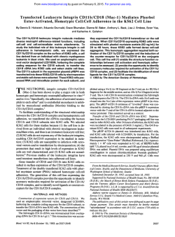
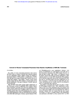
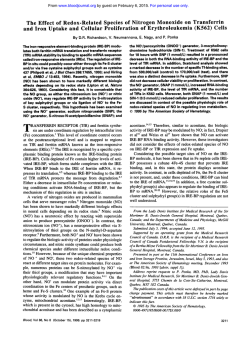
From www.bloodjournal.org by guest on February 6, 2015. For personal use only.
Expression of RHD and RHCE Gene Products Using Retroviral Transduction
of K562 Cells Establishes the Molecular Basis of Rh Blood Group Antigens
By J.S. Smythe, N.D. Avent, P.A. Judson, S F . Parsons, P.G. Martin, and D.J. Anstee
Retroviral-mediated gene transfer using cDNA transcripts of
the RHD and RHCE genes resulted in the isolation of K562
clones expressing D and G or c and E antigens, respectively.
These results represent the first direct demonstration that
the RHD gene encodes the D and G antigens and the RHCE
gene encodes the c and E antigens. Both c and E antigens
were expressed after transduction of K562 cells with a single
cDNA, indicating that thec antigen does not arise by alternative splicing (exon skipping) of the product of the RHCE gene,
as has been suggested.
0 1996 by The American Society of Hematology.
T
merase chain reaction (RT-PCR)
of total RNA purified from reticulocytes" of Rh phenotype CDe. RT-PCR was performed in a PerkinElmer DNA thermal cycler (Perkin-Elmer, Norwalk, using
CT) avian
myeloblastosisvirusreversetranscriptase,oligodTzc,(Pharmacia,
Uppsala, Sweden) and Taq polymerase (Perkin-Elmer) essentially
as described." The primers were based on the cE cDNA 5' and 3'
noncoding nucleotides -32 to -5 (exon 1 sense) and 1,300 to 1,327
(exon10anti-sense),respectively.Transformation
of Escherichia
coli XL-l Blue competent cells (Invitrogen) with pCRII and subsequentvectorDNApreparation(Qiagen,Dorking,UK)wasdone
according to the manufacturers' instructions. A colony of cells containing only DcDNAwasselectedfollowingthesequencing
of
several vectorhnsert DNA preparations. The D cDNA sequence differed from that d e ~ c r i b e din
' ~ that it contained a C,,, to G (IleIIqto
Met)changeinthecodingregion(previouslyreportedi5)andan
to G change in the 3' noncoding region. The Fyh cDNA( 1,062
bp) wasclonedintoBluescriptvector(Stratagene,LaJolla,CA)
following amplification of genomic DNA from an Epstein-Barr
virus
(EBV) lymphoblastoid cell line derivedfrom an individual of phenotype Fy(a-b+) using primers based on the 5' and 3' Fyh noncoding
regions as described."
The D and cE cDNAs were subcloned separately into the pBabe
puro retroviral vector (kindly provided by Dr H. Land, ICRF, Lonsite." TheFyhcDNAwas
don, UK) using theEcoRIrestriction
subcloned into pBabe puro using the
BamHI and Sal I restriction
sites. The pBabe puro vector
is based on the Moloney murine leukaemia virus (MoMuLV). Expression of inserted genes is driven by the
MoMuLV long terminal repeat while the puromycin resistance gene
is expressed from the SV40 early promoter 3' of the cloning site.
Vector DNA preparation was as described above. CorrectRh cDNA
orientation in pBabe was established by BamHI and Kpn I restriction
and DNA sequence analysis.
Trunsfection of the packaging cell line and retroviral supernatant
GP + env
production. Amphotropicretroviralpackagingcells,
AM12,IX from Genetix Pharmaceuticals (Rye, NY),
were cultured
in Iscove's modified Eagle's medium supplemented with 10% fetal
bovineserum(IMEMIFBS)andincubatedat
37°C in a 5% CO2
humidified incubator. Cells (S X IO') were transfected with pBabe
constructs (10 to 25 pg) using calcium phosphate precipitation essentially as described.'?
Four hours after the addition of DNA the culture
medium was replaced with 15% glycerol in phosphate-buffered saline (PBS) for 3 minutes and this in turn was replaced with IMEM/
FBS. After 2 days the medium was replaced with fresh IMEMIFBS
supplemented with puromycin (3 pg mL l ; Sigma, St Louis, MO).
Approximately 2 weeks later individual puromycin resistant colonies
were transferred to culture flasks using trypsin (Sigma) to detach
the cells. The supernatants were collected from near-confluent cells
incubated overnight at 3 3 T , filtered (0.45 pm), and stored frozen.
Retroviral transduction of K562 cells. K562 cells were obtained
fromtheEuropeanCollection
of AnimalCellCultures(Porton
Down, Salisbury, Wiltshire,UK). Cells (IO') were incubated at37°C
in IMEMFBS supplemented with viral supernatant (1.0% vol/vol)
andpolybrene (8 pg mL.~'; Sigma). After 4 hoursthecells were
centrifuged at 400g for S minutes and resuspended in IMEWFBS.
HE RH SYSTEM is the most complex of the 23 blood
group systems found on human red blood cells.' Antibodies to Rh system antigens (especially anti-D) are of clinical significance because they may cause hemolytic disease
of the newborn or transfusionreactions.' In recentyears,
there have been considerable advances in our understanding
of the biochemistry and geneticsof Rh antigens.'-5 Available
evidence suggests that the Rh antigens result from at least
two highly homologous genes (RHD and RHCE) located at
chromosome 1 p34-p36. The RHD gene is deleted in most
white individuals who lack the D antigen.h TheRHCE gene
gives rise to the
allelic antigens C/c and E/e. It has been
proposed that C/c and E/e are located on different polypeptides which arise by alternative splicing of the primary transcript of the RHCE
Formal proof that the RHD and RHCE genes encode for
the antigens of the Rh system and that alternative splicing
of the RHCE gene product gives rise to separate
polypeptides
with C/c or E/e antigens, respectively, has been lacking because previous attempts to expressthese genes in eukaryotic
cells have been unsu~cessful.~~'"
In this report we describe the use
of retroviral gene transfer
to generate stable clonesof K562 cells expressing theD and
G, or c and E blood group antigens. The results provide the
first direct evidence that the putative RHD gene gives rise
to D and G antigens and that the putative RHCE gene gives
rise to c and E antigens. However,the results refutethe
hypothesis that the c and E antigensarise by alternative
splicing of the product of the RHCE gene.
MATERIALS AND METHODS
Cloning of Rh D and cE cDNAs into the pBabe puro retroviral
vector. The Rh cE cDNA (1,463 bp)wasclonedintoBluescript
vector as previously described." It contained 41 nucleotides of 5'
and 171 nucleotides of 3' noncoding sequence and terminated with
an A,, tract. The Rh D cDNA
(1,359 bp) was cloned into pCRII
vector (Invitrogen, San Diego, CA) after reverse transcriptase-poly-
From the International Blood Group Reference Laboratory, Bristol, UK.
Submitted August 22, 199.5; accepted November 7, 1995.
Address reprint requests to J.S. Smythe, MPhil, International
Blood Group Reference Laboratory, Southmead Rd, Bristol BSI0
5ND, UK.
The publicationcosts of this article were defrayed in part by page
charge payment.This article must therefore be hereby marked
"advertisement" in accordance with l 8 U.S.C. section 1734 solely to
indicate this fact.
0 1996 by The American Society of Hematology.
0006-4971/96/8707-0033$3.00/0
2968
Blood, Vol 87, No 7 (April l), 1996: pp 2968-2973
From www.bloodjournal.org by guest on February 6, 2015. For personal use only.
EXPRESSION OF RHD AND RHCE GENES
Two days later the cells were recultured in IMEM/FBS supplemented
with puromycin (3 pgd - l ) and transferred to a 96-well microplate
(0.2 mL/well). After 2 to 3 weeks puromycin-resistant stable-transduced clones from wells containing only a single discrete colony
were transferred to 24-well plates and expanded before flow cytometric analysis. The clones exhibiting the highest levels of Rh expression
were selected using BRIC 69 and those with the highest levels of
Fyb expression using CBC-512 (vide infra).
Antibodies and red blood cells. BRIC 69 is a murine monoclonal
antibody that binds to Rh polypeptides.” Murine monoclonal antiFy3, (CBC-512, IgG) and human monoclonal anti-E (H4-1 G-4,
IgG) were provided by Dr M. Uchikawa (Japanese Red Cross, Shibuya-ku, Japan). Purifiedhuman
monoclonal anti-D, BRAD 5
(IgGI”) was used at 50 pg/mL (diluted in PBS/l% bovine serum
albumin [BSA], 0.1% NaN, [PBS-A]). Human monoclonal anti-C
(MS252, IgG3), anti-c (MS47, IgG3), anti-e (MS70, IgG3), and
Anti-G (MSI, IgG3) were provided by Dr K. Thompson (IGRI,
Oslo, Norway). Red blood cells of known Rh phenotype were available from the National Blood Service, Bristol.
Flow cytometric analysis. K562 clones transduced with D cDNA
(K562/D) or cE cDNA (K562/cE) were tested for antigen expression
by flow cytometry (FACStar Plus; Becton Dickinson, Mountain
View, CA). Mean fluorescence intensity (FLI) was used asa measure
of antibody binding. The specificity of the antibodies used was confirmed using red blood cells of the appropriate phenotype (data not
shown). A K562 clone transduced with cDNA corresponding to the
Fyb blood group gene (K562/Fyb)and untransduced K562 cells were
usedas controls. K562 cells (2 X IO5) or redblood cells (0.5%
suspension) in PBS-A (50 pL) were incubated with antibody (50
pL) for 1 hour at 37°C. Cells were washed once inPBS-Aand
incubated with (Fab’), fragments of rabbit-antihuman IgG or rabbitantimouse IgG affinity-purified fluorescein isothiocyanate (F1TC)labeled (1/20, 50 pL, DAKO, Glostrup, Denmark) for 1 hour at
room temperature. The sample volume was adjusted to 300 pL with
PBS-A before analysis.
mRNA preparation and RT/PCR amplification of pBabe specijc
sequences from K562 cells. Oligo d(T,z.z8)magnetic beads were
used to prepare mRNA according to the manufacturers instructions
(Dynal, Oslo, Norway). Synthesis of cDNA was from 1 pg of mRNA
as described above using oligo d(Tlz.la)(Pharmacia). Approximately
100 ng of cDNA was used as the template for PCR (94°C 1 minute,
60°C l minute. 72°C 2.5 minutes, 35 cycles) with 50 pmol of each
primer. Two pairs of primers were used. One set (PI and P2) corresponded to pBabe sequences 5’ and 3‘ of the cloning site (P1 sense
5’-CCC TTT ATC CAG CCC TCA CTC CT-3’ and P2 anti-sense
5’-CCC TAA CTG ACA CAC A’M CCA CAG-3’), respectively.
The second set consisted of a primer (P3) which corresponded to
pBabe sequence 5’ of the insert site (P3 sense 5’-GCC TCG ATC
CTC CCT TTA TCC-3‘) and the antisense primer to cE cDNA 3’
noncoding region (exon 10 antisense, described above).
Sequencing of cDNA. Amplified Rh-specific PCR products were
sequenced using dye-labeled terminator chemistry on a 373A Applied Biosystems automated sequencer (Warrington, UK). Set 1 and
Set 2 primers and primers complementary to sequences common to
both the cE and D coding regions were used for sequencing.
Rh typing of genomic DNA. Genomic DNA was prepared using
the proteinase Wsodium dodecyl sulfate (SDS)/EDTA method essentially as de~cribed.’~
Rh D-specific PCR was performed using
primers specific for intron 4 and exon 10 of the RHD
Allele-specific PCR for c and E alleles was performed as described.”
RESULTS
Flow cytometric analysisof K562 clones transduced with
putative D a n d CE genes. K562fDandK562/cE clones,
2969
prepared as described in Materials and Methods, were tested
for Rh antigen expression byflow cytometric analysis. A
W62/Fyb clone and untransduced K562 cells were used as
controls. The selected clones were examined using human
monoclonal antibodies to Rh system antigens (D, C, E, C, e,
and G). The results for a K562fD clone are shown in Fig 1A
and Table 1. Anti-D and anti-G bound much morestrongly to
this clone than to the K562/Fyb clone with increases in mean
fluorescence intensity (FLI) from 3.3 to 15.2 and 4.5 to 11.0
for anti-D and anti-G, respectively. Antibodies to C, E, C,
and e antigens did not detect comparable changes in antigen
expression, although anti-c and anti-e did show very minor
increases in binding to K562/D cells (Table 1).
The results obtained for a K562/cE clone are shown in
Fig 1B. Anti-c and anti-E boundmuchmore
strongly to
K562/cE cells than to K562/Fybcells with increases in FLI
from 4.7 to 24.4 (anti-c) and from 2.4 to 38.8 (anti-E), respectively (Table 1). Antibodies to D, G, C, and e antigens
did not detect comparable changes in antigen expression,
although anti-D, anti-G, and anti-e didshowveryminor
increases in binding to K562kE cells.
The FLI ofRh antibody binding to untransduced K562
cells and KS62/Fyb cells was compared with that of tissue
culture medium (TCM, Table 1). Antibodies to C, E, and e
antigens gave almost identical FLI values to TCM with both
untransduced K562 cells and K562/Fyb cells. Antibodies to
D, G, and c antigens gave slightly higher FLI values than
TCM with both cell preparations (Table 1).
In a separate experiment, murine monoclonal anti-Fy3
gave almost identical F L I values to those obtained with TCM
with K562/D, K562/cE, and untransduced K562 cells (range
2.2 to 2.5) while K562/Fyb gave an FLI value of 24.2 (data
not shown).
Demonstration of D and cE rnRNAs in the K542# clone
and
the
K562kEclone,respectively.
The presence of
mRNAs arising from retroviral transduction in K562/D and
K562kE clones was determined by isolation of mRNA followed by RT-PCR and DNA sequence analysis. Two sets
of oligonucleotide primers were used. Primer set 1 (P1 and
P2, see Materials and Methods) wasspecific for vector
(pBabe puro) sequences flanking the multiple cloning site.
Using these primers a product of 1S 5 kb was amplified from
K562/D cDNA and a product of 1.15 kb from the K562/Fyb
cDNA. No products were obtained from untransduced K562
cells or from the K562kE clone (Fig 2). Primer set 2 (P3
and an Rh specific exon 10 antisense primer, see Materials
and Methods) amplified a product of 1.5 kh from the K562/
CE cDNA and from the K562/D cDNA, butno products
were obtained using cDNA from untransduced K562 cells
or the K562/Fyb clone (Fig 2).
The PCR product of 1.S5 kb obtained from K562/D cDNA
with primer set 1 was sequenced (see Materials and Methods). It contained the expected 5’ and 3’ pBabe vector flanking sequences and a D cDNA sequence (from nucleotide
-32 to 1327) identical to that of the cDNA originally subcloned into pBabe. The PCR product of 1.5 kb obtained
from K562kE cDNA with primer set 2 was sequenced (see
Materials and Methods). It contained the expected pBabe 5’
flanking sequence and a cE cDNA sequence corresponding
From www.bloodjournal.org by guest on February 6, 2015. For personal use only.
2970
SMYTHE ET AL
A
B
IDI
11
IC
10
'
L
rn
J
Io;
n
4
4
Fig 1. Flow cytometric analysis of transduced K562 cells using monoclonal Rh antibodies. (A) (-1, K562/D; i--4,
K562/Fyb. (B) (-1, K562/cE;
of cells is plotted on the ordinate and the fluorescence intensity on the abscissa. Experimental details are in
Materials and Methods.
(---l, K562/Fyb. The number
to nucleotides -41 to 1327 of that originally subcloned into
pBabe. A product of the expected sizeI6(1.15 kb) was amplified from the K562/Fyb cDNA with primer set 1 (Fig 2).
Rh typing of genomic DNA from untransduced and transduced K562 cells. Analysis of genomic DNA derived from
untransduced K562 cells, K562/D, and K562/Fyb showed
the presence of D- and c- (data not shown), but not E-specific
sequences (Fig 3). The presence of an E-antigen-specific
product (149 bp) in genomic DNA derived from the K562/
Table 1. Rh Antigen Expression on Untransduced K562 Cells and
K562 Clones Transduced With D, cE, and Fyb cDNA
~
Rh Antibody
TCM
Cells
K562
K562/Fyb
K562/D
K562/cE
2.8
2.8
2.7
2.8
Anti-D
Anti-C
Anti-G
Anti-E
Anti-c
Anti-e
3.8
3.3
15.2
4.5
2.8
2.5
2.6
2.9
4.2
4.5
11.0
5.9
2.5
2.4
2.7
38.8
5.2
4.7
6.1
24.4
2.8
2.6
3.8
3.4
Mean fluorescence intensity (FLI) was used as a measure of antibody binding. Antibody binding is compared with background fluoin tissue culture
rescence obtained when the cells were incubated
medium (TCM).
cE clone established that integration of the cE cDNA had
occurred (Fig 3).
DISCUSSION
The purpose of the present work was to investigate K562
cells as a model in vitro system for the study of RH gene
expression. Previous attempts to express Rh cDNAs by transfection of K562 cells have metwith little succe~s.~~'"
In
the present study we have used retroviral gene transfer to
transduce K562 cells with cDNAs corresponding to the putative RHCE gene (syn RhIXb=; Rh30A1'), and the putative
RHD gene (syn RhXIIIl4; Rh13I5;RhPIIz5).The levels of D,
c, E, C, e, and G antigen expression on these transduced
cells were compared with cells transduced with cDNA corresponding to Fyb glycoprotein gene16.26-z8
and with untransduced K562 cells. K562/D cells gave much higher FLI valueswith anti-D and anti-G in comparison with K562/Fyb
cells (Fig lA), K562kE cells, or untransduced K562 cells
(Table 1). In contrast, K562/cE cells gave much higher FLI
values with anti-c and anti-E than the other cells tested (Fig
1B and Table 1). The levels ofRh antigen expression on
K562/Fyb cells were virtually identical to those on untransduced cells (Table 1). The expression of D, c, and E antigens
From www.bloodjournal.org by guest on February 6, 2015. For personal use only.
2971
EXPRESSION OF RHD AND RHCE GENES
1 2
3 4 5
6 7
8
9 10 I I 1213
kb
3.0
2.0
1.6
1.o
0.5
Fig 2. Demonstration of D and c€ mRNAs in K562/D and K562/cE
cells. mRNA preparation and RT-PCR was as described in Materials
and Methods. Lanes 2 through 6, PCR performed with set 1 primers;
lanes 8 through 12, PCR performed with set 2 primers; 2,8, no template control; 3,9, cDNA from untransduced K562 cells; 4, 10, cDNA
from K562/Fybcells; 5,11, cDNA from K562/D cells; 6,12, cDNA from
K562/cE cells; 1, 7, 13, l-kb ladder (GIBCO-BRL, Paisley, UK).
that we observed is consistent with previous predictions
based onsequence analysis of putative RH genes in individuals of known Rh-phenotype.'
The G antigen is an Rh antigen that is expressed on red
blood cells carrying a D and/or C antigen.29Our experiments
involving expression of the R H D gene showed that the same
gene also gives rise to the G antigen (Fig I A). This observationis consistent with recent studies"" suggesting that G
antigen expression is dependent on the amino acid sequence
deriving from exon 2 of the R H D gene.
Previous studies have indicated that K562 cells express
low levels of mRNA corresponding to Rh polypeptides and
that Rh antigens can be detected on the cell surface.3'." Our
flow cytometric results suggest the possibility that there are
Fig 3. Rh E typing of genomic
DNA
from
untransduced and
transduced K562 cells. PCR was
performed using primer pairs
specific for E and exon 4 of the
RHCEgene as described in Materials and Methods. Lane2, no
template control; 3, gDNA from
the lymphocytes of an individual
ofRh phenotype c€; 4, gDNA
from untransduced K562 cells; 5,
gDNA from K562/Fyb cells; 6,
gDNA from K562/D cells; 7,
gDNA from K562/cEcells;
1.8.
100-bp ladder (GIBCO-BRL).
1
low levels ofRh antigen expression on the untransduced
K562 cells used in this study (compare FLI in the presence
of TCM with that in the presence of Rh antibodies, Table
l ) . In addition. the slightly increased binding of anti-c and
anti-e to K562/D cells and anti-D, anti-G, and anti-e binding
to K562kE cells (Table 1) may indicate enhanced expression
of existing Rh antigens on K562 cells after transduction with
Rh cDNAs. However, the increases in antibody binding are
small and may be artifactual. K562 cells express FcRII receptors that are known to bind oligomeric human antibodies.33Therefore, it is possible that the weak binding of some
human monoclonal antibodies to K562 cells is nonspecific,
a problem we and others have found associated with polyclonal alloimmune sera.34
Confirmation that the cDNA corresponding to R H D and
RHCE genes hadbeeninserted
into the K562 clones and
was responsible for antigen expression was obtained by amplification and sequencing of pBabe-specific cDNA followingRT-PCRwithpurifiedmRNA.
Primers specific for
pBabe amplified only the D-specific cDNA from cells transduced with D cDNA. The amplification of pBabe/cE specific
cDNA from K562/cE cells required a 5' pBabe primer and
an anti-sense primer to the cE cDNA 3' noncoding region.
This suggests the pBabe/cE RNA transcript was terminated
by the poly A tract at the 3' end of the cE cDNA and did
not include pBabe sequence 3' of the cE cDNA insert. RNA
transcripts generated from the pBabe template are usually
terminated by the pBabe Poly A signal positioned approximately 2.5 kb 3' of the cloning site.
Typing of genomic DNA from untransduced K562 cells
provided evidence for D- and c- but not E-antigen-specific
sequence (vide supra). Confirmation that the putative RHCE
gene product was inserted into the genome of the transduced
K562kE cells was obtained by the use of allele-specific
PCR whichdemonstrated the presence of E-antigen-specific
sequence (Fig 3). It was not possible to analyze genomic
DNA for C- and e-antigen-specific sequences in the presence of the R H D gene because the R H D gene contains homologous sequences. Differences in the conformation of the
proteins encoded by the R H D and RHCE genes are thought
to ensure that the C and e antigens are only expressed when
encoded by the RHCE gene.'
The Rh polypeptides encoded by the R H D and RHCE
genes are known to associate with a glycoprotein of =50 kD
2
3
4
5
6
7
8
bp
-300
Rh E specific
PCR
-200
-
exon 4 specificPCR product
I
-loo
From www.bloodjournal.org by guest on February 6, 2015. For personal use only.
2972
SMYTHE ET AL
(Rh
and evidence obtained from analysis of
the rare
erythrocytes which lack all Rh antigens has
led to suggestions that other red blood cell proteins (glycophorin B, CD47, LW glycoprotein, FY glycoprotein) are
also part of an R
' h complex.' Available evidence indicates
that untransduced K562 cells express Rh glycoprotein3*(and
unpublished observations, November 1992), CD47,37and
glycophorin B,38but not LW3' or FY,Z6and therefore K562
cells provide an attractive model for the study of Rh antigen
expression in vitro. However, previous attempts to express
Rh antigens in K562 cells using plasmid expression vectors
have been unsuccessful?," Our results indicate that the use
of retroviral delivery of the gene is of critical importance in
achieving expression of Rh antigens on K562 cells.
We considered the possibility that the process of retroviral
transduction may, of itself, activate existing RH genes. The
results with K562/Fyb clones show quite clearly that we are
not observing a phenomenon related to the retrovirus but the
genuine expression of D and cE cDNAs inserted into the
genome of K562 cells. These results provide the first direct
evidence that the proposed RHD and RHCE gene products
do indeed code for the D and cE antigens, respectively. The
results also suggest that FY is not vital for the assembly and
expression ofRh because D, G, c, and E antigens are expressed in its absence.
Our experiments involving expression of the cE cDNA
are relevant to the hypothesis that Clc and Ele antigens are
expressed on different proteins which derive from alternative
splicing of the product of the RHCE gene.7 Mouro et a
'l
proposed that the full-length mRNA product of the RHCE
gene produces a protein that expresses E or e antigen but
not C or c antigen and that spliceoforms of this mRNA,
giving rise to products that lack exon 5 (which encodes the
critical residues for E/e antigen activity), are translated into
proteins that only express C/c antigens.
The validity of this hypothesis has been questioned since
the spliceoforms have not been detected in Northern blotting
studies and it has not been established that they give rise to
proteins which are expressed in a stable form in the red blood
cell
Our results clearly show
that
alternative
splicing of hnRNA is not a prerequisite for c antigen expression and suggest that a single polypeptide is able to express
both c and E antigens.
Progress in elucidating the molecular basis of the Rh system antigens, the role of Rh-associated proteins in antigen
expression, and the assembly and transport of the 'Rh complex' to the membrane has been severely hampered by the
lack of an in vitro expression system. These results suggest
that retroviral-mediated gene transfer into K562 cells provides such an in vitro system.
Rh.,,
ACKNOWLEDGMENT
We thank H. Land for the pBabe pur0 vector, Genetix Pharmaceuticals (Rye, NY) for the Gp + env AM12 packaging cell line, K.
Thompson and M. Uchikawa for monoclonal antibodies, and John
Bridgewater and Mary Collins for helpful discussions.
REFERENCES
1. Daniels GL, Anstee DJ, Cartron J-P, Dahr W, Issitt PD: Blood
group terminology 1995. Vox Sang 69:265, 1995
2. Mollison PL, Engelfriet CP. Contreras M: Blood Transfusion
in Clinical Medicine. Oxford, UK, Blackwell Scientific, 1993
3 . Cartron J, Agre P: Rh blood group antigens: Protein and gene
structure. Semin Hematol 30:193, 1993
4. Cartron J-P: Defining the Rh blood group antigens. Blood Rev
8: 199, 1994
5. Anstee DJ, Tanner MJ: Biochemical aspects of the blood group
Rh (rhesus) antigens. Baillieres Clin Haematol 6:401, 1993
6. Colin Y, Cherif Zahar B, Le van Kim C, Raynal V, Van Huffel
V, Cartron J: Genetic basis of the RhD-positive and RhD-negative
blood group polymorphism as determined by Southern analysis.
Blood 78:2747, 1991
7. Mouro I, Colin Y, Cherif Zahar B, Cartron J-P, Le van Kim
C: Molecular genetic basis of the human Rhesus blood group system.
Nat Genet 5:62, 1993
8. Le van Kim C, Cherif Zahar B, Raynal V, Mouro I, Lopez M,
Cartron J, Colin Y: Multiple Rh messenger RNA isofoms are produced by alternative splicing. Blood 80:1074, 1992
9. Hermand P, Mouro 1, Huet M, Bloy C, Suyama K, Goldstein
J, Cartron J, Bailly P Immunochemical characterization of rhesus
proteins with antibodies raised against synthetic peptides. Blood
82:669, 1993
10. Suyama K, Roy S , Lunn R, Goldstein J: Expression of the
32Kd polypeptide of the Rh antigen. Blood 821006, l993
1 1. Avent ND, Ridgwell K, Tanner MJ, Anstee DJ: cDNA cloning of a 30 kDa erythrocyte membrane protein associated with Rh
(Rhesus)-blood-group-antigen expression. Biochem J 27 1:821, 1990
12. Temple GF, Chang JG, Kan YW: Authentic beta-globin
mRNA sequences in homozygous beta-0-thalassemia. ProcNatl
Acad Sci USA 74:3047, 1977
13. Sambrook J, Fritsch EF, Maniatis T: Molecular Cloning: A
Laboratory Manual. Cold Spring Harbor, NY, Cold Spring Harbor
Laboratory, 1989
14. Le van Kim C, Mouro I, Cherif Zahar B, Raynal V, Cherrier
C, Cartron J , Colin Y: Molecular cloning and primary structure of
the human blood group RhD polypeptide. Proc Natl Acad Sci USA
89: 10925, 1992
15. Arce M, Thompson ES, Wagner S, Coyne KE, Ferdman BA,
Lublin DM: Molecular cloning of Rh D cDNA derived from a gene
present in Rh D positive but not Rh D-negative individuals. Blood
8 2 6 5 1, 1993
16. Mallinson G, So0 KS, SchaH TJ,Pkacka M, Anstee DJ:
Mutations in the erythrocyte chemokine receptor (Duffy) gene: The
molecular basis of the Fy"iFyhantigens and identification of a deletion in the Duffy gene of an apparently healthy individual with the
Fy(a-b-) phenotype. Br J Haematol 90:823, 1995
17. Morgenstern JP, Land H: Advanced mammalian gene transfer: High titre retroviral vectors with multiple drug selection markers
and a complementary helper free packaging cell line. Nucleic Acids
Res18:3587, 1990
18. Markowitz D, Goff S, Bank A: Construction and useof a
safe and efficient amphotropic packaging cell line. Virology 167:400,
1988
19. Avent ND, Judson PA, Parsons SF, Mallinson G, Anstee
DJ, Tanner MJ, Evans PR, Hodges E, Maciver AG, Holmes C:
Monocional antibodies that recognize different membrane proteins
that are deficient in Rh,,,,human erythrocytes. One group of antibcdies reacts with a variety of cells and tissues whereas the other group
is erythroid-specific. Biochem J 251:499, 1988
20. Hadley AG, Zupanska B, Kumpel BM, Leader KA: The functional activity of Fc gamma RI1 and Fc gamma RI11on subsets of
human lymphocytes. Immunology 76:446, 1992
21. Bennett PR, Le van Kim C, Colin Y, Warwick RM, Cherif
Zahar B, Fisk NM, Cartron J-P: Prenatal determination of fetal Rh
D type by DNA amplification. N Engl J Med 329507, 1993
From www.bloodjournal.org by guest on February 6, 2015. For personal use only.
EXPRESSION OF RHD AND RHCE GENES
22. Avent ND, Martin PG, Canitt B: Improved Rh D genotyping
assay for prenatal determination of fetal Rh D status: The exon 101
intron 4 multiplex assay. Transfus Med 520, 1995 (abstr)
23.LevanKimC,
M o m I,Brossard Y, ChavinieJ,CartronJP,
Colin Y: PCR-based determination of Rhc and RhE status of fetuses at
risk of Rhc and RhE haemolytic disease. Br J Haematol 88:193, 1994
24. Cherif Zahar B, Bloy C, Le van Kim C, Blanchard D, Bailly
P, Hermand P, Salmon C, Cartron J, Colin Y: Molecular cloning
and protein structure of a human blood group Rh polypeptide. Proc
Natl Acad Sci USA 87:6243, 1990
25. Kajii E, Umenishi F, Isamoto S, Ikemoto S: Isolation of a
new cDNA clone encoding an Rh polypeptide associated with Rh
blood group system. Hum Genet 91:157, 1993
26. Chaudhuri A, Polyakova J, Zbrzezna V, Pogo AO: The coding
sequence of Duffy blood group gene in humans and simians: Restriction fragment length polymorphism, antibody and malarial parasite
specificities, and expression in nonerythroid tissues in Duffy-negative individuals. Blood 85:615, 1995
27. Iwamoto S, Omi T, Kajii E, Ikemoto S: Genomic organization
of the glycoprotein D gene: Duffy blood group Fya/Fyballoantigen
system is associated with a polymorphism at the 44-amino acid
residue. Blood 85:622, 1995
28. Tournamille C, Le vanKim C, Cane P, Cartron J-P, Colin
Y: Molecular basis and PCR-DNA typing of the Fya/Fybblood group
polymorphism. Hum Genet 95:407, 1995
29. Allen FH, Tippett P: A new Rh blood type which reveals the
Rh antigen G. Vox Sang 3:321, 1958
30. Rouillac C, Le van Kim C, Blancher A, Roubinet F, Cartron
J-P, Colin Y: Lack of G blood group antigen in DIIIb erythrocytes
is associated with segmental DNA exchange between RH genes. Br
J Haematol 89:424, 1995
2973
31. Wiener E, Shiels A, Wickramasinghe SN, Anstee DJ, Avent
ND: Ara-C treatment of K562 cells increases the expression of Rh
polypeptides. Transfus Med 4:44, 1994 (abstr)
32. Suyama K, Lunn R, Haller S, Goldstein J: Rh(D) antigen
expression and isolation of a new Rh(D) cDNA isoform in human
erythroleukemia K562 cells. Blood 84:1975, 1994
33. van de Winkel JGJ, Anderson CL: Biology of human immunoglobulin G Fc receptors. J Leukoc Biol49:511, 1991
34. Russo DCW, Lee S, Reid M E , Redman CM: Topology of
Kell blood group protein and the expression of multiple antigens by
transfected cells. Blood 84:3518, 1994
35. Moore S, Green C: The identification of specific rhesus polypeptidehlood group ABH-active glycoprotein complexes in the human red cell membrane. Biochem J 244:735, 1987
36. Ridgwell K, Spurr NK, Laguda B, MacGeoch C, Avent ND,
Tanner MJ: Isolation of cDNA clones for a 50 kDa glycoprotein of
the humanerythrocyte membrane associated with Rh (rhesus) bloodgroup antigen expression. Biochem J 287:223, 1992
37. Anstee DJ, Holmes CH, Judson PA, Tanner MJ: Use of monoclonal antibodies to determine the distribution of red cell surface
proteins on human cells and tissues, in Agre PC, Cartron J (eds):
Protein Blood Group Antigens of the Human Red Cell: Structure,
Function and Clinical Significance. Baltimore, MD, Johns Hopkins
University, 1992, p 170
38. Siebert PD, Fukuda M: Molecular cloning of human glycophorin B cDNA: Nucleotide sequence and genomic relationship to
glycoprotein A. Proc Natl Acad Sci USA 84:6735, 1987
39. Ikemoto S, Umenishi F, Iwamoto S, Tsuchida S, Oxamada
T, Kajii E: Present situation of the analysis of Rh genes. Biochem
J 309:695, 1995
From www.bloodjournal.org by guest on February 6, 2015. For personal use only.
1996 87: 2968-2973
Expression of RHD and RHCE gene products using retroviral
transduction of K562 cells establishes the molecular basis of Rh
blood group antigens
JS Smythe, ND Avent, PA Judson, SF Parsons, PG Martin and DJ Anstee
Updated information and services can be found at:
http://www.bloodjournal.org/content/87/7/2968.full.html
Articles on similar topics can be found in the following Blood collections
Information about reproducing this article in parts or in its entirety may be found online at:
http://www.bloodjournal.org/site/misc/rights.xhtml#repub_requests
Information about ordering reprints may be found online at:
http://www.bloodjournal.org/site/misc/rights.xhtml#reprints
Information about subscriptions and ASH membership may be found online at:
http://www.bloodjournal.org/site/subscriptions/index.xhtml
Blood (print ISSN 0006-4971, online ISSN 1528-0020), is published weekly by the American
Society of Hematology, 2021 L St, NW, Suite 900, Washington DC 20036.
Copyright 2011 by The American Society of Hematology; all rights reserved.


© Copyright 2026