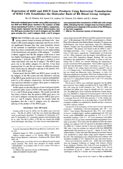
The Effect of Redox-Related Species of Nitrogen Monoxide
From www.bloodjournal.org by guest on February 6, 2015. For personal use only.
The Effect of Redox-Related Species of Nitrogen Monoxide on Transferrin
and Iron Uptake and Cellular Proliferation of Erythroleukemia (K562)Cells
By D.R. Richardson, V. Neurnannova, E. Nagy, and P. Ponka
The iron-responsiveelement-bindingprotein (IRE-BP)modulates both ferritin mRNA translation and transferrin receptor
(TfR) mRNAstability by binding t o specific mRNA sequences
called iron-responsive elements
(IRES). The
regulation of IREBP in situ auld possibly occur either through its Fe-S cluster
and/or via free cysteine sulphydryl groups such as cysteine
437 (Philpott et al, J Biol Chem 268A7655.1993; and Hirling
et al, EMBO J 13:453, 1994). Recently, nitrogen monoxide
(NO) has been shown t o have markedly different biologic
effects depending on its redox state (Lipton et al. Nature
364626, 1993). Considering this fact, it is conceivable that
the NO group, as either the nitrosonium ion (NO') or nitric
oxide (NO), may regulate IRE-BP activity by S-nitrosylation
of key sulphydryl groups or via ligation of N O t othe FeS cluster, respectively. This hypothesis has been examined
using the NO+ generator, sodium nitroprusside (SNP); the
N O generator, S-nitroso-N-acetylpenicillamine(SNAP); and
the NO/peroxynitrite (ONOO-) generator, 3-morpholinosydnonimine hydrochloride (SIN-l). Treatment of K562 cells
for 18 hours with SNP (1mmol/L) resulted in a pronounced
decrease in boththe RNA-bindingactivity of IRE-BP and the
level of TfR mRNA. In addition, Scatchard analysis showed
a marked decrease in the number of specific
Tf-binding sites,
from 590,00O/cell (control) t o 170,000/cell (test), and there
was also adistinct decrease in Fe uptake. Furthermore, SNP
did not decrease cellular viability or proliferation. In contrast,
the N O generator, SNAP(1 mmol/L), increased RNA-binding
activity of IRE-BP, the level of TfR mRNA, and the number
of TfRs in K562 cells. Moreover, both SNAP (1mmol/L) and
SIN-l (0.5 mmol/L) reduced cellularproliferation. The resutts
are discussed in context of the possible physiologic role of
redox-related species of NO in regulating iron metabolism.
0 1995 by The American Society of Hematology.
T
aconitase.'4.'5 Therefore, similar to aconitase, the biologic
activity of IRE-BP may be modulated by NO; in fact, Drapier
et a l l 6 and Weiss et al" have shown that NO can activate
IRE-BP RNA-binding activity. However, these investigators
did not consider the effects of redox-related species of NO
on IRE-BP or TW expression and Fe uptake.
Considering the possible target sites of NO on the IREBP molecule, it has been shown that in Fe replete cells IREBP possesses a cubane 4Fe-4s cluster that prevents IRE
binding, and, in this state, the protein displays aconitase
activity. In contrast, in cells depleted of Fe, the Fe-S cluster
is not present, and, under these conditions, IRE-BP can bind
to the IRE of mRNA.'53'S*'9
In addition, a free cysteine sulphydryl group(s) also appears to regulate the binding of IREBP to mRNA.'@'* However, the relative roles of the FeS
cluster and sulphydryl group(s) in IRE-BP regulation are not
well understood.
RANSFERRIN RECEPTOR (TW) and ferritin synthesis are under coordinate regulation by intracellular iron
(Fe) concentration.' This level of coordinate control occurs
at the posttranscriptional level and is mapped to regions
on TW and ferritin mRNA known as the iron-responsive
elements (IREs).**~ The
IREis recognized by a specific cytoplasmic binding protein known as the IRE-binding protein
(IRE-BP). Cells depleted of Fe contain higher levels of activated IRE-BP, which forms stable complexes with the IRE.
When IRE-BP binds to the IRE of ferritin mFWA, it represses its tran~lation?~
whereas IRE-BP binding to the IRE
of TfR mRNA protects the message from degradati~n.~.'
Either a decrease in intracellular Fe concentration or reducing conditions activate RNA-binding ofIRE-BP, but the
mechanism of this regulation in situ is unclear.
A variety of nitrogen oxides are produced in mammalian
cells that serve messenger roles.8 Nitrogen monoxide (NO)
has been shown to have markedly different biologic effects
in neural cells depending on its redox state.9 Nitric oxide
(NO) has a neurotoxic effect by reacting with superoxide
anion to produce peroxynitrite (ONOO-). In contrast, the
nitrosonium ion (NO+), has a neuroprotective effect via Snitrosylation of thiol groups on the N-methyl-D-aspartate
r e ~ e p t o rFurthermore,
.~
both NO+ and N O have been shown
to regulate the biologic activity of proteins under physiologic
circumstances, and nitric oxide synthase could produce both
chemical species under different intracellular redox conditions." However, because of the unique chemical properties
of NO and NO, these two redox-related species ofNO
react at different target sites on protein molecules. For example, numerous proteins can be S-nitrosylated by NO+ via
their thiol groups, a modification that may have important
physiologically relevant regulatory functions.9-" On the
other hand, N O can modulate protein activity via direct
coordination to the Fe centers of prosthetic groups, such as
heme and Fe-S clusters.'o One example of an Fe-S protein
whose activity is modulated by NO is the Krebs cycle enzyme, mitochondrial
Interestingly, IRE-BP,
which is present in the cytosol, has high homology to mitochondrial aconitase and has been described as a cytoplasmic
Blood, Vol 86, No 8 (October 15). 1995: pp 3211-3219
From the Lady Davis Institute for Medical Research of the Sir
Mortimer B. Davis-Jewish General Hospital, Montreal, Quebec,
Canada; and the Departments of Medicine and Physiology, McGill
University, Montreal, Quebec, Canada.
Submitted July 5, 1994; accepted June 12, 1995.
Supported by an operating grant from the Medical Research
Council of Canada. D.R.R. is the recipient of a Medical Research
Council of Canada Postdoctoral Fellowship. V.N. is the recipient
of a Bertha Mime Fellowshipfrom the Sir-Mortimer B. Davis Jewish
General Hospital, Montreal, Canada.
Presented in part at the 11th International Conference on Iron
and Iron Storage Proteins, Jerusalem, Israel, May 5, 1993, and also
at The American Society of Hematology meeting, December 1993
(Blood 82:9a, 1993 [abstr, suppl l ] ) .
Address reprint requests toP. Ponka, MD, PhD, Lady Davis
Institute for Medical Research, Sir Mortimer B. Davis-Jewish General Hospital, 3755 Chemin de la Cote-Ste-Catherine, Montreal,
Quebec, H3T IE2 Canada.
The publication costs of this article were defrayed in part by page
charge payment. This article must therefore be hereby marked
"advertisement" in accordance with 18 U.S.C. section 1734 solely to
indicate this fact.
0 1995 by The American Society of Hematology.
0006-4971/95/8608-0017$3.OO/0
321 1
From www.bloodjournal.org by guest on February 6, 2015. For personal use only.
3212
RICHARDSON ET AL
It is conceivable that NO, as either NO+ or N O , may
regulate IRE-BP activity via S-nitrosylation of key sulphydryl groups or via ligation of N O to the Fe-S center of the
protein. This hypothesis has been examined in the present
study using the NO+ generating drug, sodium nitroprusside9*’’; the N O generator, S-nitroso-N-acetylpenicillamine
(SNAP)23; and the NO/ONOO- generator, 3-morpholinosydnonimine hydrochloride (SIN-l).9 The effects of these
agents on IRE-BP activation, TW expression, Fe uptake, and
cellular proliferation have been examined in K562 erythroleukemia cells.
MATERIALS ANDMETHODS
Chemicals. Iron-59 chloride (as ferric chloride in 0.1 m o m
HCI), iodine-l25 (as sodium iodide), and [a-3zP]-CTP (800 Ci/
mmol) were purchased from Dupont (NEN Products, Boston, MA).
Human transfemn and T7 RNA polymerase was purchased from
Boehringer Mannheim (Mannheim, Germany). RNase TI was obtained from Calbiochem (San Diego, CA). Penicillin-streptomycin
was obtained from GIBCO Laboratories Ltd (Grand Island, NY).
N-2-hydroxyethylpiperazine-N’-2-ethanesulfonic
acid (HEPES), bovine liver catalase (EC 1.1 1.1.6), bovine erythrocyte superoxide dismutase (EC 1.15.1.1), N-acetyl-D-penicillamine(NAP), sodium pyruvate, and L-glutamine were obtained from Sigma Chemical CO
(St Louis, MO). RPMI-1640 medium and fetal calf serum (FCS)
were obtained from ICN Biomedicals Inc (Costa Mesa, CA). Desferrioxamine (DFO) was obtained from Ciba-Geigy Pharmaceutical CO
(Summit, NJ). All other chemicals were of analytical reagent quality.
Nitrogen monoxide generating compounds. SNAP was synthesized from NAP according to the method of Field et al.z4 SIN-l and
its inactive analogue, SIN-IC, were kind gifts from Dr Rainer Henning (Cassella, A.G., Frankfurt, Germany). Sodium nitroprusside
was obtained from Sigma Chemical Co. All chemicals were added
to RPMI medium immediately before an experiment and all solutions
were prepared in containers wrapped in aluminium foil. This latter
procedure was adopted to prevent photolysis of the compounds in
solution.23For each of the experiments listed below, cells were exposed for 1 to 18 hours with the NO donors dissolved in RPMI
medium without 10% FCS.
Cells. Human K562 erythroleukemia cells were obtained from
the American Type Culture Collection (Rockville, MD) and were
used inthe present study because their Fe metabolism is well characterized.zs-27
Cells were grown in 25-cmZplastic culture flasks (Costar,
Cambridge, MA) in an humidified atmosphere of 95% air and 5%
COn at 37°C in RPMI-1640 containing 10%FCS, extra L-glutamine
(300 pg/mL), sodium pyruvate (110 pg/mL), HEPES (15 mmoVL;
pH 7.2), penicillin (100 U/mL), and streptomycin (100 pg/mL). The
cells were maintained in log-phase growth at approximately 5 to 8
x io5 cells/mL.
Human transferrin. Human apoTf was prepared and labeled with
59Feand In5I, as described previously.z8
Gel-rerardntion assay. The gel-retardation assay wasusedto
measure the interaction between IRE-BP and the IRE using established te~hniques.4’~
Briefly, after incubation with RPMI alone (control) or various NO’/NO producing agents, 5 X IO6 cells were
washed with ice-cold phosphate-buffered saline (PBS) and lysed at
4°C in 100 pL of extraction buffer (10 mmoVL HEPES, pH 7.5, 3
mmol/L MgCln,40 mmol/L KCI, 5% glycerol, 1 mmol/L dithiothreitol, and 0.2% Nonidet P-40). After lysis, the samples were then
centrifuged at 10,000g for 3 minutes to remove nuclei. Samples of
cytoplasmic extracts were diluted to a protein concentration of 100
pg/mL in lysis buffer without Nonidet P-40, and 2 pg aliquotes
were analyzed for IRE-BP by incubation with 0.1 ng of 32Plabeled
pSPT-fer RNA transcript.” RNA was transcribed in vitro from linearized plasmid templates using T7 RNA polymerase in the presence
of [w3’P CTP]. TO form RNA-protein complexes, cytoplasmic extracts were incubated for IO minutes at room temperature with 0.1
ng of labeled RNA. Unprotected probe was degraded by incubation
with l U of RNAse TI for I O minutes. Heparin (5 mg/mL) was
then added for another IO minutes to exclude nonspecific binding.
RNA-protein complexes were analyzed in 6% nondenaturing polyacrylamide gels as described by Konarska and Sharp.3”In parallel
experiments, samples were treated with 2% 2-mercaptoethanol before the addition of the RNAprobe. Autoradiographs were quantified
by scanning densitometry.
Northern blot analysis. Briefly, Northern blot analysis was performed by isolating total RNAvia cell solubilization in a buffer
containing acid guanidinium thiocyanate, followed by phenol extraction and ethanol precipitation.” RNA wasquantitated by spectrophotometry at a wavelength of 260 nm and its quality was checked by
ethidium bromide staining on nondenaturing I % agarose gels. RNA
samples were denatured by heating at 65°C for 10 minutes and 15
pg ofRNAwas
then loaded onto 1.2% agarose gels containing
formaldehyde. After electrophoresis, RNA was transferred to nitrocellulose filters using the capillary blotting method. The filters were
air-dried, bakedin a vacuum oven at 80°C for 2 hours, andthen
hybridized with the human TW probe (pCDTR1; obtained from Dr
Frank Ruddle, Yale University, New Haven, CT) and P-actin probe
(cDNA obtained from Dr Lee Wall, Institut du Cancer de Montreal,
Hopital Notre Dame, Montreal, Quebec, Canada). Hybridizations
were performed as described by Meinkoth and Wahl” at 42°C overnight in hybridization solutions (50% [voUvol] formamide, 0.1%
polyvinylpyrrolidone, 0.1% Ficoll400, 0.1% bovine serum albumin
[BSA], 0.1% sodium dodecyl sulfate [SDS], 5X SSPE [lX SSPE is
0.15 molL NaC1,O.Ol mol/L NaH2POCHz0,and 1 mmom EDTA,
sodium salt], 10% [wt/vol] dextran sulphate, and200 pglmL of
denatured hemng sperm DNA) containing 3zP-labeledprobes prepared usingthe random primed DNA labeling kit (Boehringer Mannheim). The filters werethenwashed according tothemethod of
Maniatis et al.” Densitometric data were collected withanLKB
2222-020 UltroScan XL Laser Densitometer and data analyzed by
GelScan XL Software (Uppsala, Sweden).
Transferrinand iron uptake. For Tf and Fe uptake studies, K562
cells were collected by centrifugation at 500g for 5 minutes and
then washedinbasicRPM1medium
containing no serum. After
this procedure was performed, the cells were exposed to the NO’
generator, SNP (1 mmom), or the N O generator, SNAP (1 mmol/
L), for 18 hours at37°C. Transferrin andFe uptake studies were
then performed by standard techniques described previously.’*
Briefly, after exposure to NO-generating agents, the medium was
replaced and the cells were resuspended in RPMI containing 59Fe‘”I-transfemn (Tf; 1 to 40 pg/mL). Cells (3 X 10‘ per Tf concentration) were incubated with doubly labeled Tf for 2 hours at37°C
and then washed three times at 4°C with ice-cold PBS to remove
nonspecifically bound 59Fe-1251-Tf.
In no case was FCS added to the
preincubation medium containing NO-generating agents or tothe
labeling solution. Scatchard analysis was performed using standard
techniques.2RIt is important to note that, because the incubations
were performed at 37”C, the calculated number of Tf-binding sites
represents total binding. The ’Z51-Tf-labeledK562 cells were also
further processed to estimate the numbers of internalized transfemnbinding sites. This was achieved by incubating the cells for 30
minutes at 4°C with pronase ( 1 mg/mL), followed by centrifugation
to separate membrane-bound (supernatant) from internalized radioactivity in the cell elle et.'^.^^ However, after treatment with pronase,
the cell pellet became gelatinous, thus preventing complete separation of the supernatant from the cells. Hence, this latter technique
was not used further.
OF
From www.bloodjournal.org by guest on February 6, 2015. For personal use only.
EFFECT
NO ON CELLULAR IRON METABOLISM
Radioactivity was measured in a gamma scintillation counter with
appropriate correction for overlap of "Fe activity into the '"I channel.
Effect of NO' and NO' donors on cellular proliferation. The
effect of NO' and NO' donors on cellular proliferation was assessed
after 18 hours of incubationwith these agents usingviable cell
counts.
RESULTS
The effect of NO', NO' and NO'/ONOO- donors on IREBP RNA-binding activity and T P mRNA. Initial studies
examined the effect of incubating K562 cells for 18 hours
with the NO' donor, SNP. Increasing concentrations of SNP
(0.1,1,and 5 mmol/L; lanes 2, 3, and 4, respectively, of
Fig1A) decreased IRE-BP RNA-binding activity compared
with that ofthe control (lane 1, Fig 1A), but did not decrease
total IRE-BP (data not shown). In addition, the decrease
in IRE-BP binding activity was accompanied by a marked
decrease in the level of TfR mRNA (Fig 1B).As shown
previously by other^,^^.^^ DFO (50 pmol/L, lane 5) increased
IRE-BP RNA-binding activity (Fig 1A) and TfR mRNA
levels (Fig 1B). The addition of increasing concentrations
of SNP (0.1, 1, and 5 mmol/L; lanes 6,7, and 8, respectively)
to DFO (50 pmoVL) substantially prevented the increase in
the RNA-binding activity of IRE-BP seen in the presence
of DFO alone and also prevented the DFO-mediated increase
in TfR mRNA. In addition, it should be noted that SNP only
totally overcame the inducing effects of DFO when the molar
ratio of SNP to DFO was greater than two (see also Fig 4,
lanes 6 and 7). Further experiments examined the effect of
SNP (1 mmoVL) compared with DFO (1 mmol/L) on TfR
mRNA after 0 to 18 hours of incubation with these agents
(Fig 2). The effect of SNP at decreasing TfR mRNA was
evident already after only 1 hour of incubation with this
compound, with the result becoming more pronounced as
the incubation was continued up to 4, 8, and 18 hours (Fig
Fig 1. Changes in (A) the RNA-binding activity of IRE-BP and (B)
TfR mRNA levels in K562 cells incubated for 18 hours with RPM1
(untreated control, lane 1); the nitrosonium ion (NO') generator, SNP
(0.1,1, and 5 mmol/L; lanes 2,3,and 4, respectively); DFO (50 pmoll
L, lane 5); or DFO (50 pmollL) plus SNP (0.1. 1, and 5 mmollL; lanes
6,7, and 8, respectively).
3213
2). Importantly, when compared with the relevant control
(C) at each time point, SNP had no significant effect on pactin mRNA levels (Fig 2).
The decomposition of SNP (Na2[Fe(CN)sNO].2H20)in
solution leads to the production ofNO', ferricyanide, and
cyanide (CN-).9.'".37Hence, control experiments weredesigned to investigate whether either CN- or ferricyanide
could result in a decrease in IRE-BP RNA-binding activity
and a decrease in TfR expression. Treatment of K562 cells
with increasing concentrations of ferricyanide (0.5, 1, and 5
mmoI/L; lanes 2, 3, and 4, respectively, Fig 3) had little
effect on IRE-BP binding activity (Fig 3A) but resulted in
an increase in the level of TfR mRNA (Fig 3B). In contrast
to the results obtained with ferricyanide, cyanide ( 1 mmol/
L) hadno effect on IRE-BP RNA-binding activity or the
level of TfR mRNA (results not shown).
Because SNP is an Fe complex, it was also possible that
treatment of cells with SNP could lead to the donation of
Fe and result in a decrease in IRE-BP binding activity and
TfR mRNA. However, because ferricyanide did not decrease
the RNA-binding activity of IRE-BP or TfR mRNA levels
(Fig 3A and B), Fe donation to the cells via SNP could not
explain the observed decrease in the RNA-binding activity
of IRE-BP. Moreover, the addition of the impermeable Fe
chelator, EDTA (1 mmoVL), to SNP (1 mmol/L) didnot
prevent the decrease in IRE-BP RNA-bindingactivity or
TfR mRNA (compare lane 3 [SNP] with lane 4 [SNP +
EDTA]; Fig 4A and B), further suggesting that the effect of
SNP was not due to Fe donation to the cell.
The effect of the NO' donor, SNP (1 mmoVL), on IREBP binding activity and TfR mRNA was compared with that
of the NO/ONOO- donor, SIN-l (Fig 4). SIN-l (lane 2, Fig
4A) only slightly reduced IRE-BP RNA-binding, whereas
SIN-IC, a SIN-l analogue without the N-NO group, had no
effect on IRE-BP binding activity when compared with the
control.
After treatment of K562 cells with SIN-l (1 mmoVL),
TfR mRNA was decreased (lane 2, Fig 4B). However, SIN1 generates both N O and superoxide: resulting in the formation of ONOO-, which has been proposed to initiate lipid
peroxidation and cytotoxicity.9.38 Hence,the results obtained
with this compound may be due to these effects.
Considering the cytotoxicity of ONOO- generated by
SIN- 1, wecompared the effects of the N O generator, SNAP,
with those of SIN-l alone or SIN-l added in combination
with superoxide dismutase (SOD; 500 U/mL) and catalase
(CAT; 500 U/mL). The addition of SOD to SIN-l prevents
the generation of ONOO- by scavenging superoxide, and,
under these conditions, SIN-l acts as an N O generator? We
conducted six independent experiments of this type, all of
which yielded similar results; one representative experiment
is shown in Fig 5. Treatment of K562 cells with increasing
concentrations of SNAP (0.1. 0.5, and 1 mmoVL, lanes 2,
3, and 4, Fig 5A and B) resulted in an increase in the IREBP RNA-binding activity (Fig 5A) and TfR mRNAlevel
(Fig 5B) at SNAP concentrations of 0.5 mmol/L and 1 mmol/
L. In contrast, N-acetylpenicillamine (NAP), the precursor
of SNAP that does not contain the S-NO group, did not affect
the RNA-binding activity of IRE-BP (results notshown).
From www.bloodjournal.org by guest on February 6, 2015. For personal use only.
3214
ET
08 h
C
S
4 h
D
l h
C
RICHARDSON
18 h
S
D C
~
S
D C
S
D C
S
Fig 2. The effectofincubation time(0,1,4,8,
and 18 hours)
with RPM1 (untreatedcontrol; C),
D
~~
Tf R
?mm9
Moreover, none of the agents tested in our study decreased
total IRE-BP levels.
As seen previously in Fig 4A and B (lane 2), when the
NO/ONOO- generator SIN- I was incubated withK562 cells
at a concentration of I mmol/L, it slightly inhibited IRE-BP
activation andmoremarkedly
inhibited the level of TfR
mRNA (lane 7; Fig 5A and B). In contrast, SIN-l at a
concentration of 0.1 mmol/L and 0.5 mmol/L had little effect
on IRE-BP activation but slightly increased TfR mRNA levels (lanes 5 and 6, respectively, Fig 5A and B). The addition
of SOD and CAT to SIN-l ( I mmol/L; lane IO, Fig 5B)
prevented the decrease in TfR mRNA level thatwas observed in the presence of 1 mmol/L SIN-l alone (lane 7, Fig
5B). These results suggest that the decrease in TfR mRNA
observed in the presence of SIN-l alone was due to the
production of ONOO-. However, SOD and CAT added to
SIN-l (1 mmol/L; lane IO, Fig 5A) didnot eliminate the
slight inhibition of IRE-BP activation seen with SIN-l only
(lane 7, Fig 5A). At present, we do not understand the lack
of effect on IRE-BP activation of SIN-l added in combination with SOD and CAT. This is because under these condi-
1
AL
2
3
4
5
P-Actin
the nitrosonium ion (NO') generator, sodium nitroprusside (S;
1 mmol/L), or desferrioxamine
(D;1 mmollL1 on TfR and p-actin
mRNA levels in K562 erythroleukemia cells.
tions SIN-l should be mainly generating NO,' and therefore
the results obtained should be similar to those found with
SNAP. It is possible that ONOO- produced by SIN-l may
effect IRE-BPattwo possible target sites on the protein,
namely the regulatory FeS cluster and the sulphydryl group
of cysteine 437."." Indeed, Mohr et a13' have recently shown
that ONOO- canmediatethe S-nitrosylation of a critical
sulphydryl group of another protein. There is no reason to
assume that the reaction rates of ONOO- at both sites on
IRE-BP will be equally efficient or that the addition of SOD
and CAT completely prevents ONOO- generation from SIN1. Taken together, these findings could explain the lack of
effect of SIN- I on IRE-BP activation in the presence of SOD
and CAT. It is also relevant to point out that, in contrast to
SNP and SNAP, SIN-l added to cells resulted in a marked
decrease in cellular viability (see below). Again, these data
suggest that some of the effects of SIN-l are primarily due
to toxicity associated with ONOO- production.
The efect of NO+ and N O donors on Tfl expression and
iron uptake. The decrease in IRE-BP binding activity and
the level of TfR mRNA after treatment with the NO' donor,
SNP, suggested that a decrease in TfR expression and Fe
uptake may also be expected. To examine this, K562 cells
were incubated for 18 hours with the NO* generator, SNP,
and the N O donor, SNAP. After this preincubation, the cells
A
1
3 1 4 2-
5
-.--,
B
Fig 3. (A) Active IRE-BP levels and (B) TfR mRNA levels in K562
cells incubated for 18 hours with RPM1 only (control, lane l), potassium ferricyanide (0.5, 1, and 5 mmol/L; lanes 2, 3, and 4, respectively), or DFO (50 pmol/L, lane 5).
B
2
3
4
5
6
-
7
1
TfR
8-Actin
Fig 4. Effect ofthe nitricoxide (NOIlperoxynitrite(ONO07 generator, SIN-l, and the nitrosonium ion (NO') generator, SNP, on (AI
active IRE-BP levels and (B1 TfR mRNA levels
in K562 cells. Cellswere
incubatedfor 18 hours with RPM1 (control, lane l ) , SIN-l (1 mmollL;
lane 2). SNP (1 mmollL; lane 31, SNP (1 mmol/L) and EDTA (1 mmoll
L, lane 4). EDTA alone (1 mmol/L, lane 5). SNP (1 mmollL) and DFO
(1 mmol/L; lane 61, and DFO alone (1 mmollL, lane 71.
From www.bloodjournal.org by guest on February 6, 2015. For personal use only.
EFFECTOF
B
NO ON CELLULAR IRON METABOLISM
1
2
3
4
5
6
7
B
9
3215
l01112
-TfR
Fig 5. Effect of the N O generator, SNAP, and the NOlperoxynitrite (ONOO-l generator, SIN-l, on (A) active IRE-BP levels and (B)
transferrin receptor mRNA levels in K562 cells. Cells were incubated
for 18 hours with RPM1 only (control, lane l),
SNAP (0.1, 0.5, and 1
mmol/L; lanes 2,3,and 4, respectively), SIN-l (0.1.0.5, and 1.0 m m o l l
L; lanes 5, 6, and 7 , respectively), SIN-l (0.1, 0.5, and 1.0 m m o l l
L) plus SOD (500 UlmL) and CAT (500 UlmL) (lanes 8, 9, and 10,
respectively), DFO (0.1 mmollL, lane 11).and SOD plus CAT (lane 12).
were incubated with '9Fe-12'I-Tf (1 to 40 pg/mL) and the
uptake of"Fe and I2'I-Tf was then assessed. Both '*'I-Tf
and '9Fe uptake were substantially decreased after 18 hours
of incubation with 1 mmol/L SNP (Fig 6A and B). Scatchard
analysis of'''I-Tf
binding showed that SNP had no effect
on the affinity of Tf for the TfR (control kd, 1.70 2 0.01 X
lo-' m o m ; test kd, 2.10 2 0.44X IO-' m o m ) but substantially decreased the level of TfR from 590,000/cell (control)
to 170,000/cell (test). The number of Tf-binding sites and
the kd calculated in the present study for control K562 cells
was in agreement with that found in a previous investigation
(5 X 10' to 1 X 10" sitedcell; kd,1.25 to 2 X IO-' mol/
L)?'In contrast to "'I-Tf uptake by control K562 cells,
which plateaued off after a Tf concentration of 15 pg/mL
I
I
I
I
0 '
I
I
I
(Fig 6A), "Fe uptake continued to increase (Fig 6B). These
results suggest that, as found for melanoma ~ e l l s ' ' ~and
~~
hepatocytes,'" K562 cells may have a second mechanism of
Fe uptake that increases after saturation of the TfR. In contrast to the effect of SNP, the N O donor, SNAP (1 mmoll
L), only slightly increased both'2'I-Tf (Fig 7A) and '9Fe
uptake (Fig 7B). After treatment of K562 cells with SNAP,
Scatchard analysis ofI2'I-Tf binding in three experiments
showed a 10%. 29%, and 39% increase in TfR number compared with the control; a representative experiment is shown
in Fig 7A and B. It is of interest that the effects of the NO
donors on Tf-binding were far more pronounced than the
effects on Fe uptake. This finding may reflect the fact that
Tf and Fe uptake are two processes that are controlled independently?' In addition, as described above, K562 cells have
two mechanisms of Fe uptake from Tf. Therefore, it is not
surprising that Fe and Tf uptake are not closely coupled in
these cells.
Effect of NO', NO', and NO'/ONOO- producing agents
on cellular proliferation. There was a marked difference
in the effect of the NO' generating agent compared with
N O donors on cellular proliferation (Fig 8). The NO' donor,
SNP (1 mmol/L), had noappreciable effect on cellular proliferation, whereas the NO/ONOO- donor, SIN-l (0.5 mmol/
L), and the N O donor, SNAP (1 mmol/L), decreased cellular
proliferation to 65% and 71% of the control value, respectively (Fig 8). These studies are in agreement with previous
work that showed that the NO' donor, SNP, had no appreciable effect on 'H-thymidine uptake, whereas the N O donor,
SNAP, and the NO/ONOO- donor, SIN-l, markedly decreased 'H-thymidine incorporation?' In the present study,
the addition of 1 mmol/L ascorbate to 1 mmol/L SNP resulted in a distinct decrease in cellular proliferation to a level
comparable to that seen with SIN-l and SNAP, viz to 63%
I
0
I
I
10
20
30
40
Transferrin Concentration (pg/ml)
0
I
I
I
10
20
30
40
Transferrin Concentration (pglml)
Fig 6. Effect of the nitrosonium ion
(NO') generator, SNP (1 mmol/L), on (A)'%ransferrin uptake and (B) "Fe uptake by K562 cells. Cells
were incubatedfor18 hours at 37°C in the presence of SNP. After thisincubation, the medium was removed andcells
thewere thenreincubated
for 2 hours at 37°C with medium containing "Fe-'~I-transferrin(1 t o 40 pglmL). The cells were thenwashed three times at4°C with ice-cold
PBS t o remove nonspecifically bound 59Fe-'XI-transferrin.Results are the means of duplicate determinations froma typical experiment.
From www.bloodjournal.org by guest on February 6, 2015. For personal use only.
RICHARDSON ET A t
10.0 I
I
/.
I
I
SNAP
7.5
5.0
2.5
0.0
0
10
20
30
40
Transfemn Concentration (pg/ml)
0
10
20
30
40
Transferrin Concentration (pg/ml)
Fig 7. Effect of the nitric oxide (NO) generator, SNAP (1 mmollL), on (A) 1Z51-transferrin
and (B) 59Feuptake by human K562 cells. Cells
were incubated for 18 hours at 37°C in the presence of SNAP. After this incubation, the medium was removed and the cells were then
reincubated for 2 hours at 37°C with medium containing "Fe-"l-tranoferrin
(1t o 40 pglmL). The cells were then washed three times at 4°C
with ice-cold PBS t o remove nonspecifically bound "Fe-1z51-transferrin. Results are the means of duplicate determinations from a typical
experiment.
of the control (Fig 8). In contrast, ascorbate (1 mmol/L)
added alone had no appreciable effect. These studies confirm
previous reports suggesting that SNP is a nitroso compound
with strong NO' character that requires the presence of
ascorbate or another reducing agent to convert it to an N O
generat~r.~,'~+',~~
The addition of SOD (100 U/mL) and CAT
(l00 U/mL) to SIN-l (0.5 mmol/L) substantially prevented
its inhibitory effect on cellular proliferation (Fig 8). These
results suggest that the generation of superoxide by SIN-l
resulted inthe production of cytotoxic ONOO-?3x which
may have been responsible for the decrease in cellular proliferation and viability. It should be noted that, in contrast to
SIN-l, neither SNP nor SNAP reduced cellular viability.
IO0
75
50
25
0
DISCUSSION
0
v)
3
U
0
0
v)
2
+
0
>
-I
Fig 8. Effect of the nitrosonium ion (NO') generator, SNP; the
nitricoxide(NO)
generator, SNAP; andtheNOlperoxynitrite
(ONOO-) generator, SIN-l, on the proliferationof human K562 cells.
Cells were incubatedfor 18 hours a t 37°C in the presence of SNP (1
mmollL), SNP (1 mmollL) plus ascorbate (1 mmollL), ascorbate (1
mmollL), SIN-l (0.5 mmol/L), SIN-l (0.5 mmol/L) plus CAT (100 U/
mL1 and SOD (100 UlmL), SOD (100 UlmLl plusCAT (100 UlmLl, or
SNAP (1 mmollL). Rwuttsare the means of triplicate determinations
in a typical experiment.
Many of the biologic effects of NO are mediated through
its binding to Fe in the active centers of proteins that are
essential for cellular proliferation, such as ribonucleotide
reductase,?' aconita~e,'~.'~
NADH ubiquinone oxidoreductase, and succinate-ubiquinone oxidoreducta~e.~~
In addition,
the tumoricidal effect of NO may be attributed to the release
of Fe from
and NO has also been shown to mobilize
Fe from ferritin.49Drapier et all6 and Weiss et all7 have
recently shown that NO increases IRE-BP activation. Furthermore, endogenously produced NO can stabilize TfR
mRNA against targeted degradati~n.~'
However, these latter
investigators did not consider the effects of congeners of
NO on IRE-BP RNA-binding activity or the effects of these
species on TW expression and Fe uptake from Tf.
In the present investigation, we have shown that the NO'
producing agent, SNP, has a very different effect on cellular
Fe metabolism than the N O generator, SNAP. Considering
that IRE-BP is a major regulator of intracellular Fe homeostasis and that NO has a marked effect on IRE-BP RNA-
From www.bloodjournal.org by guest on February 6, 2015. For personal use only.
EFFECTOF
NO ON CELLULAR IRON METABOLISM
binding a~tivity,’~*’’
we suggest that some of the effects of
NO in the present study may be due to its interaction with
IRE-BP.
Regulation of IRE-BP via NO could occur at two different
sites on the protein. One target site may be the sulphydryl
group of cysteine 437, which has been shown to regulate
the binding of IRE-BP to the IRE?’.22A second control site
may be the dynamic Fe-S cluster, because only the apoprotein can bind to the IRE.’8.’y.22
Regarding these two possible
regulatory sites, recent studies have suggested that both NO+
and N O can regulate the biologic activity of proteins under
physiologic conditions, and nitric oxide synthase could produce both chemical forms under different redox condition~.’,’~
Moreover, these two chemical forms of NO react
at different sites on protein molecules: N O via direct coordination to Fe centers (such as those found in Fe-S proteins)
and NO+ via S-nitrosylation of thiol groups.” Hence, it is
conceivable that these redox forms of NO may have distinct
roles in regulating the RNA-binding activity of IRE-BP. Furthermore, Drapier et a1,I6 studying the effect of NO on both
RNA-binding and aconitase activities of IRE-BP, have
shown that low doses of NO inhibit the aconitase activity
of this protein, whereas higher doses are required to substantially activate RNA-binding activity. These investigators
suggest that, at low concentrations, NO may coordinate to
the Fe-S center and prevent interaction with cis-aconitate.16
In contrast, higher NO concentrations may completely disrupt the Fe-S center.I6
Based on our investigation, we suggest that the two redox
forms of NO ( N O and NO+) have different effects on IREBP RNA-binding activity. Recent studies on IRE-BP have
suggested that cysteine 437 must remain in its free and reduced form to allow the protein to bind to the IRE.2’.22Indeed, modification of this residue via the use of alkylating
agents prevents mRNA binding.*’,’’In addition, diamidecatalyzed formation of a disulphide bond between cysteine
437 and cysteine 503 or 506 results in decreased binding of
IRE-BP to the IRE.22From these data it was concluded that
cysteine 437 must remain in its reduced state to obtain an
IRE-BP-IRE interaction.” Considering this fact, it has been
reported that S-nitrosylation of cysteine SH groups with NO+
may aid disulphide bond f o r m a t i ~ n . Therefore,
~~~’
NO+ may
react similarly to diamide, where S-nitrosylation of cysteine
437 results in disulphide bridging between critical cysteine
residues that inhibit IRE-BP binding to the IRE. Such an
interaction of NO+ could explain the decrease in RNA-binding activity of IRE-BP observed with the NO+ generator,
SNP (Fig 1A). However, further studies are required to directly determine the possible target sites of NO+ on this
protein.
In contrast to the results obtained with the NO+ producing
agent SNP, the N O generator SNAP increased the RNAbinding activity of IRE-BP, the level of TfR mRNA, and
the number of TfRs in K562 cells. It has been suggested
that N O may react directly with the 4Fe-4S cluster of IREBP, leading to a loss of the cluster and an increase in RNAbinding a c t i ~ i t y . ’ ~However,
.’~
two recent st~dies’~.’~
have
shown that it is not N O but ONOO- that reacts with the
4Fe-4S cluster of mitochondrial aconitase and IRE-BP.
3217
Hence, the effect of N O described in the present work may
be due to the reaction of N O with intracellular 0; to produce
ONOO-.52,53In this context, it is relevant that Drapier et a l l 6
used SIN-1, an N O generator that also produces large
amounts of ONOO-? to show an increase in RNA-binding
activity of recombinant IRE-BP. However, similar IRE-BP
activation was also observed when these investigators used
NO gas.I6 In our present study, when SIN-l was added to
K562 cells, it did not activate IRE-BP as expected, and
actually decreased both the RNA-binding activity of this
protein and also the level of TfR &A
(Figs 4 and 5).
This effect may be due to limitations in the permeability of
ONOO- through the cell membrane andor the cytotoxic
activity of this a n i ~ n . Indeed,
~ . ~ ~ it is known that ONOOreacts avidly with membrane lipids resulting in peroxidat i ~ nTherefore,
.~~
this reaction at the level of the cell membrane may effectively quench ONOO- and prevent it from
reaching intracellular compartments. It is also pertinent to
note that, after incubation of K562 cells with SIN-l, there
was a marked decrease in cellular proliferation (Fig 8) and
viability. Moreover, this effect could be prevented by the
addition of SOD, suggesting that ONOO- was the cytotoxic
agent. An alternative explanation for the decrease in the
RNA-binding activity of IRE-BP after exposure to SIN-l
could be due to the reaction of ONOO- with critical cysteine
residues on this molecule.3y
It is evident from our present experiments that the changes
in IRE-BP activity observed using the N O generator, SNAP,
were not as pronounced as those found by Weiss et al,I7 in
which cells were transfected with NOS to act as an intracellular generator of NO. However, one must expect that the cell
membrane will hinder the diffusion of N O to some extent
and that the effect will not be as great as an intracellular
N O source. More importantly, as described above, it is probable that N O must be converted intracellularly to ONOObefore it can affect the regulatory Fe-S cluster of IRE-BP.’2’3
Interestingly, there are significant quantitative discepancies between the effects of pharmacologic effectors on IREBP activity and TfR &A
levels in K562 cells. This finding
is clearly shown by examining the effects of SNP and DFO
on IRE-BP activity and TfR mRNA in Fig 4, in which only
relatively minor changes in IRE-BP activity are seen in contrast to larger changes inmRNA levels. It is relevant to
discuss that Weiss et all7have also shown that the Fe regulation of IRE-BP in K562 cells is less pronounced than that
found in another cell type. Moreover, it can be suggested
that, in addition to changes in active IRE-BP levels, changes
in the rate of transcription of the TW may occur after exposure to DFO or NO congeners. Indeed, there is evidence
of transcriptional regulation of the TfR in K562 cells after
exposure to DF0,36and transcriptional regulation of the TfR
has been shown to occur in erythroid ~ e l l s . Furthermore,
~~~’~
it is intriguing that NO can influence gene trans~ription:~and
additional studies examining the effects of NO-generating
agents on the transcription rate of the TfR gene maybe
valuable.
Finally, we suggest that NO congeners could indirectly
modulate IRE-BP RNA-binding activity by affecting other
metabolic pathways. Recent work has suggested that phos-
From www.bloodjournal.org by guest on February 6, 2015. For personal use only.
3218
RICHARDSON ET AL
phorylation of IRE-BP by protein kinase C can modulate the
activity of this protein.58NO via its interaction with soluble
guanylate cyclase can activate both cGMP-dependent and
CAMP-dependent protein kinases that could possibly phosphorylate IRE-BP.59Hence, it cannot be excluded that NO
congeners may be acting on this or another system to control
IRE-BP RNA-binding activity, rather than directly interacting with the protein as suggested above. Our proposal
that NO may also regulate IRE-BP activity by affecting other
metabolic pathways concurs with recent work by Pantopoulos and Hentze."
Further studies will investigate the direct interaction of
NO congeners with recombinant IRE-BP and also the possible physiologic role of the NO+/NO equilibrium in controlling IRE-BP RNA-binding activity, TfR expression, and Fe
uptake.
ACKNOWLEDGMENT
The authors thank Dr Rainer Henning (Casella, A.G., Frankfurt,
Germany) for the kind gifts of SIN-I and SIN-IC, Dr L.C. Kiihn
for the pSF'T-fer plasmid, Dr F. Ruddle for transferrin receptor
cDNA, and Dr L. Wall for p-actin cDNA. We also gratefully acknowledge the excellent technical assistance of Mrs A. Wilczynska.
REFERENCES
l . Kiihn LC, Schulman HM, Ponka P: Iron-transferrin requirements and transferrin receptor expression in proliferating cells, in
Ponka P, Schulman HM, Woodworth RC (eds): Iron Transport and
Storage. Boca Raton, FL, CRC, 1990, p 149
2. Kuhn LC: m-RNA-protein interactions regulate critical pathways in cellular iron metabolism. Br J Haematol 47: 183, 1992
3. Klausner RD, Rouault TA, Harford JB: Regulating the fate of
mRNA: The control of cellular iron metabolism. Cell 72:19, 1993
4. Leibold EA, Munro HN: Cytoplasmic protein binds in vitro to
a highly conserved sequence in the 5' untranslated region of ferritin
heavy- and light-subunit mRNAs. Proc Natl Acad Sci USA 85:2171,
1988
5 . Walden WE, Patino MM, Gaffield L: Purification of a specific
repressor of ferritin mRNA translation from rabbit liver. J Biol Chem
264:13765, 1989
6. Casey JL, Hentze MW, Koeller DM, Caughman SW, Rouault
TA, Klausner RD, Harford JB: Iron responsive elements: Regulatory
RNA sequences that control mRNA levels and translation. Science
240:924, 1988
7. Miillner EW, Kilhn LC: A stem-loop inthe3' untranslated
region mediates iron-dependent regulation of transferrin receptor
mRNA stability in the cytoplasm. Cell 53:815, 1988
8. Moncada S, Palmer RMJ, Higgs EA: Nitric oxide: Physiology,
pathophysiology, and pharmacology. Pharmacol Rev 43:109, 1991
9. Lipton SA, Choi Y-B, Pan Z-H, Lei SZ, Chen H-SV, Sucher
NJ, Loscalzo J, Singel DJ, Stamler JS: A redox-based mechanism
for the neuroprotective and neurodestructive effects of nitric oxide
and related nitroso-compounds. Nature 364:626, 1993
10. Stamler JS, Singel DJ, Loscalzo J: Biochemistry of nitric
oxide and its redox-activated forms. Science 258:1898, 1992
11. Stamler JS, Simon DI, Osbome JA, Mullins ME, Jaraki 0,
Michel T, Singel DJ, Loscalzo J: S-nitrosylation of proteins with
nitric oxide: Synthesis and characterisation of biologically active
compounds. Proc Natl Acad Sci USA 89:444, 1992
12. Drapier J-C, Hibbs JB Jr: Murine cytotoxic activated macrophages inhibit aconitase in tumor cells. Inhibition involved the ironsulphur prosthetic group and is reversible. J Clin Invest 78:790, 1986
13. Drapier J-C, Hibbs JB Jr: Differentiation of murine macrophages to express nonspecific cytotoxicity for tumor target cells
results in L-arginine-dependent inhibition of mitochondrial iron-sulfur enzymes in the macrophage effector cells. J Immunol 140:2829,
1988
14. Kaptain S , Downey WE, Tang C, Philpott C, Haile D, Orloff
DG, Harford JB, Klausner RD: A regulated RNA-binding protein
also possesses aconitase activity. Proc Natl Acad Sci USA 88: 10109,
1991
15. Haile DJ, Rouault TA, Tang CK, Chin J, Harford JB, Klausner
RD: Reciprocal control of RNA-binding and aconitase activity in
the regulation of the iron-responsive element binding protein: Role
of the iron-sulfur cluster. Proc Natl Acad Sci USA 89:7536, 1992
16. Drapier J-C, Hirling H, Wietzerbin J, KaldyP,KiihnLC:
Biosynthesis of nitric oxide activates iron regulatory factor in macrophages. EMBO J 12:3643, 1993
17. Weiss G, Goossen B, Doppler W, Fuchs D, Pantopoulos K,
Werner-Felmayer G, Wachter H, Hentze MW: Translational regulation via iron-responsive elements by the nitric oxide/NO-synthase
pathway. EMBO J 12:3651, 1993
18. Haile DJ, Rouault TA, Harford JB, Kennedy MC, Blondin
GA, Bienert H, Klausner RD: Cellular regulation of the iron-responsive element binding protein: Disassembly of the cubane iron-sulfur
cluster results in high affinityRNA binding. Proc NatlAcad Sci
USA 89:11735, 1992
19. Emery-Goodman A, Hirling H, Scarpellino L, Henderson B,
Kiihn LC: Iron regulatory factor expressed from recombinant baculovirus: Conversion between the RNA-binding apoprotein and Fe-S
cluster containing aconitase. Nucleic Acids Res 21:1457, 1993
20. Hentze MW, Rouault TA, Harford JB, Klausner RD: Oxidation-reduction and the molecular mechanism of a regulatory RNAprotein interaction. Science 244:357, 1989
21. Philpott CC, Haile D, Rouault TA, Klausner RD: Modification
of a free Fe-S cluster cysteine residue in the active iron responsive
element-binding protein prevents RNA binding. J Biol Chem
268:17655, 1993
22. Hirling H, Henderson BR, Kiihn LC: Mutational analysis of
the [4Fe-4S]-cluster converting iron regulatory factor from its RNAbinding form to cytoplasmic aconitase. EMBO J 13:453, 1994
23. Feelisch M: The biochemical pathways of nitric oxide formation from nitrovasodilators: Appropriate choice of exogenous NO
donors and aspects of preparation and handling of aqueous NO
solutions. J Cardiovasc Pharmacol 17:S25, 1991
24. Field L, Dilts RV, Ravichandran R, Lenhert PG, Carnahan
GE: An unusually stable thionitrite from N-acetyl-D,L-penicillamine: X-ray crystal and molecular structure of 2-(acetylamino)-2carboxy-l ,l-dimethylethyl thionitrite. J Chem Soc Chem Commun
249, 1978
25. Schulman HM, Wilczynska A, Ponka P: Transfenin and iron
uptake by human lymphoblastoid and K562 cells. Biochem Biophys
Res Commun 100:1523, 1981
26. Klausner RD, Ashwell G, van Renswoude J, Harford JB,
Bridges KR: Binding of apotransferrin to K562 cells: Explanation
of the transferrin cycle. Proc Natl Acad Sci USA 80:2263, 1983
27. Bottomley S S , Wolfe LC, Bridges KR: Iron metabolism in
K562 erythroleukemia cells. J Biol Chem 260:6811, 1985
28. Richardson DR, Baker E: The uptake of iron and transferrin
by the human melanoma cell. Biochim Biophys Acta 1053:1, 1990
29. Miillner EW,Neupert B, Kuhn LC: A specific mRNA binding
factor regulates the iron-dependent stability of the cytoplasmic transferrin receptor mRNA. Cell 58:373, 1989
30. Konarska MM, Sharp PA: Electrophoretic separation of complexes involved in thesplicing of precursors to mRNAs.Cell 46:845,
1986
31. Chomczynski P, Sacchi N: Single-step method of RNA isola-
From www.bloodjournal.org by guest on February 6, 2015. For personal use only.
EFFECT OF NOON CELLULAR IRON METABOLISM
tion by acid guanidinium thiocyanate-phenol-chloroform extraction.
Anal Biochem 162:156, 1987
32. Meinkoth J, Wahl G: Hybridisation of nucleic acids imrnobilized on solid supports. Anal Biochem 138:267, 1984
33. Maniatis T, Fritsch EF, Sambrook J: Molecular Cloning: A
Laboratory Manual. Cold Spring Harbor, NY, Cold Spring Harbor
Laboratory, 1982
34. Karin M, Mintz B: Receptor-mediated endocytosis of transferrin in developmentally totipotent mouse teratocarcinoma stem
cells. J Biol Chem 256:3245, 1981
35. Richardson DR, Baker E: Two mechanisms ofiron uptake
from transferrin by melanoma cells. The effect of desferrioxamine
and ferric ammonium citrate. J Biol Chem 267:13972, 1992
36. Rao K, HarFord, JB, Rouault T, McClelland A, Ruddle FH,
Klausner RD: Transcriptional regulation by iron of the gene for the
transferrin receptor. Mol Cell Biol 6:236, 1986
37. Butler AR, Glidewell C: Recent chemical studies on sodium
nitroprusside relevant to its hypotensive action. Chem SOCRev
16:361, 1987
38. Beckman JS, Beckman TW, Chen J, Marshall PA, Freeman
BA: Apparent hydroxyl radical production by peroxynitrite: Implications for endothelial injury from nitric oxide and superoxide. Proc
Natl Acad Sci USA 87:1620, 1990
39. Mohr S, Stamler JS, Briine B: Mechanism of covalent modification of glyceraldehyde-3-phosphate dehydrogenase at its active
site thiol by nitric oxide, peroxynitrite and related nitrosating agents.
FEBS Lett 348:223, 1994
40. Richardson DR, Baker E: Two saturable mechanisms of iron
uptake from transferrin by melanoma cells. The effect of transferrin
concentration, chelators and metabolic probes on transferrin and iron
uptake. J Cell Physiol 161:160, 1994
41. Page M, Baker E, Morgan EH: Transfenin and iron uptake
by rat hepatocytes in culture. Am J Physiol 246:G26, 1984
42. Bomford A, Young SP, Nouri-Aria K, Williams R: Uptake
and release of transferrin and iron by mitogen-stimulated human
lymphocytes. Br J Haematol 55:93, 1983
43. Richardson DR, Neumannova V, Nagy E, Ponka P Effect of
nitric oxide species on cellular proliferation and transferrin and iron
uptake by erythroleukemia (K562) cells. Blood 82:9a, 1993 (abstr,
SUPPI 1)
44. Zumft WG, Frunzke K: Discrimination of ascorbate-dependent non-enzymatic and enzymatic, membrane-bound reduction of
nitric oxide in denitrifying pseudomonas perfectomarinus. Biochim
Biophys Acta 681:459, 1982
45. Bates JN, Baker MT, Guerra R Jr, Harrison DG: Nitric oxide
generation from nitroprusside by vascular tissue. Evidence that reduction of the nitroprusside anion and cyanide loss are required.
Biochem Pharmacol 42:s 157, 1991 (suppl)
3219
46. Lepoivre M, Fieschi F, Coves J, Thelander L, Fontecave M:
Inactivation of ribonucleotide reductase by nitric oxide. Biochem
Biophys Res Commun 179:442, 1991
47. Hibbs JB Jr, Taintor RR, Vavrin Z, Granger DL, Drapier JC,
Amber IJ, Lancaster J Jr: Synthesis of nitric oxide from a terminal
guanidino nitrogen atom of L-arginine: A molecular mechanism
regulating cellular proliferation that targets intracellular iron, in
Moncada S, Higgs EA (eds): Nitric Oxide from L-Arginine: A Bioregulatory System. New York, NY, Elsevier Science, 1990, p 189
48. Hibbs JB Jr, Taintor RR, Vavrin Z Iron depletion. Possible
cause of tumor cell cytotoxicity induced by activated macrophages.
Biochem Biophys Res Commun 123:716, 1984
49. Reif DW, Simmons RD: Nitric oxide mediates iron release
from ferritin. Arch Biochem Biophys 283:537, 1990
50. Pantopoulos K, Hentze MW: Nitric oxide signalling to ironregulatory protein: Direct control of ferritin mRNA translation and
transfenin receptor mRNA stability in transfected fibroblasts. Proc
Natl Acad Sci USA 92:1267, 1995
51. Lei SZ, Pan Z-H, Agganval SK, Chen H-SV, Hartman J,
Sucher NJ, Lipton SA: Effect of nitric oxide production on the redox
modulatory site of the NMDA receptor-channel complex. Neuron
8:1087, 1992
52. Hausladen A, Fridovich I: Superoxide and peroxynitrite inactivate aconitases, but nitric oxide does not. J Biol Chem 269:29405,
1994
53. Castro L, Rodriguez M, Radi R: Aconitase is readily inactivated by peroxynitrite, butnot its precursor, nitric oxide. J Biol
Chem 269:29409, 1994
54. Radi R, Beckrnan JS, Bush KM, Freeman BA: Peroxynitriteinduced membrane lipid peroxidation: The cytostatic potential of
superoxide and nitric oxide. Arch Biochem Biophys 288:481, 1991
55. Chan L-NL, Gerhardt EM: Transferrin receptor gene is hyperexpressed and transcriptionally regulated in differentiating erythroid
cells. J Biol Chem 267:8254, 1992
56. Chan RYY, Seiser C, Schulman HM, KiihnLC, Ponka P:
Regulation of transferrin receptor mRNA expression. Distinct regulatory features in erythroid cells. Eur J Biochem 220:683, 1994
57. Peunova N, Enikolopov G: Amplification of calcium-induced
gene transcription by nitric oxide in neuronal cells. Nature 364:450,
1993
58. Eisenstein RS, Tuazon PT, Schalinske KL,Anderson SA,
Traugh JA: Iron-responsive element-binding protein. Phosphorylation by protein kinase C. J Biol Chem 268:27363, 1993
59. Schmidt HHHW, Lohman SM, Walter U: The nitric oxide
and cGMP signal transduction system: Regulation and mechanism
of action. Biochim Biophys Acta 1178:153, 1993
From www.bloodjournal.org by guest on February 6, 2015. For personal use only.
1995 86: 3211-3219
The effect of redox-related species of nitrogen monoxide on
transferrin and iron uptake and cellular proliferation of
erythroleukemia (K562) cells
DR Richardson, V Neumannova, E Nagy and P Ponka
Updated information and services can be found at:
http://www.bloodjournal.org/content/86/8/3211.full.html
Articles on similar topics can be found in the following Blood collections
Information about reproducing this article in parts or in its entirety may be found online at:
http://www.bloodjournal.org/site/misc/rights.xhtml#repub_requests
Information about ordering reprints may be found online at:
http://www.bloodjournal.org/site/misc/rights.xhtml#reprints
Information about subscriptions and ASH membership may be found online at:
http://www.bloodjournal.org/site/subscriptions/index.xhtml
Blood (print ISSN 0006-4971, online ISSN 1528-0020), is published weekly by the American
Society of Hematology, 2021 L St, NW, Suite 900, Washington DC 20036.
Copyright 2011 by The American Society of Hematology; all rights reserved.
© Copyright 2026
