
Structure and Expression of the RH Locus in the Rh
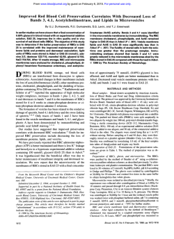
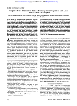
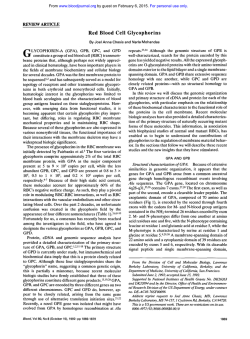
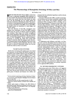
From www.bloodjournal.org by guest on February 6, 2015. For personal use only.
Structure and Expression of the RH Locus in the Rh-Deficiency Syndrome
By B. Ch6rif-Zahar, V. Raynal, C. Le Van Kim, A.-M. D'Ambrosio, P. Bailly, J.-P. Cartron, and Y. Colin
Red blood cell deficiency of R h proteins is associated with
morphologic and functional abnormalities of erythrocytes
and with a chronic hemolytic anemia of varying severity.
Rh-deficiency may be the result of homozygosity either for
a silent allele at the RH locus (Rh,
amorph type) or for a
recessive inhibitor gene(s) at an autosomal locus unlinked
t o RH locus (Rh,
regulator and Rh,,,,,,,). In this report, we
investigated the RH locus structure of Rh-deficient individuals by Southern analysis using cDNA and exon-specific
probes deduced from the recent cloning of Rh genes (CcEe
and 0).As expected from family studies indicating that
R
,h,
and Rh,,
regulator individuals are unable t o express
Rh antigens but are able to convey functional Rh genes
from one generation t o another, no alteration of the Rh
genes was detected in these variants. Although Rh,
of
the amorph type arose by inheritance of a pair of silent
alleles at the RH locus, the general organization of the
unique CcEe gene in the genome of the particular individual
under examination was apparently normal and indistinguishable from a Rh-negative chromosome. More surprisingly, no mutation could be detected by sequencing the
polymerase chain reaction (PCR)-amplified reticulocyte
mRNAs, suggesting that the RH locus of this patient might
be altered in its transcriptional activity. Through hybridization with exon-specific probes, w e were also able t o determine the zygosity for the D gene in DNA samples from individuals of known genotypes; using this approach, w e
found that Rh,
regulator variants could be either of the
DD, Dd, or dd genotypes. These findings suggest that the
postulated inhibitor gene(s) can negatively suppress the
RH locus expression from chromosomes carrying either
one or two of the Rh genes.
0 1993 by The American Society of Hematology.
T
of Rh,,,, is much less common than other Rh-deficient
types. Of 42 recently listed Rh-deficient phenotypes, only 5
are the amorph type of Rh,,,, .9
All types of Rh deficiency show the same clinical abnormalities associated with a chronic hemolytic anemia, stomatocytosis and spherocytosis, reduced osmotic fragility,
and increased cation permeability. In addition, Rh,,,, membranes have characteristically hyperactive membrane adenosine triphosphatases (ATPases), reduced RBC cation and
water contents, and a relative deficiency of membrane choIesteroI.'.2.'0.1I Rh,,,, RBCs show an abnormal membrane
phospholipid distribution characterized by an increased exchangeability of phosphatidylcholine and phospholipase accessibility of phosphatidylethanolamine.'2 Rh,,,, cells also
lack several membrane proteins and glycoproteins (GPs),
including the 30- to 32-Kd proteins carrying the Rh antig e n ~ ' ~and
, ' ~ the Rh-related GPs recognized by murine
monoclonal antibodie~.'~,''-'~
In addition, glycophorin B,
the carrier of Ss antigens, is reduced to approximately 30%
of the normal level?' certain other blood group antigens,
including LW, Duffy (Fy5), U, and Duclos antigens, are
also missing or severely decreased in these cells." One important and still unresolved question is to explain how a
single genetic alteration at the RH locus itself or at an independent locus controlling its expression (XOr, XQ) may result in so many membrane abnormalities. To address this
issue we have examined the structure and expression of the
RH locus in these patients using the recently available information deduced from the cloning of the two Rh genes that
compose the RH ~ O C U S . ~ ~ ~ ~ ~
HE Rh DEFICIENCY syndrome'-3 is a rare hemolytic
anemia of varying severity caused by the defect or severe decrease of membrane proteins carrying the Rh blood
group antigens. It is associated with morphologic and functional abnormalities of erythrocytes, thus indicating that Rh
proteins are important for normal red blood cell (RBC)
membrane structure and f ~ n c t i o nCells
. ~ deficient in all Rh
antigens are subdivided into Rh,,,, and Rh,,,
which totally
lack Rh structures or have severely depressed Rh antigens,
respectively.' Family studies have shown that the Rh,,,, phenotype may arise from two distinct genetic backgrounds.
The most common type of Rh,,,,, called the "regulator
type" occurs by an inhibition mechanism. According to Levine et a1,6 this phenotype is caused by homozygosity for an
autosomal recessive suppressor gene (Xor)that is genetically
independent of the RH locus. The second type of Rh,,,,,
which was first noted in a Japanese family,7 is called
"amorph" or "silent type" and arose by homozygosity for a
silent allele at the RH locus. The Rh,, phenotype was discovered more recently,' and was found to arise from the
homozygous state of a suppressor gene (XQ) at an autosomal locus independent of RH. However, the X"r and XQ
nomenclature is inappropriate, because these genes are not
located on the X chromosome and because it is not known
whether they are alleles at the same locus. The amorph type
From the Unit6 INSERM U76, Institut National de Transfusion
Sanguine, Paris, France.
Submitted January 8, 1993; accepted February 25, 1993.
Supported in part by North Atlantic Treaty Organization Grant
No. 0556/88 and by Caisse Nationale dlssurance Maladie des
Travailleurs Salari6s.
Address reprint requests to Jean-Pierre Cartron. PhD, Unite INSERM U76, Institut National de Transfusion Sanguine, 6 rue Alexandre Cabanel, 75015 Paris, France.
The publication costs of this article were defayed in part by page
charge payment. This article must therefore be hereby marked
"advertisement" in accordance with 18 U.S.C. section 1734 solely to
indicate this fact.
0 1993 by The American Society OfHematology.
0006-49 71/93/8202-0016$3.00/0
656
MATERIALS AND METHODS
Patients. Blood samples from Rh-deficient patients were collected on heparin ( IO U/mL) and shipped to Paris. Three samples of
Rh,,,, of the regulator type were investigated: A.L., provided by L.
Lebeck (Loma Linda, CA); Y.T., provided by C . Hyland (Brisbane,
Australia); and T.B., provided by Dr M.J. Stelling (Geneva, Switzerland). One sample of Rh,,,, of the amorph type (D.A.A.) was
from Dr C. Perez-Perez26(Linares, Spain) and one sample of Rh,,
(S.S.) from P. Hartnett (Miami, FL). Blood samples from individuals with common Rh phenotypes were from the National Blood
Transfusion Service in Paris.
Blood, VOI 82,NO2 (July 15), 1993:pp 656-662
From www.bloodjournal.org by guest on February 6, 2015. For personal use only.
657
RH-DEFICIENCY SYNDROME
-
03
Q)
L
a
Q)
U
u
0
*
Q)
Q)
U
0
U
3
C
c
ce
P
i2
c
ce
Anti-Rh proteins
MPC 1
(N-ter)
MPC 8
(C-ter)
-Rh
(30-32kDa)
-Rh
(30-32kDa)
Anti-LW
BS46
-1W
(42 k De)
Fig 1. lmmunostaining of RBC membrane proteins f" Rh-deficient erythrocytes. One hundred
to 1 5 0 pg of total membrane proteins from A.L.
(Rh,
reg, U-negative), S.S. (Rh,
U-negative),
and D.A.A. (Rh,,, silent, U-positive) separated by
SDS-PAGE on a 12% polyactylamideseparating gel
were transfered to nitrocellulose sheets. Each was
incubated with rabbit anti-Rh protein antibodies
(MPC1 and MPCI, 1 :4,000dilution), with a murine
MoAb anti-LWb (BS46; 1 0 pg/mL), with the murine MoAb 2D10 (10 pg/mL), and with the rabbit
antiprotein 7.2b antiserum, respectively. Antibodies bound were detected by the alkaline phosphatase conjugate substrate method. Arrows on the
left side indicate the migration position of the Rh,
LW, 2D10, and 7.2b polypeptides.
2D10
Membrane protein anal.vsis. For immunostaining analysis, sodium dodecyl sulfate (SDS)-lysates from RBC membrane preparations prepared as described2'were separated by discontinuous SDS
polyacrylamide gel electrophoresis (PAGE):' transferred to nitrocellulose sheets:9 and incubated as described26*M
with rabbit polyclonal antibodies raised against synthetic Rh peptides (MPCI and
MPCI reacting with the NH2- and COOH-terminal regions of the
Rh proteins. respectively) or with the murine MoAbs BS46 (gift of
Dr H.H. Sonnebom, Dreieich, Germany) and 2DIO (gift of Dr
A.E.G. von dem Bome, Amsterdam, The Netherlands) that react
with blood group LW3' or Rh-related18.'9membrane GPs, respectively. A rabbit polyclonal antibody against the membrane protein
7.2b (gift of Dr R. Johnson, Detroit, MI) was also used as control.
Bound antibodies were detected with alkaline phosphatase-labeled
goat antirabbit IgG or rabbit antimouse diluted (1:800) followed by
revelation with the alkaline-phosphatase conjugate substrate kit
(Biorad Laboratories, Rockville Centre, NY).
Genomic DNA anal.vsis. Human leukocyte DNA extracted
from peripheral leucocytes was digested with restriction enzymes
(Appligene, Strasbourg, France; IO U/mg DNA), resolved by elec-
(45 -7 5 k Da 1
trophoresis in 0.8%agarose gel, and transferred onto nitrocellulose
membrane (BA85; Schleicher & Schiill, Dassel, Germany) as described.32DNA probes used for hybridizations were: ( I ) the complete RhlXb cDNA encoding the Cc/Ee proteins:2 (2) exon-specific sequencesobtained by PCR from exon I, exon 4, and exon 9 +
IO ofthe CcEegene (Cherif-Zahar et al. unpublished data). Hybridization with DNA probes (IO6 cpm/mL) was performed for 16
hours at 65°C in 5 X SSPE ( I X SSPE = 0.15 mol/L NaCI, 0.01
mol/L NaH,PO,, 0.001 mol/L EDTA, pH 7.4), 5 X Denhardt ( 1 X
Denhardt = 0.02%wt/vol each of Ficoll, polyvinylpyrolidone,and
bovine serum albumin [BSA]),0.1% SDS, and 100 mg/mL sonicated salmon-sperm DNA. Final washes were performed at 65°C
for I5 minutes in 0. I X SSPE, 0. I % SDS.
Reverse transcription coirpled with PCR amplijication. Total
RNA was extracted by the acid-phenol-guanidinium method3'
from 5 mL of peripheral blood (PB)and 0.5 pg was incubated for 40
minutes at 42°C in a reaction mixture (50 pL) containing 100
mmol/L Tris (pH 8.3). 140 mmol/L KCI, IO mmol/L MgCI,, 30
mmol/L &mercaptoethanol, I mmol/L of each deoxynucleoside
triphosphate (dNTP), 40 U of ribonuclease inhibitor (RNasine),
From www.bloodjournal.org by guest on February 6, 2015. For personal use only.
CHfRIF-ZAHAR ET AL
658
B
kb
25
21
7
6.7
. 3-@mi@
-
5.1
4.2 -
uv
m
m
.. c)
.
-
-
-
7.3
6.7
-
5.1
-
4.2
-
3.5
-
2.5
Hindlll
BamHl
Fig 2. Southem blot analysis of the RH locus in the Rh-deficient patients. DNA from RhD-positive and RhD-negative donors with the
indicated phenotypeand from Rh-deficient individualswere digested by Hindlll or BamHl restriction enzyme and hybridizedon Southem blot
with the exon-specific Rh probes (A) as indicated or (E) with the full-length Rh cDNA probe.
and IO U of avian myeloblastosis virus reverse transcriptase (Promega Biotec. Madison, wl). The cDNA products were then subjected to PCR in 50 mmol/L KCI, IO mmol/L Tris(pH 8.3). 0.01%
gelatin, 0.2 mmol/L of the four dNTPs, 50 pmol of each primer,
and 2.5 U of T h m i t s acqitariciis DNA polymerase(Perkin-Elmer,
Norwalk, CT).All primers were chosen from the RhlXb cDNA
sequence. PCR amplifications (30 cycles) were performed under
the following conditions: I minute of denaturation at 94OC, I .5
minutes of primer annealingat 55”C, and 2 minutes ofextension at
72°C. Amplification products were purified using agarose gels electrophoresis, phosphorylated with polynucleotide kinase, and subcloned in pUC I8 vector.
DNA sequencing Inserts from recombinant pUC18 vectors
were sequenced on both strands using the dideoxy chain termination method.”
RESULTS AND DISCUSSION
Protein defects. In preliminary studies membrane preparations from the regulator (reg), the amorph (silent), and the
Rh,, types of Rh-deficient erythrocytes that are unreactive
(or very weak reactive for Rh,,) with all anti-Rh antibodies
of different specificities(D, C, c, E, e, etc) were separated by
SDS-PAGE and examined by immunostaining with antibodies raised against synthetic peptides and directed against
the Rh proteins (30 to 32 Kd), the LW G P (42 Kd), and
Rh-related GPs (45 to 75 Kd). Neither the MPCl nor the
MPC8 antibodies that recognize the NH2-terminal (residues 34 to 46) and the COOH-terminal (residues 409 to
41 7) ofthe Rh
respectively, from RhD-positive
(DCCee)and RhD-negative (ddccee) samples, reacted with
the membrane proteins of Rh-deficient cells (Fig I). Because the Cc/Ee and D proteins have conserved NH2- and
COOH-terminal domains?’ it is concluded that the Rhde-
ficient cells lack both the Cc/Ee and the D proteins. This
conclusion is consistent with the finding that the polyclonal
antipeptide antibodies react with RBCs from homozygous
D - - individuals who cany the D but not the Cc and Ee
protein^.^' The present immunoblotting analysis of Rh-deficient cells, together with those reported by Suyama and
Goldstein,” support the conclusions of previous studies
that have suggested the lack of Rh proteins in RhnUllcells
using the indirect method ~flabeling.”*’~
Although Rhdeficient erythrocytes and RBCs from patients with hereditary
stomatocytosis show similar morphologic abnormalities,
both conditions are distinct at the molecular level36because
the antiprotein 7.2b antiserum that selectively recognized a
3 I -Kd protein that was deficient in patients with hereditary
stomatocyt~sis~’reacted normally with all Rh-deficient
erythrocytes (Fig 1).
Rh-deficient cells also lack the LW and Rh-related
The BS46 (anti-LW) antibody reacted only with
RBC membrane proteins from common Rh phenotypes
and not with those from Rh-deficient individuals (Fig I).
The murine MoAb 2D IO also recognizes membrane GPs of
45 to 75 Kd that are absent from most Rh,,,, or Rh,, cells
lacking the blood group U, but are weakly expressed on
some Rh,,,, cells that are U - p o s i t i ~ e . ’Accordingly,
~,~~
2D IO
did not react with the U-negative A.L. and S.S. samples (Fig
1) but did react weakly with the U-positive D.A.A. sample.26
These multiple protein abnormalities have not been explained on a molecular basis. A currently favored hypothesis is that Rh polypeptides are required for the correct transport or cell-surface expression of certain GPs or,
alternatively but not exclusively, that a G P missing in Rh,,,,
cells is necessary for the cell-surface expression of Rh pro-
From www.bloodjournal.org by guest on February 6, 2015. For personal use only.
RH-DEFICIENCY SYNDROME
659
El2.7 //
2.5
Hind1I I
Fig 3. Determination of the zygosity for the RhD gene by Southem blot analysis. Genomic DNA from donors with the indicated genotype for the D gene and from the Rh-deficient patients Y.T., T.B.,
A.L. (Rhd reg); S.S. (Rh,);
and D.A.A. (Rh,,,, silent) were digested with the Hindlll restriction enzyme and hybridized with the
exon 4 and exon 1 probes. Gene dosage effects were estimated by
determination of the relative intensity of 6.7/7.3-kb fragments
(exon 4 probe) and from 2.7/2.5-kbfragments (exon 1 probe) corresponding to the D and CcEe genes, respectively, after densitometry
analysis of the autoradiogram.
teins. This explanation suggests that the Rh antigen structure is a large membrane complex containing the Rh polypeptides, Rh-related GPs. and possibly other unrelated
GPs.~
Southern blot analjais qftlie R H locia in Rh,,,,l and Rh,,
phenotypes. Genomic DNA from the Rh,,,, reg Y.T., the
Rh,,,, silent D.A.A., and the Rh,, S.S. were digested with
HindIII and BamHI restriction enzymes and subjected to
Southern blot analysis with Rh-specific probes. DNAs from
RhD-positive (DCCee) and RhD-negative (ddccee) donors
were used as a control. Figure 2A shows the hybridization
patterns obtained with probes for exon- I , exon-4. and exon9+ IO of the CcEe gene (Cherif-Zahar et al. unpublished
data) that correspond to residues ( 1-48. 162-2 I I , and 4084 16, respectively) of the mature CcEe-encoded proteins.
Two hybridizing bands were detected with each exon-specific probe after llindlll and BarnHI digestion of the DCCee
sample (in the IfindIII profile, ie, exon-I: 2.7 kb and 2.5 k b
exon-4: 7.3 kb and 6.7 k b exon-9+10: 21 kb and 18 kb).
However, only one band was detected (Hind111 profile,
exon-I: 2.5 kb; exon-4: 7.3 k b exon-9+10: 18 kb) with the
ccddee sample, as expected from recent studies showing that
the RH locus is composed of two homologous genes (Dand
CcEe) in RhD-positive individuals, but of only one (CcEe)
in RhD-negative individual^.^^
The hybridization patterns of Y.T. (Rh,,,, reg) and S.S.
(Rh,,) did not differ from those obtained with the RhDpositive control, and those from D.A.A. (Rh,,,, silent) were
identical with those of the control RhD-negative phenotype
(Fig 2A). No additional or missing band could be detected
with the Rh-deficient samples suggesting a large sequence
insertion and/or deletion at the RH locus of these individuals. This inference was confirmed by hybridization with
the full length RhlXb cDNA probe (Fig 2B). It is not surprising that no defect of the Rh genes occurs in Rh deficiency caused by a postulated recessive suppressor gene independent of the RH locus. Studies of families have shown
that these individual family members carry the normal RI?
genes but fail to express Rh antigen^.^ Our results provide
the first direct proof that confirms this hypothesis.
Hybridization with exon-specific probes was also performed to determine whether the Rh-deficient individuals
carrying the D gene were homozygous or heterozygous for
this gene. Gene dosage effects were obvious when Hind111
digests of DNA from individuals of known DD.Dd.and dd
genotypes, which all carry two copies of the CcEegene, were
estimated by densitometric analysis of autoradiograms (Fig
3). As expected from the presence of two copies of the D and
CcEegenes in DD genome, the D-specific and CcEe-specific
fragments24were detected with the same intensity as with
the exon 4 probe (6.7 kb and 7.3 kb, respectively) and the
exon 1 probe (2.7 kb and 2.5 kb. respectively). On the other
hand, a I :2 gene dosage effect was observed between the D
and CcEe fragments in the heterozygous Dd genome. Only
the CcEe gene was detected in the Rh-negative (dd) DNA.
Examination of the hybridization pattern of the Rh-deficient samples (Fig 3) showed that S.S. (Rh,,)
and Y.T.
(Rh,,,, reg) were homozygous for the D gene, whereas A.L.
(Rh,,,, reg) was heterozygous for the D gene. T.B. (Rh,,,, reg)
and D.A.A. (Rh,,,, silent) carried only the CcEe gene as
Rh-negative donors. Although the Rh genotypes of the Rhdeficient individuals in this study were not known. these
findings agree with studies of families that suggest that Rh
haplotypes carrying or not carrying the D gene are segregating in Rhnirll reg and Rhrnod pedigree^.^'.^' These data also
show that the regulatory genes that determine the Rh,,,, reg
and Rh,, phenotypes are effective both on chromosomes
that carry either one or two Rh genes (CcEeor CcEeand D).
Moreover, our findings show for the first time that zygosity
for the D gene (DDor Dd)can be easily determined without
studying families by using exon-specific probes on a DNA
sample. Together with the possibility of detecting the D
gene,2sthis may have applications, for instance, in the prediction of the D genotype of a father in couples where there
is an RhD-negative woman at risk of fetal alloimmunization.4'
Soirthern blot analwis ofthe R H Iocirs in the Rh,,, silent
jamilv. Genomic DNA isolated from Epstein-Barr virus
(EBV)-immortalized lymphoblastoid cell lines derived from
each family member of the Rh,,,, silent D.A.A.26 was digested with Hind111 and probed with the RhlXb cDNA on
Southern blot (Fig 4). No obvious alteration of the restriction profiles from either D.A.A. (genotype denoted - - -/
- - - on Fig 4, indicating two copies of the silent Rh allele)
or her parents and children could be detected. Comparison
of the Rh genotypes and the restriction profiles (best shown
with the 7.3-kb and 6.7-kb hybridization bands) is consis-
From www.bloodjournal.org by guest on February 6, 2015. For personal use only.
CHERIF-ZAHAR ET AL
660
kb
21
5.1
3.5
2.0
-
-
-
.
4.2
Fig 4. Pedigree of family
ALA showing the inheritance of
a silent Rh gene and Hindlll restriction patterns of genomic
DNA. Solid symbols refer to homozygous (the prepwita D.A.A.)
or heterozygous individuals for
the silent Rh gene. Open symbols refer to wild-type Rh
genes. ( -)
Refers to
the silent Rh chromosome
segregating in the family. DNA
from each family member was
digested by Hindlll enzyme and
hybridized with the Rh cDNA
probe on Southern blot. Arrows
on the right side indicate the
7.3-kb and 6.7-kb hybridization
bands corresponding to the
CcEe and D genes, respectively.
uu--
U
Hlndl I I
tent with the inheritance of the silent Rh allele (---) with a
chromosome carrying only the CcEe gene at the RH locus
(hybridization band of 7.3 kb), as found in the RhD-negative condition (ddccee sample). In this experiment, the silent chromosome could not be distinguished from a normal
RhD-negative chromosome. Obviously, a large deletion at
the RH locus cannot account for this particular example of
the amorphic type of Rh,,,,, but it is not to be concluded
that this may not occur in other individuals with a similar
phenotype.
Analvsis of Rh transcripts in the RhnuIlamorph phenotype. To determine whether the lack of expression of the
Rh polypeptides in the RBCs of D.A.A. might result from a
mutation in the coding sequence of the CcEe gene, overlap
ping regions ofthe reticulocyte Rh cDNA ofthis patient and
of an Rh-positive control were amplified in vitro (not
shown). Amplification products of expected size obtained
from the D.A.A. samples were cloned in a pUCl8 vector,
and several clones were sequenced to detect possible errors
caused by mutations that could have occurred during amplification. No difference was seen between the nucleotide
sequence of the Rh transcript from D.A.A. and the previously characterized transcript of the CcEe gene from the
individual with common Rh phenotypes.22*2s
Therefore, it
is concluded that the absence of Rh antigens at the RBC
surface of the Rh,,,, amorph phenotype is neither the result
of a gene rearrangement at the RH locus nor the result of
point mutations(s) in the Rh coding region.
w
. h
This conclusion raised the hypothesis that the transcrip
tional activity ofthe Rh,,,gene or the message stability may
have been affected. To estimate the relative level of the Rh
transcript in Rh,,,, and normal cells, Northern blot analysis
was performed on RNA preparations from circulating reticulocytes of D.A.A. and the Rh-negative donor. Although
the Rh mRNA species were easily detected in the control
K562 and HEL human erythroleukemic cell lines,22the
level of hybridization of the Rh probe in reticulocyte RNA
preparations was too low to ascertain whether the observed
reduction ofsignal in the Rh,,,, sample, compared with normal reticulocytes, signified an altered Rh gene transcription
(not shown). However, it is assumed that mutation(s) in
regulatory elements of the Rh gene (CcEe in this patient)
may have caused a drastic reduction of mRNAs encoding
the Rh polypeptides. Under this hypothesis, it cannot be
ruled out that the Rh,,,, silent gene could direct the synthesis of a very low amount of Rh polypeptides, and this might
account for the characterization of a so called homologous
Rh-like polypeptide in Rh,,,, erythrocytes recently reported
by Connor et aL4*However, we and others have been unable
to detect such a polypeptide, either by immunostaining or
by radioactive surface labeling'3~'4.26~30~35
(and this work).
This apparent discrepancy might result from a difference in
the sensitivity of the techniques used. Further comparison
of the present data cannot be done because neither the regulator nor amorph status ofthe Rh,,,, sample studied, nor the
level of expression and the membraneous/cytosolic nature
From www.bloodjournal.org by guest on February 6, 2015. For personal use only.
RH-DEFICIENCY SYNDROME
of the Rh-like polypeptide, were clearly discussed by Connor et al." Because Rh,,,, cells have a normal aminophospholipid translocase a ~ t i v i t y ,but
~ ~lack
, ~ ~or are severely defective in R h proteins, it is unlikely that R h polypeptides
and the translocase are identical molecules as proposed earIier.43
Because the silent and regulator types of Rh-deficiencies
occur under different genetic background, distinct mechanisms are expected to explain the very low or nonexpression
of R h proteins in each instance. However, as discussed
above, the R h membrane complex hypothesis may provide
a simple explanation for the absence of the R h polypeptides
and other membrane proteins in both instances, assuming
that either one of the R h protein(s) or some as yet unidentified component(s), respectively, might be necessary for the
correct transport or cell-surface expression of all these proteins as a stable complex in the membrane.
ACKNOWLEDGMENT
We thank L. Lebeck (Loma Linda, CA), C. Hyland (Brisbane,
Australia), M. J. Stelling, MD (Geneva, Switzerland), C. PerezPerez, MD (Linares, Spain), and P. Hartnett (Miami, FL) for the
generous gift of Rh-deficient samples. We are also grateful to Dr
H. H. Sonneborn (Dreieich, Germany) and Dr A. E. G. von dem
Borne (Amsterdam,The Netherlands)for providing us with murine
MoAbs, and to Dr R. Johnson for the antiprotein 7.2b antiserum.
REFERENCES
1. Schmidt PJ, Vos GH: Multiple phenotypic abnormalities associated with Rh-null (- - -/- - -). Vox Sang 13:18, 1967
2. Sturgeon P: Hematological observations on the anemia associated with blood type Rh-null. Blood 36:310, 1970
3. Marsh W L Deleted antigens of the Rhesus and Kell blood
groups: Association with cell membrane defects, in Garratty G (ed):
Blood Group Antigensand Disease. Arlington, VA, American Association of Blood Banks, 1983, p 165
4. Agre P, Cartron JP: Molecular biology of the Rh antigens.
Blood 78551, 1991
5. Race RR, Sanger R: The Rh blood Groups, in Blood Groups
in Man (ed 6). Oxford, UK, Blackwell, 1975, p 78
6. Levine P, Celano MJ, Falkowski F: A second example of
- - -/- - - blood or Rh-null. Nature 2045392, 1964
7. Ishimori T, Hasekura H: A Japanese with no detectable Rh
blood group antigens due to silent Rh alleles or deleted chromosomes. Transfusion 7234, 1967
8. Chown B, Lewis M, Kaita H, Lowen B: An unlinked modifier
of Rh blood groups: Effects when heterozygous and homozygous.
Am J Hum Genet 24:623, 1972
9. Nash R, Shojania AM: Hematological aspect of Rh deficiency
syndrome: A case report and review ofthe literature. Am J Hematol
24:267, 1987
10. Lauf PK, Joiner CH: Increased potassium transport and
ouabain binding in human Rh-null red blood cells. Blood 48:457,
1976
1 I . Ballas S, Clark MR, Mohandas N, Colfer HF, Caswell MS,
Bergen MO, Perkins HA, Shohet SB: Red cell membranes and cation deficiency in Rh-null syndrome. Blood 63:1046, 1984
12. Kuypers F, van Linde-Sibenius-Trip M, Roelofsen B, Tanner MJA, Anstee DJ, Op den Kamp JAF: Rh-null human erythrocytes have an abnormal membrane phospholipid organization. Biochem J 221:931, 1984
13. Ridgwell K, Roberts SJ, Tanner MJA, Anstee DJ: Absence
66 1
of two membrane proteins containing extracellular thiol groups in
Rh-null human erythrocytes. Biochem J 2 13:267, 1983
14. Gahmberg CG: Molecular characterization of the human
red-cell Rho(D) antigen. EMBO J 2:223, 1983
15. Miller YE, Daniels GL, Jones C, Palmer D K Identification
of a cell-surface antigen produced by a gene on human chromosome 3 (cenq22) and not expressed by Rh-null cells. Am J Hum
Genet 41:1061, 1987
16. Avent ND, Judson PA, Parsons SF, Mallinson G, Anstee DJ,
Tanner MJA, Evans PR, Hodges EE, Maciver AG, Holmes C:
Monoclonal antibodies that recognize different membrane proteins
that are deficient in Rh-null human erythrocytes. Biochem J
251:499, 1988
17. Sonneborn HH, Ersat M, Tills D, Lomas CG, Gorick BD,
Hughes-Jones NC: Comparison of the reactions of the Rh-related
murine monoclonal antibodies BS58 and R6A. Vox Sang 58:219,
1990
18. Mallinson G, Anstee DJ, Avent ND, Ridgwell K, Tanner
MJA, Daniels GL, Tippett P, von dem Borne AEG Murine monoclonal antibody MB-2D 10 recognizes Rh-related glycoproteins in
the human red cell membrane. Transfusion 30:222, 1990
19. von dem Borne AEG, Bos MJE, Lomas C, Tippett P, Bloy C,
Hermand P, Cartron JP, Admiraal LG, van de Graaf J, Overbeeke
MAM: Murine monoclonal antibodies against a unique determinant of erythrocytes related to Rh and U antigens. Br J Haematol
15:254, 1990
20. Dahr W, Kordowicz M, Moulds J, Gielen W, Lebeck L,
Krueger J: Characterization of the Ss sialoglycoproteinand its antigens in RhnUll
erythrocytes. Blut 54:13, 1987
2 1. Tippett P: Regulator genes affecting red cell antigens. Trans
Med Rev 456, 1990
22. ChCrif-Zahar B, Bloy C, Le Van Kim C, Blanchard D, Bailly
P, Hermand P, Salmon C, Cartron JP, Colin Y: Molecular cloning
and protein structure of a human blood group Rh polypeptide. Proc
Natl Acad Sci USA 87:6243, 1990
23. Avent ND, Ridgwell K, Tanner MJA, Anstee DJ: cDNA
cloning of a 30 kDa erythrocyte membrane protein associated with
Rh (Rhesus)-blood-group-antigenexpression. Biochem J 27 1:82 1,
1990
24. Colin Y, ChCrif-Zahar B, Le Van Kim C, Raynal V, Van
Huffel V, Cartron JP: Genetic basis of the RhD-positive and RhDnegative blood group polymorphism. Blood 78:2747, 1991
25. Le Van Kim C, Mouro I, ChCrif-Zahar B, Raynal V, Chemer
C, Cartron JP, Colin Y: Molecular cloning and primary structure of
the human blood group RhD polypeptide. Proc Natl Acad Sci USA
89:10925, 1992
26. Perez-Perez C, Taliano V, Mouro I, Huet M, Salat-Marti A,
Martinez A, Rouger P, Cartron JP: Spanish Rh,,,, family caused by
a silent Rh gene: Hematological, serologicaland biochemical studies. Am J Hematol40:306, 1992
27. Bloy C, Blanchard D, Lambin P, Goossens D, Rouger P,
Salmon C, Cartron JP: Human monoclonal antibody against
Rh(D) antigen: Partial characterization of the Rh(D) polypeptide
from human erythrocytes. Blood 69: 1491, 1987
28. Laemmli U K Cleavage of structural proteins during the assembly of the head of bacteriophage T4. Nature 227:680, 1970
29. Towbin H, Stachelin T, Gordon J: Electrophoretic transfer
of proteins from polyacrylamide gels to nitrocellulose sheets. Proc
Natl Acad Sci USA 76:4350, 1979
30. Hermand P, Mouro I, Huet M, Bloy C, Suyama K, Golstein
J, Cartron JP, Bailly P: Immunochemical characterization of Rhesus proteins with antibodies raised against synthetic peptides. Blood
82:669, 1993
3 I. Bloy C, Hermand P, Blanchard D, Chtrif-Zahar B, Goossens
D, Cartron J P Surface orientation and antigen properties of Rh
From www.bloodjournal.org by guest on February 6, 2015. For personal use only.
662
and LW polypeptides of the human erythrocyte membrane. J Biol
Chem 265:21482, 1990
32. Southern E: Detection of specific sequences among DNA
fragments separated by gel electrophoresis. J Mol Biol98:503, 1975
33. Izraeli S, Pfleiderer C, Lion T: Detection of gene expression
by PCR amplification of RNA derived from frozen heparinized
whole blood. Nucleic Acids Res 19:605 I , 1991
34. Sanger F, Nicklen S, Coulson AR: DNA sequencing with
chain-terminating inhibitors. Proc Natl Acad Sci USA 74:5463,
1977
35. Suyama K, Goldstein J: Antibody produced against isolated
Rh(D) polypeptide reacts with other Rh-related antigens. Blood
72:1622, 1988
36. Bailly P, Cartron JP, Wang D, Johnson RM: Hereditary stomatocytosis and Rh-deficient patients exhibit distinct molecular
defects. Blood 80: 1624, I992
37. Wang D, Mentzer WC, Cameron T, Johnson RM: Purification of band 7.2b, a 31-kDa integral phosphoprotein absent in hereditary stomatocytosis. J Biol Chem 266: 17826, I99 I
38. Mallinson G, Martin PG, Anstee DJ, Tanner MJA, Mery
CHERIF-ZAHAR ET AL
AH, Tillis D, Sonneborn HH: Identification and partial characterization of the human erythrocyte membrane component($ which
express the antigens of the LW blood group system. Biochem J
234:649, 1986
39. Levine P, Celano MJ, Falkowski F, Chambers JW, Hunter
OB, English CT: A second example of - - -/- - - or Rh,,,,
blood. Transfusion 5:492, 1965
40. Chown B, Lewis M, Kaita H, Lowen B: An unlinked modifier of Rh blood groups: Effects when heterozygous and when homozygous. Am J Hum Genet 24:623, 1972
41. Mollison PL, Engelfriet CP, Contreras M: Blood Transfusion in Clinical Medicine. London, UK, Blackwell, 1987, p 328
42. Connor J, Bar-Eli M, Gillum KD, Scroit AJ: Evidence for a
structurally homologous Rh-like polypeptide in Rh,,,, erythrocytes.
J Biol Chem 267:26050, 1992
43. Schroit AJ, Bloy C, Connor J, Cartron J P Involvement of
Rh blood group polypeptides in the maintenance of aminophospholipid asymmetry. Biochemistry 29: 10303, 1990
44. Smith RE, Daleke D L Phosphatidylserine transport in Rhnull erythrocytes. Blood 76: 1021, 1990
From www.bloodjournal.org by guest on February 6, 2015. For personal use only.
1993 82: 656-662
Structure and expression of the RH locus in the Rh-deficiency
syndrome
B Cherif-Zahar, V Raynal, C Le Van Kim, AM D'Ambrosio, P Bailly, JP Cartron and Y Colin
Updated information and services can be found at:
http://www.bloodjournal.org/content/82/2/656.full.html
Articles on similar topics can be found in the following Blood collections
Information about reproducing this article in parts or in its entirety may be found online at:
http://www.bloodjournal.org/site/misc/rights.xhtml#repub_requests
Information about ordering reprints may be found online at:
http://www.bloodjournal.org/site/misc/rights.xhtml#reprints
Information about subscriptions and ASH membership may be found online at:
http://www.bloodjournal.org/site/subscriptions/index.xhtml
Blood (print ISSN 0006-4971, online ISSN 1528-0020), is published weekly by the American
Society of Hematology, 2021 L St, NW, Suite 900, Washington DC 20036.
Copyright 2011 by The American Society of Hematology; all rights reserved.


© Copyright 2026