
INVESTIGATING ACID SULFATE ALTERATION PROCESSES IN
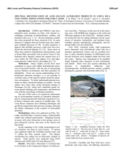
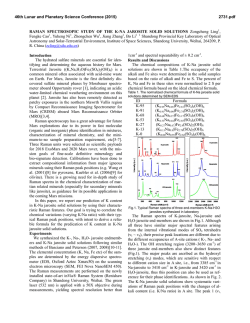
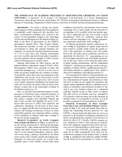
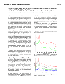
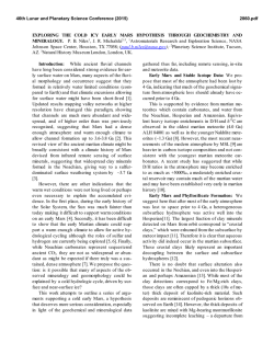
46th Lunar and Planetary Science Conference (2015)
2129.pdf
INVESTIGATING ACID SULFATE ALTERATION PROCESSES IN COSTA RICA AS TERRESTRIAL
ANALOGS FOR EARLY MARS. L. G. Beckerman1, B. M. Hynek1,2, and G. E. Alvarado3, 1Dept. of Geological
Sciences, University of Colorado-Boulder ([email protected]), 2Laboratory for Atmospheric and Space
Physics, 3Instituto Costarricense de Electricidad – ICE.
Introduction: Orbiters have identified abundant
sulfate deposits across the Martian surface, the presence of which has been confirmed with in situ observations by landers and rovers. For example, Spirit identified sulfite-rich soils at Gusev Crater that have been
attributed to acid sulfate alteration in a hydrothermal,
fumarolic system [e.g. 1]. Constraining mineral assemblages formed from acid sulfate alteration processes is
crucial to aiding interpretations of similar assemblages
on Mars. Different water/rock ratios, pH, temperature,
rock composition, and other environmental parameters
affect the pathways by which alteration occurs, and
produce varied end products. By characterizing the
mineral assemblage formed via a specific pathway,
similar mineral assemblages on Mars can be attributed
to specific processes.
Motivation: The presence of jarosite on Mars dramatically constricts the range of possible environmental conditions at those locales, if produced abiotically.
Alternatively, jarosite could be evidence for sulfuroxidizing microbes if aqueous conditions persisted
post-deposition [e.g. 2].
Recent work, however, suggests that some or all of
the jarosite identified on Mars may actually be ironrich natroalunite [3-5]. Alunite and jarosite are endmembers of a mineral group with idealized formulae
of AB3(SO4)2(OH)6, where jarosite has K+ in its A site
and Fe3+ in its B site while natroalunite has Na+ in its A
site and Al3+ in its B site. Although previous research
suggested that minerals do not naturally form between
the end-members of jarosite and alunite [e.g. 6],
McCollom et al. [4] found natroalunite at several Nicaraguan volcanoes displaying Fe substitution for Al in
the B site. This discovery resulted in the following
questions that this work aims to address: (1) Is Fe-rich
natroalunite a common alteration product in hydrothermal, acid fog systems? (2) What causes iron substitution in natroalunite? (3) What implications does iron
substitution have for understanding early Martian surface processes?
Alteration Mineralogy: To address these questions, we analyzed mineral assemblages from terrestrial analogs to early Mars at Poás and Turrialba volcanoes in Costa Rica. Basalt produced from recent eruptions at both sites is similar in composition to that of
sites on Mars [7]. Minerals were analyzed using a
portable Terra x-ray diffraction (XRD) instrument
analogous to CheMin on Curiosity, a Bruker D2 Phaser
XRD, and Raman spectroscopy. Poás and Turrialba
volcanoes in Costa Rica display similar acid sulfate
alteration products to those from Nicaraguan volcanoes
[8]. Major alteration minerals across different settings
at Poás and Turrialba include cristobalite, natroalunite,
amorphous silica, both gypsum and anhydrite, and
elemental sulfur (Figure 1).
Figure 1 : XRD diffractograms for two samples from a gully
on Turrialba at ~100°C showing major alteration minerals
with representative photos from the site. na – natroalunite, s
– sulfur, am – amorphous silica, g – gypsum.
Iron Substitution in Natroalunite: Concentrations of iron in natroalunite grains were determined
with a scanning eletron microscope (SEM) equipped
with an EDS detector and an electron microbe (EMP).
Natroalunite grains across different settings at Poás
and Turrialba contain varying concentrations of iron in
their B site and display no clear trend for environmental parameters that could be causing iron substitution.
Grains from the same cm-scale hand sample show significant variation in extent of iron substutition, suggesting that highly localized processes may be responsible for sequestration of iron in natroalunite (Table 1,
Figure 2). In some samples, iron is present as iron oxide patches and spherules rather than in natroalunite
(Figure 3).
Evaluation of Methodology: Capturing mineral
assemblages on Mars requires effective detection tools
on rovers and accurate interpretation of their data. Ra-
46th Lunar and Planetary Science Conference (2015)
man spectrometers are relatively new on rovers and
Curiosity is the first rover equipped with an XRD.
Studying the efficacy of these methods and others that
could be used in future missions will improve geochemical interpretations on Mars.
Sample
N
Fe#
Std. Dev.
Ttop6_1
3
7.82
1.49
Table 1: Four
different grains
(1,4,5 and 7)
Ttop6_4 3
3.38
0.42
from the same
Ttop6_5 3
1.92
0.19
hand
sample
(Ttop6) from a
Ttop6_7 5
17.1
2.03
gully on Turrialba at ~100°C display a wide range of iron substitution. N =
number of analyses with EMP, Fe# = [Fe]/([Fe]+[Al]) x 100
with [Fe] and [Al] in atomic wt%.
Figure 2: (left) Natroalunite grain
from Ttop6 with average Fe# of
11.8. Iron is evenly distributed
throughout the grain, unlike in
Figure 3.
Figure 3: (below) Natroalunite
grain covered with iron oxide
spherules, reflecting continued
acid sulfate alteration.
XRD: Terra XRD effectively captures major mineralogy of crystalline samples, including clays, and
serves as an effective analog for CheMin [9], but misses many minor and trace elements that can be identified with a benchtop XRD. Although some shifting of
natroalunite peaks due to iron substitution occurs [5],
Fe# cannot be effectively calculated with XRD data.
XRD also cannot identify all amorphous material when
used independently. As significant portions of the Martian crust have undergone weathering to form amor-
2129.pdf
phous material [e.g. 10,11], this is a key challenge for
understanding mineral assemblages on Mars via XRD.
Raman: While Raman spectroscopy can be used on
a bulk sample to obtain overall mineralogy, Raman can
also be used to map mineralogy across a sub-mm region in a thin section. Shifts in peak locations due to
iron substitution are more clearly visible in Raman
than in XRD diffraction patterns, but like XRD, Raman cannot identify amorphous or poorly ordered material. Remote Raman spectroscopy combined with
LIBS is already in use on Curiosity, and Raman spectroscopy is planned for both ExoMars and the 2020
rover, although all have, or will have, Raman for bulk
mineral analyses only.
SEM: Energy dispersive spectroscopy (EDS) coupled to SEM can enable direct imaging of natroalunites
at sub-micron scales, including spatial relations between Fe-rich and traditional natroalunites and the
distribution of Fe-rich natroalunite and/or iron oxides
within or on grains. EDS detectors are less accurate
than WDS detectors, which may result in less accurate
Fe#s. Ideally samples cached by the 2020 rover could
be evaluated with SEM once returned to Earth.
EMP: Electron microprobe analyses provide elemental and oxide weight abundances that can be used
not only to identify Fe-rich natroalunite, but also to
determine precise chemical formulae and Fe#s at a
sub-micron scale. However, beam damage easily occurs in natroalunite samples. Like with SEM, EMP
analyses of samples cached by the 2020 rover would
be invaluable.
Implications: A water-limited, acid fog setting,
such as that found at Poás and Turrialba in Costa Rica,
may have been widespread on Mars > 3.7 Ga. Characterizing the mineralogy of these sites in the context of
other terrestrial analogs for acid sulfate alteration on
Mars will improve interpretations of Martian mineral
assemblages. Furthermore, distinguishing between Ferich natroalunite and jarosite on Mars could help constrain the environmental parameters present during
deposition, and has implications for the possibility of
microbial life on ancient Mars.
References: [1] Yen A.S. et al. (2008) JGR 113, doi :
10.1029/2007JE002978. [2] Norlund, K.L.I, et al. (2010)
Chemical Geology 275, 235-242. [3] McCollom T.M. et al.
(2013) JGR:Planets 118, 577-614. [4] McCollom T.M. et al.
(2013) JGR:Planets 118, 1719-1751. [5] McCollom T.M. et
al. (2014) Am. Mineralogist 99, 948-964. [6] Papike J.J. et al.
(2006) Am. Mineralogist 91:7, 1197-1200. [7] Hynek B.M.
et al. (2014) LPS XXXXV, Abstract #2172 [8] Hynek B.M. et
al. (2013) JGR: Planets 118, 1-22. [9] Blake D. et al. (2012)
Space Sci Rev 170, 341–399. [10] Bish D.L. et al. (2013)
Science 341, doi : 10.1126/science.1238932. [11] Vaniman
D.T. et al. (2014) doi : 10.1126/science.1243480.
© Copyright 2026