
Download PDF
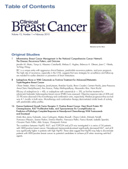
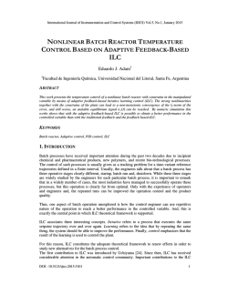
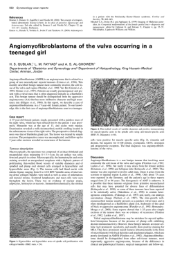
Reed et al. Breast Cancer Research (2015) 17:12
DOI 10.1186/s13058-015-0519-x
REVIEW
Invasive lobular carcinoma of the breast:
morphology, biomarkers and ’omics
Amy E McCart Reed1†, Jamie R Kutasovic1†, Sunil R Lakhani1,2,3 and Peter T Simpson1,3*
Abstract
Invasive lobular carcinoma of the breast is the most
common ‘special’ morphological subtype of breast
cancer, comprising up to 15% of all cases. Tumours
are generally of a good prognostic phenotype, being
low histological grade and low mitotic index, hormone
receptor positive and HER2, p53 and basal marker
negative, and with a generally good response to
endocrine therapy. Despite this, clinicians face countless
challenges in the diagnosis and long-term management
of patients, as they encounter a tumour that can be
difficult to detect through screening, elicits a very
invasive nature, a propensity for widespread metastatic
colonisation and, consequently, in some studies a
worse long-term poor outcome compared with invasive
carcinoma of no special type. Here we review the
morphological and molecular features that underpin
the disparate biological and clinical characteristics of
this fascinating tumour type.
Introduction
Invasive lobular carcinoma (ILC) is the most common
‘special’ type of breast cancer and presents with a distinct morphology and clinical behaviour compared with
invasive carcinoma of no special type (IC-NST). Typically, ILC tumours display features associated with a good
prognosis, being low grade and oestrogen receptor positive; however, the tumour can be highly metastatic [1]
and several studies demonstrate that the overall longterm outcome for patients diagnosed with ILC may be
similar or worse than for patients diagnosed with IC-NST
[2,3]. E-cadherin loss is responsible for the inherently
* Correspondence: [email protected]
†
Equal contributors
1
The University of Queensland, UQ Centre for Clinical Research, Herston, QLD
4029, Australia
3
School of Medicine, The University of Queensland, Herston, QLD 4006,
Australia
Full list of author information is available at the end of the article
discohesive phenotype associated with ILCs, and changes
at the genomic level account for this loss. Recent technological advances have generated masses of genomic and
transcriptomic data, some of which is further illuminating
the natural history of ILCs. We present here a review of
lobular carcinoma, paying particular attention to the morphological and immunophenotypic features of pre-invasive
and invasive lesions, the importance of E-cadherin dysfunction in tumour biology, transcriptomics, genomics and
diagnostic aspects that aid patient management.
Morphological characteristics of lobular neoplasia
and invasive lobular carcinoma
Diagnostic criteria for lobular neoplasia (LN) and ILC
(Figure 1) are now well established and described [4]
and so are only briefly outlined below. The term ‘lobular
neoplasia’ was introduced [5] to encompass a spectrum
of in situ neoplastic proliferations including atypical
lobular hyperplasia (ALH) and lobular carcinoma in situ
(LCIS), which describe different levels of involvement of
individual lobular units. The descriptions ALH and LCIS
are widely used to classify these lesions since they confer
different relative risks (4- to 5-fold and 8- to 10-fold, respectively) for the patient to subsequently develop invasive cancer compared with the general population [6].
By definition, neoplastic cells of LN remain confined to
the terminal duct-lobular unit, but they may exhibit
pagetoid spread in which cells can migrate along the
ductal system between the basement membrane and
normal epithelial cell population (Figure 2).
The cells of LN and ILC are typically small, monomorphic and lack cohesion, with round or notched ovoid
nuclei and a thin rim of cytoplasm. Intra-cytoplasmic
lumen, containing a central inclusion of mucin, may be
present and in some cells this may be large enough to
create a signet ring cell-type appearance (Figure 1). Cells
of classic LCIS or ILC may vary in appearance and have
been referred to as type A cells (classic) or the larger
type B cells (vesicular nuclei) that may show mild pleomorphism. Cells of the pleomorphic type of LCIS (PLCIS)
© 2015 Reed et al.; licensee BioMed Central. The licensee has exclusive rights to distribute this article, in any medium, for 6
months following its publication. After this time, the article is available under the terms of the Creative Commons Attribution
License (http://creativecommons.org/licenses/by/4.0), which permits unrestricted use, distribution, and reproduction in any
medium, provided the original work is properly credited. The Creative Commons Public Domain Dedication waiver (http://
creativecommons.org/publicdomain/zero/1.0/) applies to the data made available in this article, unless otherwise stated.
Reed et al. Breast Cancer Research (2015) 17:12
Figure 1 Morphological characteristics of invasive lobular
carcinoma and its variants. (A) Low power view of a terminal duct
lobular unit colonised by lobular carcinoma in situ. Classic invasive
lobular carcinoma is seen diffusely infiltrating the whole specimen
as single cells and single files of cells. The characteristic targetoid
growth pattern is evident on the left-hand side (see also Figure 2).
(B-G) Morphological variants of the classic type: (B) alveolar type,
with globular aggregates of approximately 20 cells; (C) solid type with
discohesive tumour cells growing in solid sheets; (D) a pleomorphic
variant - note the pink, foamy cytoplasm typical of an apocrine
phenotype and irregular nuclei; (E) pleomorphic invasive lobular
carcinoma with prominent signet ring cells; (F) invasive lobular
carcinoma showing mucinous/histiocytoid morphology; (G) mixed
ductal-lobular carcinoma.
may be larger still and exhibit marked nuclear pleomorphism, akin to that observed in high-grade ductal carcinoma
in situ (DCIS) [7]. Extensive or florid LCIS is also important to recognize. These lesions are characterized by the
proliferation of the same type A or type B cells, but there is
Page 2 of 11
marked expansion of involved lobular units and areas of
necrosis and microcalcification [8].
In classical ILC, the characteristic pattern of growth
involves the infiltration of single cells or single files of
cells through the stroma, with little disturbance of normal tissue architecture. The invading tumour cells are
frequently arranged in a concentric (targetoid) pattern
around normal ducts or structures (Figure 2). There are
a series of morphologically recognised variants that
demonstrate either cytological or architectural variation
of the characteristic features of classic ILC. Pleomorphic
lobular carcinoma (PLC) retains the distinctive growth
pattern of classic ILC but, as in its in situ counterpart
(PLCIS), there is marked cellular atypia and nuclear
pleomorphism relative to classic LN and ILC. PLC may
also have an increased mitotic rate, be composed of
signet ring cells (Figure 1) and/or show apocrine or histiocytoid differentiation. Conversely, the solid and alveolar variants are both characterized by classic ILC cells
(small, regular sized and lacking cohesion) that are
arranged in sheets (solid type) or in aggregates of at least
20 cells (alveolar type, Figure 1) rather than in single
cords of cells. Solid ILC may also be more frequently
pleomorphic and mitotically active relative to classic
ILC. Classic ILC may be admixed with one or more of
these morphological variants or with tumour cells of a
tubular growth pattern (tubulo-lobular carcinoma). Furthermore, around 5% of all invasive breast tumours exhibit
mixed features of both ductal and lobular differentiation
[4,9] (Figure 1).
Histological grading is an important part of breast
tumour classification, and is performed using the Nottingham histological grading system. There is debate,
however, as to the relevance of this system for the ‘special types’ including lobular carcinomas and some studies suggest it is of limited value since tubule formation is
rare (except in the tubulo-lobular variant), there is limited nuclear pleomorphism and the mitotic count is
frequently low. Consequently, most ILCs, including variants, are grade 2. Nevertheless, other studies report that
grade is indeed an independent prognostic factor in ILC,
as it is in breast cancer in general, with mitotic count
being the most useful component for predicting poor
outcome [10]. Furthermore, while several studies report
that morphological variants are aggressive subtypes associated with poor outcome, particularly relative to classic
type [11], evidence suggests that a nuclear pleomorphism score of 3 (which would indicate a classification of
PLC), in an overall grade 2 tumour does not add prognostic value, the most important discriminator being
overall grade and/or mitotic count [12].
ALH, LCIS and PLCIS can be frequently found colocalised in the same specimen, also alongside other
non-obligate precursors such as columnar cell lesions,
Reed et al. Breast Cancer Research (2015) 17:12
Page 3 of 11
Figure 2 Immunohistochemical staining of E-cadherin and its binding complex in invasive lobular carcinoma. Lobular carcinoma in situ
(LCIS) and invasive lobular carcinoma (ILC); note invasive neoplastic cells of ILC (arrows) growing in a targetoid fashion around the in situ
component. (A) E-cadherin and (B) β-catenin staining is negative in both LCIS and ILC, although positive staining is observed in the myoepithelial
cells surrounding the LCIS. (C) Pagetoid spread (asterisks) is also observed in this case, whereby the neoplastic cells (negative for E-cadherin) are
growing and invading between the luminal and myoepithelial cells of a normal ductal structure). (D) In the absence of E-cadherin, there is a
strong re-localisation of p120-catenin to the cytoplasm of neoplastic cells in LCIS and ILC.
atypical ductal hyperplasia and low-grade DCIS as part
of the ‘low-grade’ family of breast precursor lesions [13].
LNs frequently co-exist with invasive carcinomas of
lobular type, including classic ILC (Figures 1 and 2) and
tubulo-lobular carcinomas (in 90% and 57% of cases, respectively [13]), supporting a common evolutionary origin of these lesions. Indeed, the overlapping cytological
appearance and frequent co-localisation of LN and ILC,
combined with concordant immunophenotypic and molecular characteristics, supports the notion that LCIS
and PLCIS are clonal and non-obligate precursor lesions
for ILC and PLC, respectively [14,15].
Immunophenotyping invasive lobular carcinoma
Classic ILCs are almost always hormonally regulated. Up
to 95% of cases express oestrogen receptor (ER)α and 60
to 70% of cases express progesterone receptor [2,16,17],
whereas only 60 to 70% of IC-NST express these two
biomarkers. ERα is always expressed in the alveolar variant (100%) yet is less frequently found in pleomorphic
ILC (10 to 76%) [10,18]. The androgen receptor and ERβ
are also expressed in approximately 90% of ILCs [10,19].
The interplay between these receptors in ILC is yet to be
fully elucidated, though it is clear the high frequency of
hormone receptor expression reflects the overall good
response to endocrine-based therapies [2].
Biomarkers associated with poor clinical behaviour are
rarely expressed in ILC, including the HER2, p53 and
basal/myoepithelial markers (cytokeratins 14 and 5/6,
epidermal growth factor receptor, smooth muscle actin
and p63) [10,16,17]. Generally the proliferation index
(measured by Ki67 staining) is low in ILC, reflecting the
low mitotic count (see above) and this likely contributes
to reduced response to chemotherapy relative to patients
diagnosed with IC-NST. Pleomorphic ILCs, on the other
hand, are more likely to exhibit HER2 amplification (in 35
to 80% of cases) and p53 expression and the proliferation
index is typically higher [10,18].
E-cadherin dysfunction - master regulator of the
lobular phenotype
The characteristic discohesive growth pattern of ILC is the
result of the dysregulation of cell-cell adhesion properties,
primarily driven by the targeted disruption of the cell
adhesion molecule E-cadherin. E-cadherin is a calciumdependent transmembrane protein that mediates cell-cell
adhesion and cellular polarity by binding to itself on
neighbouring cells in a homophilic manner. The intracellular domain of E-cadherin associates with the actin cytoskeleton via α-, β-, γ- and p120 catenins to form adherens
junctions between non-neural epithelial cells. E-cadherin
is largely regulated by its catenin-binding partners, which
anchor E-cadherin to the membrane and the actin cytoskeleton. E-cadherin-mediated cell adhesion maintains cell
viability and when this adhesion is lost the detached cells
undergo a cell death program called anoikis.
Reed et al. Breast Cancer Research (2015) 17:12
In normal breast epithelial cells and in most IC-NST,
E-cadherin and the associated catenin binding proteins
are located at the cell membrane, maintaining cellular
cohesion. In contrast, approximately 90% of LNs and
ILCs, including variants, completely lack E-cadherin
protein expression [15,20-23]. The loss of E-cadherin in
ILC also results in the loss of α-, β- and γ-catenins, and
p120-catenin becomes up-regulated and re-localised to
the cytoplasm [24]. From a biological point of view, this
re-localisation of p120 has been found to enable anoikis
resistance in lobular cells, allowing them to survive independently of attachment to neighbouring epithelial cells
and promote cell migration through activation of Rho/
Rock signalling [25]. E-cadherin expression has become
an important diagnostic feature of LN and ILC and the
cytoplasmic localisation of p120-catenin is a positive
immunohistochemistry marker for ILC [26]. In combination these biomarkers may aid in classification when it
is difficult to differentiate between lobular and ductal
lesions [26]; however, there remains confusion regarding
the interpretation and so caution is warranted. In particular it is important to remember that approximately
10% of ILCs still express E-cadherin [10,22], either with
normal membrane localisation or aberrantly distributed
as fragmented membrane and/or cytoplasmic staining.
The E-cadherin-catenin complex may be dysfunctional
in these cases due to the presence of CDH1 gene mutation (see below) or aberrant/loss of expression of the
catenin binding proteins [22], which may be detected
using β-catenin and p120-catenin immunohistochemistry. However, a diagnosis of LN or ILC based on morphologic and cytologic criteria should therefore not be
reclassified as DCIS or IC-NST based on the status of
these immunohistochemical markers [26].
E-cadherin deregulation occurs in the earliest morphological stage of lobular tumourigenesis (that is, ALH)
and is frequently and irreversibly driven by genomic
alterations targeting its gene, CDH1 (located at chromosome 16q22.1). Molecularly, the patterns of E-cadherin
loss often follow Knudsen’s two hit hypothesis for a classic tumour suppressor gene, involving CDH1 mutation,
gene methylation and/or loss of heterozygosity in the region of 16q22.1 (frequently involving the whole chromosomal arm).
Promoter hypermethylation and concomitant downregulation of CDH1 expression has been reported in 21
to 77% of ILCs [27,28] and the detection of methylated
CDH1 promoter sequences in adjacent normal tissues
and in LN implies that this is an early hit [29]. The somatic copy number loss of 16q in ILC and ER-positive,
low-grade IC-NST is extremely frequent, suggesting
these tumours share a common pathway of evolution.
We reviewed the DNA copy number status at the CDH1
gene locus in the 153 lobular tumours from The Cancer
Page 4 of 11
Genome Atlas (TCGA) data resource [30,31] and this
revealed that 12.4% of tumours show a diploid copy
number; 84.3% show a single copy loss and 3.3% show a
putative homozygous deletion. Chromosomal analysis of
LNs has shown they too lose chromosome 16q [8,32-34],
suggesting this is also an early assault on the CDH1 gene
region.
Somatic mutations are found dispersed throughout the
CDH1 coding region and are frequently truncating [21]
(Figure 3). Identical CDH1 genetic mutations have been
detected in LCIS and in their adjacent invasive counterpart [15], which is a key finding implicating LCIS as a
direct (but non-obligate) precursor for ILC. Further to
this, CDH1 mutations were detected in LCIS [35], although, surprisingly, no mutations were found in adjacent,
microdissected ALH lesions. This may be a question of
technological sensitivity and so the application of highresolution massively parallel sequencing technologies is
certainly warranted to clarify such findings.
The reported frequency of CDH1 mutation and loss of
heterozygosity is unexpectedly discrepant between studies (from 30 to 80%) [10,21,36]. Improving technologies
and increasing cohort sizes have not necessarily resolved
this. For example, TCGA [37] reported from an exome
sequencing strategy (that is, enriching for exons only)
that CDH1 mutations were very common (30/36; 83%)
within the lobular histological subtype and, expectedly,
corresponded with low E-cadherin expression. TCGA
resource has now made comprehensive ’omic data available for 958 breast cancers through the cBioPortal
[30,31] and in an investigation of these data we identified CDH1 mutations in 78 out of 155 ILCs (50%). This
latter figure is supported by an independent exome sequencing study of ER-positive tumours in the clinical
context of aromatase inhibitor response, where they identified CDH1 mutation in 20 out of 40 ILCs [38].
Mutations in CDH1 have also been identified in other
types of epithelial cancers, most notably in diffuse gastric
carcinomas, which have a very similar infiltrative growth
pattern to ILC of the breast. Hereditary diffuse gastric
carcinoma is sometimes caused by a germline mutation
in CDH1 [39] and mutation carriers have an increased
risk for developing ILC. A diagnosis of ILC may also be
enriched within breast cancer families and since LNs/
ILCs more frequently present as multifocal or bilateral
disease, it fits with a theory of a germline predisposition
to tumour development. Despite E-cadherin being the
obvious candidate for such a predisposition, early work
suggested CDH1 germline variants are rare in familial
lobular breast cancer [40] but do account for some cases
of bilateral ILC [41]. Considerable evidence arising
from studying the human disease therefore exists for
E-cadherin playing a major role in the initiation and biology of both lobular and diffuse gastric cancer. Animal
Reed et al. Breast Cancer Research (2015) 17:12
Page 5 of 11
Figure 3 Genomic architecture of invasive lobular carcinoma. (A) CIRCOS plot of an invasive lobular carcinoma (ILC) tumour profiled using
the Illumina Omni 2.5 million SNP CNV array. Note the archetypal ILC changes, including chromosome 1q gain, 8p amplification, 11q13 amplification
and 16q deletion. (B) Spectrum of somatic mutations across the E-cadherin coding region in the cBioPortal ILC data set [30,31]. Note the cadherin
prodomain in green and the cadherin cytoplasmic domain in blue; missense mutations in green and nonsense mutations in red. (C) Oncoprint
depicting the frequency of somatic mutations in key, recurrently altered cancer genes (CDH1, TP53, PIK3CA, ERBB2) affecting 75% of the 155 ILCs
in The Cancer Genome Atlas cohort [30,31]. Percentages are numbers of tumours exhibiting an alteration in the specified gene.
models of hereditary diffuse gastric cancer and lobular
breast cancer provide additional support for this concept,
whereby CDH1 germline deficiency in combination with
a second hit (carcinogen treatment or TP53 mutation) is
sufficient to initiate disease development [42,43]. (These
aspects are covered in more detail in a review in this
series [44].)
The loss of E-cadherin is also associated with the
process of epithelial to mesenchymal transition (EMT)
where cells lose polarity and adhesion to become more
migratory and invasive during embryonic morphogenesis
and wound healing. Tumour cells are believed to be able
to hijack this process to facilitate migration away from
the primary tumour microenvironment and metastatic
dissemination. The acquisition of the mesenchymal phenotype is accompanied by cadherin switching (loss of
E-cadherin and activation of N-cadherin), which is driven
by transcriptional regulators of E-cadherin, including
SNAIL and TWIST, as well as post-transcriptionally
active microRNAs (for example, the miR200 family), and
the gain in expression of mesenchymal markers, such as
vimentin. Given the loss of E-cadherin and the infiltrating growth pattern of ILC, it is tempting to speculate that
EMT plays a mechanistic role in driving this phenotype.
Indeed, a meta-analysis of microarray gene expression
data found TWIST to be highly expressed in human ILC
Reed et al. Breast Cancer Research (2015) 17:12
Page 6 of 11
samples, showing 70% had elevated TWIST mRNA expression, compared with 32% of ductal carcinomas [45].
However, immunohistochemical analysis of EMT markers
in human breast tumours demonstrated that: i) neoplastic
lobular cells retain their epithelial identity; ii) TWIST protein was expressed by the fibroblasts in the prominent stromal component of ILC; and iii) only 1 out of 24 (4%) ILCs
expressed EMT markers [46]. While EMT is traditionally
associated with late stages of tumour progression (invasion
and metastasis) and is a dynamic process, the loss of
E-cadherin in ILC is an early and typically irreversible
event in ILC. Thus, the functional role of EMT in driving
the invasive nature of ILC remains unlikely.
associated with E-cadherin dysfunction, at the individual
transcript level there was minimal overlap between all five
studies. Given the variety of platforms used for these
assays, small sample sizes and modes of analysis, this is
not altogether surprising. A meta-analysis of these studies
identified THBS4 (thrombospondin 4) as a potential ILC
biomarker, but investigations at the protein level confirmed no difference in expression between ILCs and their
ductal counterparts, and instead revealed THBS4 as a
marker of tumour-associated extracellular matrix [56].
Again, this finding is probably more associated with the
fact that ILC tumours exhibit a higher stromal content,
thus skewing the subsequent downstream analyses.
Transcriptome profiling of lobular tumours
At the turn of the century, a pivotal study used gene
expression profiling microarrays to categorise breast
cancers into a series of ‘intrinsic’ subtypes that stratified
prognosis: luminal A, luminal B, HER2, and basal-like
[47,48]. These categories have since been expanded to
include claudin-low [47,48] and normal breast-like. Due
to the nature of ILCs generally being low-grade and ERpositive, they are frequently classed as luminal A, and
owing to their commonly infiltrative histology and thus
a comparatively reduced tumour to stroma cellularity
(compared to ductal tumours), they may also be classed
as normal-like, simply as a consequence of there being
more normal cells and/or stroma in their processed
samples [49]. Ultimately though, like ductal carcinomas,
they are a heterogeneous group and have the potential
to be classed as any of the defined subtypes, including
molecular apocrine for the PLC variant [48,50], while,
interestingly, the non-lobular special types of breast cancer (for example, medullary, metaplastic, micropapillary,
tubular, apocrine and neuroendocrine carcinomas) cluster within a single subtype only, underscoring their more
inherent homogeneity.
Gene expression profiling studies have also been
undertaken to better understand the biological differences between lobular and ductal invasive tumours.
Overall, the number of lobular tumours profiled has
been considerably lower than that for ductal invasive tumours [51-55]). Korkola and colleagues [52] defined 11
genes as capable of differentiating ILCs from ductal carcinomas, but more recent studies report larger, functional groups of genes as being responsible for their
different aetiologies. Of most relevance are those functional gene groups that were identified when 20 ILCs
were compared with 91 ER-positive, grade-matched invasive ductal carcinomas (IDCs): adhesion, transforming
growth factor beta signalling; cell communication and
trafficking; actin remodelling; lipid/prostaglandin synthesis; transcription factor/immediate early genes [54].
Ultimately, other than expected transcriptional changes
The genomic landscape of lobular carcinomas
LNs and ILCs are more likely to be diploid than ductal
tumours [16]. Indeed, chromosomal and array-based
comparative genomic hybridisation (aCGH) analyses
have defined, on a gross scale, the genomic profile of
lobular carcinomas - in short, they harbour fewer
chromosomal changes than ductal carcinomas and are
generally less complex. Genomic losses, such as at 16p,
16q, 17p and 22q, and gains at 6q were detected in LN
by chromosomal CGH [33]. The key alterations identified more recently by aCGH in classic LCIS, florid/
extensive LCIS and PLCIS are 1q gain and 16q loss,
with increased genomic complexity observed in the latter
two groups of lesions, including loss of 8p, 11q and 17p
and amplifications at 11q13 (CCND1) and 17q12 (ERBB2)
[8,14,34]. Like their pre-invasive counterparts and ERpositive IC-NST, both classic and pleomorphic ILC exhibit
a high frequency of gain of chromosome 1q and loss of
16q [18,23,57,58] and it has been reported that all ILCs
lose at least part of 16q [58]. Other recurrent alterations
include losses at 8p23-p21, 11q14.1-q25, and 13q, gains of
8q and 16p, and high-level amplifications at 1q32, 8p12p11.2, and 11q13. Although some candidate genes in the
various regions have been postulated (for example, FGFR1
in 8p12-p11.2 and CCND1 in 11q13 [23]), no definitive
data confirming the drivers contained in these various
regions have been reported specifically for lobular breast
cancer. This is likely a result of the complexity of the
chromosomal changes and the context-dependent nature
of some of these alterations. Numerous candidate oncogenes have been identified in these regions but not specifically for lobular tumours - for example, ZNF703 gene
amplification at 8p12 specifies luminal B breast cancer
[59]. As mentioned above, PLC contains a similar profile
of chromosomal change, although there is increased complexity and additional amplifications are present - 8q24
(MYC), 17q12 (ERBB2/Her2) and 20q13, which are usually considered to be archetypal changes of high-grade
ductal tumours [18]. Some attempts have been made
to classify tumour genome profiles based on genomic
Reed et al. Breast Cancer Research (2015) 17:12
architecture as either simple, complex-firestorm or
complex-sawtooth. The genomes of both classic and pleomorphic ILC are generally classified as simple (in that they
frequently harbour 1q gain and 16q loss and few other alterations) or complex-firestorm (relating to the additional
presence of complex, high-level amplifications at the
stated loci) [18,23]. It is conceivable that those ILCs that
are classed as complex-firestorm have a worse prognosis,
though this has yet to be explored.
A catalogue of the transcriptomic and genomic architecture of 2,000 breast cancers, and their integration into
novel clusters was reported in 2012 [60]. The discovery
set of this large study included 148 classic ILCs, of
which 88.5% were ER positive and were classified as:
luminal A, 44.9%; luminal B, 19.7%; basal, 2.7%; HER2,
6.1%; normal, 25.9%. This study also presented an alternative categorisation algorithm combining transcriptome
and genomic copy number data to define 10 ‘integrative
clusters’ (IntClusts). ILCs were predominantly assigned
to IntClust 3 (39.7%), 4 (23.5%) and 8 (14.7%), with varying frequencies of the archetypal chromosomal changes
(1q+, 16p+, 16q-). Predictably, IntClust 3, into which
most ILCs segregated, showed overall the simplest genomes, a high frequency of 1q + and 16q- and the best
survival outcome. Similarly, tumours in IntClust 8 also
harbour a high frequency of 1q + and 16q-, but also
16p+. Conversely, tumours in IntClust 4 showed infrequent 1q + and 16q-. The groups in which lobular
carcinomas are not enriched (that is, less than approximately 6% of the ILCs) showed more recurrent gains/
amplifications on chromosomes 8q, 11q or 17q. Subtle
variation in the genomic alterations in these tumours may
therefore have a strong influence on tumour behaviour.
The data era: ‘next-generation’ sequencing
Significant technological advances in recent years have
meant that the depth of interrogation of individual
tumour genomes has increased significantly. This socalled ‘next-generation sequencing’ technology combined with the activities of several large consortia has
led to the production of masses of high quality sequence
and genomic copy number data for large numbers of
tumours. As noted above, two studies have performed
exome sequencing on ILC of any significant numbers
[37,38]. Beyond the highly recurrent mutations in
CDH1 and PIK3CA, which for PIK3CA the mutation
rate is similar to that observed overall in ER-positive
breast cancers, there is a paucity of recurrent driver
mutations in this tumour type (Table 1), supporting
the idea that heterogeneity within and between tumours
is complex.
One of the first studies to report the application of the
then novel sequencing technologies to breast cancer
samples was that of Shah and colleagues in 2009 [61].
Page 7 of 11
Table 1 Recurrent mutations in invasive lobular carcinoma
Gene
cBioPortal [30,31]
(n = 155)
Ellis et al. [38]
(n = ~40)a
Ross et al. [62]
(n = 22)b
AKT1
0.6%
NR
9.1%
ARID1A
4.5%
NR
NA
ATR
2.0%
2.5%
NA
BIRC6
2.0%
2.5%
NA
CDH1
50.0%
50.0%
100%
CDKN1B
1.3%
3.4%
NA
ERBB2
3.8%
NR
18%
GATA3
2.6%
3.4%
NA
KRAS
0.6%
NR
9.1%
MALAT1
0.0%
2.5%
NA
MAP2K4
0.6%
3.4%
9.1%
MAP3K1
5.0%
13.8%
NA
NCOR1
4.5%
9.1%
NA
NF1
3.2%
NR
NA
PIK3CA
41.0%
41.4%
36.4%
RB1
1.30%
0
9.1%
RUNX1
7.0%
5.0%
4.5%
SMAD4
1.30%
NR
NA
TP53
5.0%
12.1%
36.4%
a
The exact number of invasive lobular carcinomas in the Ellis study is difficult
to define as several cohorts were assessed using a variety of technologies.
b
The Ross study selected only CDH1 mutant cases. NA, not available; NR,
not reported.
This study sequenced a pleural effusion metastasis and
matched primary ILC diagnosed 9 years earlier and
found that 5 somatic mutations (of a possible 32 defined
for the metastasis) were present in the primary tumour,
a telling comment on the degree of clonal evolution occurring during progression from primary clone to metastasis. This patient also had an ERBB2 mutation, as did 2
of 192 ILCs in their validation set. Somatic mutations
(not including amplifications) in ERBB2 have since been
shown to be generally rare in breast cancer but interestingly were significantly enriched in the ILC subtype [37].
Consulting the cBioPortal [30,31] for an updated data
review, 6 of 155 ILCs (3.9%) harboured an ERBB2
mutation. Interestingly, in a massively parallel, targeted
amplicon sequencing of ‘actionable cancer genes’ in ILC
post-treatment relapse (that is, recurrence or metastasis),
Ross and colleagues [62] reported HER2/ERBB2 genetic
alterations in 6 of 22 (27%) cases, including 4 mutations,
one gene fusion and one amplification. HER2 is an
important clinically actionable target, indicating this
type of targeted sequencing analysis, which has greater
sensitivity than exome sequencing and is applicable to
formalin-fixed paraffin-embedded tissue, may soon aid
in the management of patients when planning primary or
secondary treatment regimes.
Reed et al. Breast Cancer Research (2015) 17:12
Diagnostic algorithms
As the era of molecular technology for subtyping disease
and identifying molecular targets takes huge leaps forward it is tempting to begin to ignore the more traditional morphological classification of disease and consider
molecular subtyping (for example, luminal, basal, HER2
subtypes) and testing (for example, OncotypeDX) for classification and management. However, the breast cancer
morphological special types remain fundamental to the
management of patients since the biological and clinical
significance of these entities provides important information regarding appropriate management strategies and
outcomes.
A diagnosis of lobular carcinoma, as a special morphological type, supports this idea, since there are clinical
and biological features that set it apart from the more
commonly diagnosed IC-NST, and despite the ‘good
prognostic features’ exhibited by ILC, some large studies
consistently demonstrate that ILCs have a similar or
worse long-term outcome compared with IC-NST [2,3].
Many of the challenging aspects in the diagnosis and
management of ILC relate to the indolent but highly
infiltrative nature of the tumour growth and the biology
of dysfunctional E-cadherin that underpins this. For instance, LNs and ILCs are not always detected as a palpable
mass and can be difficult to detect by mammography [63]
owing to the rare association with calcification and the
lack of host stromal response to the diffusely infiltrating
tumour.
Differentiating classic LCIS from its morphological
variants (that is, extensive/florid LCIS and PLCIS) may
be important from a management point of view, owing
to anecdotal evidence that these lesions have a different
clinical course and that they exhibit more genomic
instability [8,14,34]. Correct diagnostic classification of
LN is also very important because the management of
patients diagnosed with LN are different to those with
DCIS, in the setting of core needle biopsy or surgical
margin status, where further excision is required for all
cases of DCIS but not for LN. There is a significant body
of literature regarding this and readers are directed to
[10,26,64] and references therein for more information.
Briefly, differentiating LCIS and PLCIS from low- and
high-grade DCIS, respectively, or lesions with indeterminate features may be difficult in certain scenarios.
The use of ancillary immunohistochemical staining for
E-cadherin, β-catenin and p120-catenin can therefore
be helpful to aid classification [24,26]. In terms of ILC,
histological grading is considered a critical component
of classification, and description of the morphological
variants is recommended given the prognostic insight
this may provide and the potential for future epidemiological and biological studies related to tumour
subtyping [11,12,65]. As above, the use of E-cadherin,
Page 8 of 11
p120 catenin or β-catenin is appropriate to help resolve
the diagnosis of difficult cases, although it is important to
consider classification first based on morphology and
cytology and not to reclassify a bona fide ILC as IC-NST
based on ‘normal’ E-cadherin or p120-catenin staining
since around 10% of ILCs still express membranous
E-cadherin [20,22,26]. Pan-cytokeratin markers are also
utilized to differentiate small ILC cells from macrophages
in biopsies and extremely diffuse cases.
ILCs respond less well to chemotherapy compared to
IC-NST, probably resulting in part from their indolent,
low proliferative index (low mitotic count and low Ki-67
index). Many molecular tests are now available to prognosticate and inform decisions regarding the addition of
chemotherapy to a patient’s treatment program. Many
ILC tumours meet the requirements for the Oncotype
DX 21-gene clinical assay, in that they are generally
grade 2 and ER positive, and may not have spread to the
lymph nodes. The usefulness of this and other tests is
reviewed in [66], where it is also noted that many of
these signatures focus on proliferation as a mechanism
of assessing likelihood of recurrence.
Expression of ER, progesterone receptor and HER2
guide therapeutic decisions and the vast majority of
patients will receive endocrine-based therapy, for which
there is generally a good response [2]; however, de novo
or acquired resistance is an inevitable problem for some
patients. The somatic mutation profile of a tumour may
contribute to this; for example, tumours harbouring or
acquiring driver mutations in ESR1 [67] or ERBB2 [37]
or amplifications at 8p12 (FGFR1) or 11q13 (CCND1)
[23] may be less responsive to targeted endocrine therapy. In support of this, the ER-positive ILC cell line
model MDA-MB-134VI was found to be de novo tamoxifen resistant, yet cells were sensitized to anti-oestrogen
therapy when in combination with FGFR1 inhibitors [68].
Oestrogen-related receptor gamma/AP1 signalling may
also mediate tamoxifen resistance in the SUM44 cell
model system [69]. Recent research has also shown that
PIK3CA mutations are selected for during progression
from the primary ILC tumour to a local recurrence but
not through to dissemination of distant metastases [70].
While links between PIK3CA mutation and endocrine
therapy resistance have been investigated in some breast
cancers, this mechanism has not been specifically studied
in ILC; however, it is reasonable to hypothesize that this
may be the case in some endocrine-resistant ILCs [71]. A
gene expression study comparing ILC and IDC tumour biopsies in the neo-adjuvant setting suggests that letrozole
both induces near identical transcriptome changes in the
tumour types and does not interfere with histological
subtype-specific gene expression [72]. Recent data suggest
there may be an improved response to the aromatase inhibitor letrozole compared with tamoxifen in ILCs but the
Reed et al. Breast Cancer Research (2015) 17:12
biological mechanisms driving the differences in response
need to be further investigated [73]. As our understanding
of the biological mechanisms that underpin response and
resistance to anti-oestrogen therapies improves we will be
able to better predict which treatment regime would be
most effective (endocrine therapy or in combination with
other targeted therapies).
Conclusion
Lobular carcinoma is an important breast cancer subtype
with some peculiar clinical and biological characteristics
compared with the more commonly diagnosed IC-NST.
Rather surprisingly, and despite the good prognostic
features of the primary tumour and good response to
endocrine therapy, the long-term outcome for patients
diagnosed with ILC is, in some studies, worse than for
IC-NST. There remain significant challenges, therefore,
managing patients with this specific disease. Although
considered a ‘special’ histological type, the disease is heterogeneous, and so identifying patients with poor prognostic subtypes will likely provide benefit in delineating
more personalized and aggressive treatment or monitoring
for disease progression. A detailed assessment of the genomic landscape of a large cohort of ILCs with long-term
follow-up and/or in the context of treatment resistance will
no doubt be essential to moving forward with precision
medicine for patients diagnosed with this tumour type.
Note
This article is part of a series on Lobular breast cancer,
edited by Ulrich Lehmann. Other articles in this series can
be found at http://breast-cancer-research.com/series/LBC.
Abbreviations
aCGH: Array-based comparative genomic hybridisation; ALH: Atypical lobular
hyperplasia; CGH: Comparative genomic hybridisation; DCIS: Ductal
carcinoma in situ; EMT: Epithelial to mesenchymal transition; ER: Oestrogen
receptor; IC-NST: Invasive carcinoma no special type; IDC: Invasive ductal
carcinoma; ILC: Invasive lobular carcinoma; IntClust: Integrative cluster;
LCIS: Lobular carcinoma in situ; LN: Lobular neoplasia; PLC: Pleomorphic
lobular carcinoma; PLCIS: Pleomorphic lobular carcinoma in situ; TCGA: The
Cancer Genome Atlas.
Competing interests
The authors declare that they have no competing interests.
Acknowledgements
We apologise to authors whose work we were not able to discuss due to
space limitations. We thank Sarah Sim for insightful discussion. PTS was
the recipient of a fellowship from the National Breast Cancer Foundation,
Australia.
Author details
1
The University of Queensland, UQ Centre for Clinical Research, Herston, QLD
4029, Australia. 2Pathology Queensland, The Royal Brisbane & Women’s and
Gold Coast Hospitals, Herston, QLD 4029, Australia. 3School of Medicine, The
University of Queensland, Herston, QLD 4006, Australia.
Page 9 of 11
References
1. Ferlicot S, Vincent-Salomon A, Medioni J, Genin P, Rosty C, Sigal-Zafrani B,
et al. Wide metastatic spreading in infiltrating lobular carcinoma of the
breast. Eur J Cancer. 2004;40:336–41.
2. Rakha EA, El-Sayed ME, Powe DG, Green AR, Habashy H, Grainge MJ, et al.
Invasive lobular carcinoma of the breast: response to hormonal therapy and
outcomes. Eur J Cancer. 2008;44:73–83.
3. Pestalozzi BC, Zahrieh D, Mallon E, Gusterson BA, Price KN, Gelber RD, et al.
Distinct clinical and prognostic features of infiltrating lobular carcinoma of
the breast: combined results of 15 International Breast Cancer Study Group
clinical trials. J Clin Oncol. 2008;26:3006–14.
4. Lakhani SR, Ellis IO, Schnitt SJ, Tan PH, van de Vijver MJ. WHO Classification
of Tumours of the Breast. 4th ed. Lyon: IARC; 2012.
5. Haagensen CD, Lane N, Lattes R, Bodian C. Lobular neoplasia (so-called
lobular carcinoma in situ) of the breast. Cancer. 1978;42:737–69.
6. Page DL, Kidd Jr TE, Dupont WD, Simpson JF, Rogers LW. Lobular neoplasia
of the breast: higher risk for subsequent invasive cancer predicted by more
extensive disease. Hum Pathol. 1991;22:1232–9.
7. Sneige N, Wang J, Baker BA, Krishnamurthy S, Middleton LP. Clinical,
histopathologic, and biologic features of pleomorphic lobular (ductal-lobular)
carcinoma in situ of the breast: a report of 24 cases. Mod Pathol.
2002;15:1044–50.
8. Shin SJ, Lal A, De Vries S, Suzuki J, Roy R, Hwang ES, et al. Florid lobular
carcinoma in situ: molecular profiling and comparison to classic lobular
carcinoma in situ and pleomorphic lobular carcinoma in situ. Hum Pathol.
2013;44:1998–2009.
9. Rakha EA, Gill MS, El-Sayed ME, Khan MM, Hodi Z, Blamey RW, et al. The
biological and clinical characteristics of breast carcinoma with mixed ductal
and lobular morphology. Breast Cancer Res Treat. 2009;114:243–50.
10. Rakha EA, Ellis IO. Lobular breast carcinoma and its variants. Semin Diagn
Pathol. 2010;27:49–61.
11. Iorfida M, Maiorano E, Orvieto E, Maisonneuve P, Bottiglieri L, Rotmensz N,
et al. Invasive lobular breast cancer: subtypes and outcome. Breast Cancer
Res Treat. 2012;133:713–23.
12. Rakha EA, van Deurzen CH, Paish EC, Macmillan RD, Ellis IO, Lee AH.
Pleomorphic lobular carcinoma of the breast: is it a prognostically
significant pathological subtype independent of histological grade? Mod
Pathol. 2013;26:496–501.
13. Abdel-Fatah TM, Powe DG, Hodi Z, Lee AH, Reis-Filho JS, Ellis IO. High
frequency of coexistence of columnar cell lesions, lobular neoplasia,
and low grade ductal carcinoma in situ with invasive tubular carcinoma and
invasive lobular carcinoma. Am J Surg Pathol. 2007;31:417–26.
14. Reis-Filho JS, Simpson PT, Jones C, Steele D, Mackay A, Iravani M, et al.
Pleomorphic lobular carcinoma of the breast: role of comprehensive molecular
pathology in characterization of an entity. J Pathol. 2005;207:1–13.
15. Vos CB, Cleton-Jansen AM, Berx G, de Leeuw WJ, ter Haar NT, van Roy F,
et al. E-cadherin inactivation in lobular carcinoma in situ of the breast: an
early event in tumorigenesis. Br J Cancer. 1997;76:1131–3.
16. Arpino G, Bardou VJ, Clark GM, Elledge RM. Infiltrating lobular carcinoma of
the breast: tumor characteristics and clinical outcome. Breast Cancer Res.
2004;6:R149–56.
17. Sastre-Garau X, Jouve M, Asselain B, Vincent-Salomon A, Beuzeboc P, Dorval T,
et al. Infiltrating lobular carcinoma of the breast. Clinicopathologic analysis of
975 cases with reference to data on conservative therapy and metastatic
patterns. Cancer. 1996;77:113–20.
18. Simpson PT, Reis-Filho JS, Lambros MB, Jones C, Steele D, Mackay A, et al.
Molecular profiling pleomorphic lobular carcinomas of the breast: evidence
for a common molecular genetic pathway with classic lobular carcinomas.
J Pathol. 2008;215:231–44.
19. Skliris GP, Munot K, Bell SM, Carder PJ, Lane S, Horgan K, et al. Reduced
expression of oestrogen receptor beta in invasive breast cancer and its
re-expression using DNA methyl transferase inhibitors in a cell line model.
J Pathol. 2003;201:213–20.
20. Rakha EA, Patel A, Powe DG, Benhasouna A, Green AR, Lambros MB, et al.
Clinical and biological significance of E-cadherin protein expression
in invasive lobular carcinoma of the breast. Am J Surg Pathol.
2010;34:1472–9.
21. Berx G, Cleton-Jansen AM, Strumane K, de Leeuw WJ, Nollet F, van Roy F,
et al. E-cadherin is inactivated in a majority of invasive human lobular breast
cancers by truncation mutations throughout its extracellular domain.
Oncogene. 1996;13:1919–25.
Reed et al. Breast Cancer Research (2015) 17:12
22. Da Silva L, Parry S, Reid L, Keith P, Waddell N, Kossai M, et al. Aberrant
expression of E-cadherin in lobular carcinomas of the breast. Am J Surg
Pathol. 2008;32:773–83.
23. Reis-Filho JS, Simpson PT, Turner NC, Lambros MB, Jones C, Mackay A, et al.
FGFR1 emerges as a potential therapeutic target for lobular breast
carcinomas. Clin Cancer Res. 2006;12:6652–62.
24. Dabbs DJ, Bhargava R, Chivukula M. Lobular versus ductal breast
neoplasms: the diagnostic utility of p120 catenin. Am J Surg Pathol.
2007;31:427–37.
25. Schackmann RC, van Amersfoort M, Haarhuis JH, Vlug EJ, Halim VA,
Roodhart JM, et al. Cytosolic p120-catenin regulates growth of metastatic
lobular carcinoma through Rock1-mediated anoikis resistance. J Clin Invest.
2011;121:3176–88.
26. Dabbs DJ, Schnitt SJ, Geyer FC, Weigelt B, Baehner FL, Decker T, et al.
Lobular neoplasia of the breast revisited with emphasis on the role of
E-cadherin immunohistochemistry. Am J Surg Pathol. 2013;37:e1–11.
27. Droufakou S, Deshmane V, Roylance R, Hanby A, Tomlinson I, Hart IR.
Multiple ways of silencing E-cadherin gene expression in lobular carcinoma
of the breast. Int J Cancer. 2001;92:404–8.
28. Sarrio D, Moreno-Bueno G, Hardisson D, Sanchez-Estevez C, Guo M,
Herman JG, et al. Epigenetic and genetic alterations of APC and CDH1
genes in lobular breast cancer: relationships with abnormal E-cadherin
and catenin expression and microsatellite instability. Int J Cancer.
2003;106:208–15.
29. Zou D, Yoon HS, Perez D, Weeks RJ, Guilford P, Humar B. Epigenetic
silencing in non-neoplastic epithelia identifies E-cadherin (CDH1) as a target
for chemoprevention of lobular neoplasia. J Pathol. 2009;218:265–72.
30. Cerami E, Gao J, Dogrusoz U, Gross BE, Sumer SO, Aksoy BA, et al. The cBio
cancer genomics portal: an open platform for exploring multidimensional
cancer genomics data. Cancer Discov. 2012;2:401–4.
31. Gao J, Aksoy BA, Dogrusoz U, Dresdner G, Gross B, Sumer SO, et al.
Integrative analysis of complex cancer genomics and clinical profiles using
the cBioPortal. Sci Signal. 2013;6:pl1.
32. Simpson PT, Reis-Filho JS, Gale T, Lakhani SR. Molecular evolution of breast
cancer. J Pathol. 2005;205:248–54.
33. Lu YJ, Osin P, Lakhani SR, Di Palma S, Gusterson BA, Shipley JM.
Comparative genomic hybridization analysis of lobular carcinoma in situ
and atypical lobular hyperplasia and potential roles for gains and losses of
genetic material in breast neoplasia. Cancer Res. 1998;58:4721–7.
34. Chen YY, Hwang ES, Roy R, DeVries S, Anderson J, Wa C, et al. Genetic and
phenotypic characteristics of pleomorphic lobular carcinoma in situ of the
breast. Am J Surg Pathol. 2009;33:1683–94.
35. Mastracci TL, Tjan S, Bane AL, O’Malley FP, Andrulis IL. E-cadherin alterations
in atypical lobular hyperplasia and lobular carcinoma in situ of the breast.
Mod Pathol. 2005;18:741–51.
36. Cleton-Jansen AM. E-cadherin and loss of heterozygosity at chromosome 16
in breast carcinogenesis: different genetic pathways in ductal and lobular
breast cancer? Breast Cancer Res. 2002;4:5–8.
37. Network CGA. Comprehensive molecular portraits of human breast
tumours. Nature. 2012;490:61–70.
38. Ellis MJ, Ding L, Shen D, Luo J, Suman VJ, Wallis JW, et al. Whole-genome
analysis informs breast cancer response to aromatase inhibition. Nature.
2012;486:353–60.
39. Guilford P, Hopkins J, Harraway J, McLeod M, McLeod N, Harawira P, et al.
E-cadherin germline mutations in familial gastric cancer. Nature.
1998;392:402–5.
40. Schrader KA, Masciari S, Boyd N, Salamanca C, Senz J, Saunders DN, et al.
Germline mutations in CDH1 are infrequent in women with early-onset or
familial lobular breast cancers. J Med Genet. 2011;48:64–8.
41. Petridis C, Shinomiya I, Kohut K, Gorman P, Caneppele M, Shah V, et al.
Germline CDH1 mutations in bilateral lobular carcinoma in situ. Br J Cancer.
2014;110:1053–7.
42. Derksen PW, Liu X, Saridin F, van der Gulden H, Zevenhoven J, Evers B, et al.
Somatic inactivation of E-cadherin and p53 in mice leads to metastatic
lobular mammary carcinoma through induction of anoikis resistance and
angiogenesis. Cancer Cell. 2006;10:437–49.
43. Humar B, Blair V, Charlton A, More H, Martin I, Guilford P. E-cadherin
deficiency initiates gastric signet-ring cell carcinoma in mice and man.
Cancer Res. 2009;69:2050–6.
44. Christgen M, Derksen PW. Lobular breast cancer: molecular basis, mouse
and cellular models. Breast Cancer Res. in press.
Page 10 of 11
45. Yang J, Mani SA, Donaher JL, Ramaswamy S, Itzykson RA, Come C, et al.
Twist, a master regulator of morphogenesis, plays an essential role in tumor
metastasis. Cell. 2004;117:927–39.
46. Sarrio D, Rodriguez-Pinilla SM, Hardisson D, Cano A, Moreno-Bueno G,
Palacios J. Epithelial-mesenchymal transition in breast cancer relates to
the basal-like phenotype. Cancer Res. 2008;68:989–97.
47. Perou CM, Sorlie T, Eisen MB, van de Rijn M, Jeffrey SS, Rees CA, et al.
Molecular portraits of human breast tumours. Nature. 2000;406:747–52.
48. Prat A, Parker JS, Karginova O, Fan C, Livasy C, Herschkowitz JI, et al.
Phenotypic and molecular characterization of the claudin-low intrinsic
subtype of breast cancer. Breast Cancer Res. 2010;12:R68.
49. Mackay A, Weigelt B, Grigoriadis A, Kreike B, Natrajan R, A’Hern R, et al.
Microarray-based class discovery for molecular classification of breast cancer:
analysis of interobserver agreement. J Natl Cancer Inst. 2011;103:662–73.
50. Weigelt B, Horlings HM, Kreike B, Hayes MM, Hauptmann M, Wessels LF,
et al. Refinement of breast cancer classification by molecular
characterization of histological special types. J Pathol. 2008;216:141–50.
51. Gruel N, Lucchesi C, Raynal V, Rodrigues MJ, Pierron G, Goudefroye R, et al.
Lobular invasive carcinoma of the breast is a molecular entity distinct from
luminal invasive ductal carcinoma. Eur J Cancer. 2010;46:2399–407.
52. Korkola JE, DeVries S, Fridlyand J, Hwang ES, Estep AL, Chen YY, et al.
Differentiation of lobular versus ductal breast carcinomas by expression
microarray analysis. Cancer Res. 2003;63:7167–75.
53. Turashvili G, Bouchal J, Baumforth K, Wei W, Dziechciarkova M, Ehrmann J,
et al. Novel markers for differentiation of lobular and ductal invasive breast
carcinomas by laser microdissection and microarray analysis. BMC Cancer.
2007;7:55.
54. Weigelt B, Geyer FC, Natrajan R, Lopez-Garcia MA, Ahmad AS, Savage K,
et al. The molecular underpinning of lobular histological growth pattern: a
genome-wide transcriptomic analysis of invasive lobular carcinomas and
grade- and molecular subtype-matched invasive ductal carcinomas of no
special type. J Pathol. 2010;220:45–57.
55. Zhao H, Langerod A, Ji Y, Nowels KW, Nesland JM, Tibshirani R, et al.
Different gene expression patterns in invasive lobular and ductal
carcinomas of the breast. Mol Biol Cell. 2004;15:2523–36.
56. McCart Reed AE, Song S, Kutasovic JR, Reid LE, Valle JM, Vargas AC, et al.
Thrombospondin-4 expression is activated during the stromal response to
invasive breast cancer. Virchows Arch. 2013;463:535–45.
57. Loo LW, Grove DI, Williams EM, Neal CL, Cousens LA, Schubert EL, et al.
Array comparative genomic hybridization analysis of genomic alterations in
breast cancer subtypes. Cancer Res. 2004;64:8541–9.
58. Roylance R, Gorman P, Papior T, Wan YL, Ives M, Watson JE, et al. A
comprehensive study of chromosome 16q in invasive ductal and lobular
breast carcinoma using array CGH. Oncogene. 2006;25:6544–53.
59. Sircoulomb F, Nicolas N, Ferrari A, Finetti P, Bekhouche I, Rousselet E, et al.
ZNF703 gene amplification at 8p12 specifies luminal B breast cancer. EMBO
Mol Med. 2011;3:153–66.
60. Curtis C, Shah SP, Chin SF, Turashvili G, Rueda OM, Dunning MJ, et al. The
genomic and transcriptomic architecture of 2,000 breast tumours reveals
novel subgroups. Nature. 2012;486:346–52.
61. Shah SP, Morin RD, Khattra J, Prentice L, Pugh T, Burleigh A, et al.
Mutational evolution in a lobular breast tumour profiled at single
nucleotide resolution. Nature. 2009;461:809–13.
62. Ross JS, Wang K, Sheehan CE, Boguniewicz AB, Otto G, Downing SR, et al.
Relapsed classic E-cadherin (CDH1)-mutated invasive lobular breast cancer
shows a high frequency of HER2 (ERBB2) gene mutations. Clin Cancer Res.
2013;19:2668–76.
63. Hilleren DJ, Andersson IT, Lindholm K, Linnell FS. Invasive lobular carcinoma:
mammographic findings in a 10-year experience. Radiology. 1991;178:149–54.
64. Niell B, Specht M, Gerade B, Rafferty E. Is excisional biopsy required after a
breast core biopsy yields lobular neoplasia? AJR Am J Roentgenol.
2012;199:929–35.
65. Rakha EA, El-Sayed ME, Menon S, Green AR, Lee AH, Ellis IO. Histologic
grading is an independent prognostic factor in invasive lobular carcinoma
of the breast. Breast Cancer Res Treat. 2008;111:121–7.
66. Gluz O, Hofmann D, Wurstlein R, Liedtke C, Nitz U, Harbeck N. Genomic
Profiling in Luminal Breast Cancer. Breast Care (Basel). 2013;8:414–22.
67. Jeselsohn R, Yelensky R, Buchwalter G, Frampton G, Meric-Bernstam F,
Gonzalez-Angulo AM, et al. Emergence of constitutively active estrogen
receptor-alpha mutations in pretreated advanced estrogen receptor-positive
breast cancer. Clin Cancer Res. 2014;20:1757–67.
Reed et al. Breast Cancer Research (2015) 17:12
68. Sikora MJ, Cooper KL, Bahreini A, Luthra S, Wang G, Chandran UR, et al.
Invasive lobular carcinoma cell lines are characterized by unique estrogenmediated gene expression patterns and altered tamoxifen response. Cancer
Res. 2014;74:1463–74.
69. Riggins RB, Lan JP, Zhu Y, Klimach U, Zwart A, Cavalli LR, et al. ERRgamma
mediates tamoxifen resistance in novel models of invasive lobular breast
cancer. Cancer Res. 2008;68:8908–17.
70. Christgen M, Noskowicz M, Schipper E, Christgen H, Heil C, Krech T, et al.
Oncogenic PIK3CA mutations in lobular breast cancer progression. Genes
Chromosomes Cancer. 2013;52:69–80.
71. Miller TW, Balko JM, Arteaga CL. Phosphatidylinositol 3-kinase and antiestrogen
resistance in breast cancer. J Clin Oncol. 2011;29:4452–61.
72. Arthur LM, Turnbull AK, Webber VL, Larionov AA, Renshaw L, Kay C, et al.
Molecular changes in lobular breast cancers in response to endocrine
therapy. Cancer Res. 2014;74:5371–6.
73. Metzger O, Giobbie-Hurder A, Mallon E, Viale G, Winer E, Thürlimann B, et al.
Relative effectiveness of letrozole compared with tamoxifen for patients
with lobular carcinoma in the BIG 1–98 trial [abstract]. In: Thirty-Fifth Annual
CTRC-AACR San Antonio Breast Cancer Symposium, vol. 72. San Antonio, TX:
Cancer Research; 2012. p. S1–1.
Page 11 of 11


© Copyright 2026