
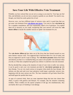
1-s2.0-S1072751514009077-main
S154
Surgical Forum Abstracts
2117Æ1080 mL occurred during HD. Mean GSV diameter was
3.0Æ1.3 mm pre-dialysis vs 2.5Æ1.2 mm post-dialysis
(p¼0.001). Using a 2.5 mm criterion for suitability, GSV diameters in 3 patients (9.7%) were adequate pre-HD but inadequate
post-HD. No individual variables predictive of diameter change
were identified. No significant changes in arm vein diameters
were identified.
CONCLUSIONS: Post-HD decreases in GSV diameter may result
in incorrect assessment of vein as inadequate, particularly when
vein mapping is performed on the same day. Timing of HD should
be considered when borderline vein diameters are identified.
Further studies are required to evaluate associations between volume status and vein diameters in other populations.
Fabrication of Durable, Perfusable Microvessels within
Tissue-Engineered Constructs
Rachel Campbell Hooper, MD, Adam Jacoby, BA,
Ope A Asanbe, MD, Hector L Osoria, BS, Tarek Elshazly,
Jason A Spector, MD, FACS
Weill Cornell Medical College, New York, NY
INTRODUCTION: The fabrication of tissue-engineered constructs
with an inherent vascular system remains challenging. Further, an
intact, healthy endothelial surface remains critical for thrombosisfree blood flow. Here we synthesize tissue-engineered endothelialized microvessels within collagen hydrogels, sustainable for up to
28 days in culture and suitable for perfusion.
METHODS: U-shaped pluronic F127 fibers were sacrificed in type
I collagen, creating a central microchannel, 1.5 mm in diameter.
After fiber sacrifice, 5 x10⁶ cells/mL human aortic smooth muscle
cells (HASMC) and 5 Â 106 cells/mL human umbilical vein endothelial cells (HUVEC) were sequentially seeded into the microchannel 24 hours apart. After 7, 14, and 28 days of culture,
constructs were fixed and processed for histology.
RESULTS: Microchannels were successfully seeded with HUVEC/
HASMC. Hematoxlyin and eosin staining demonstrated a
confluent lining by 7 days of culture, which was thickened at 14
days and maintained at the 28-day time point. Immunohistochemical analysis revealed the formation of a neointima and neomedia
comprised of CD31 expressing endothelial cells and -SMA expressing smooth muscle cells respectively. In addition after 7 and 14
days we noted deposition of critical extracellular matrix proteins
basal lamia collagen IV and fibronectin.
CONCLUSIONS: We have successfully fabricated tissue-engineered constructs with microchannels that support engraftment
and proliferation of smooth muscle and endothelial cells forming
a durable vascular network. Such constructs recapitulate the organization of vascular cells within blood vessels found in vivo,
providing a surface for thrombosis-free blood flow after in vivo
anastomosis.
J Am Coll Surg
Inhaled Carbon Monoxide Induces a Phenotypic Shift In
Macrophages
Andrew E Leake, MD, Guiying Hong, Kent R Zettel, MD,
Mostafa H Ramadan, MBCHB, Edith Tzeng, MD, FACS
University of Pittsburgh Medical Center, Pittsburgh, PA
INTRODUCTION: Inhaled carbon monoxide (CO) has potent vasoprotective actions in vivo but its mechanisms of action remain unknown. We have previously shown that peritoneal macrophages
isolated from CO treated animals secrete pro-endothelial and
angiogenic factors that cannot be reproduced by direct CO treatment. We hypothesize that inhaled CO mediates phenotypic shifts
in the macrophages to favor vasoprotective functions.
METHODS: Peritoneal macrophages were collected from rats after
inhaled CO (250 PPM for 1 hr) or from an air control rat. The
cells were cultured overnight and stimulated with and without
LPS. Levels of TNFa, IL6 and IL10 in the media were quantified
with ELISA and nitric oxide (NO) with the Griess assay. Mouse
peritoneal macrophages were similarly collected for flow cytometry
to determine CO effect on M1 (proinflammatory, CD86) and M2
(anti-inflammatory, CD206) phenotypes in CD11b macrophages.
RESULTS: Macrophages from CO treated rats had similar baseline cytokine and NO production as cells from air treated rats. After LPS treatment, CO-macrophages exhibited increased NO,
TNFa, and IL6 production vs air-macrophages. Similarly IL10
was increased in CO-macrophages. By flow cytometry there was
no difference in phenotypic markers without stimulation. However, with LPS stimulation, CO-macrophages exhibited decreased
M1 and increased M2 positive cells compared to LPS treated air
macrophages.
CONCLUSIONS: Inhaled CO does not alter baseline macrophage
phenotype, but with inflammatory stimulus, it induces a shift toward a mixed inflammatory/anti-inflammatory phenotype. The
duration and dynamic nature of these changes will be examined
in future studies.
Upregulation of Toll-Like Receptor 2 (TLR2) on
Endothelial Cells (EC) is Dependent on Extracellular
Matrix (ECM) Composition and Integrin Engagement
Jia Xu, PhD, Kelly Benabou, BA, Jun Xu, MD,
Xiangdong Cui, MD, Ulka Sachdev, MD, FACS
University of Pittsburgh Medical Center, Pittsburgh, PA
INTRODUCTION: We have demonstrated that TLR2 may be
important in angiogenesis after limb ischemia. While expression
of TLR2 on ECs grown on gelatin is very low at baseline, upregulation is TLR4-dependent. However, knockdown (KD) of TLR2
inhibits tube formation in laminin-rich Matrigel while KD of
TLR4 does not. We hypothesize that differences in TLR2 expression is ECM-specific, possibly through integrin-mediated
mechanisms.
METHODS: EC were plated on gelatin, laminin or Matrigel and
tested for TLR2 expression at 2, 4 and 6 hours using PCR. EC


© Copyright 2026