
Inhibition of CD4 Cross-Linking-Induced Lymphocytes
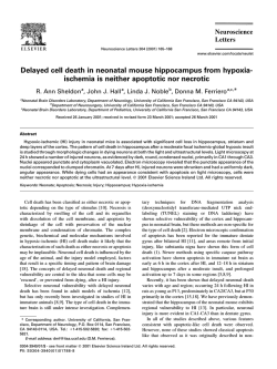
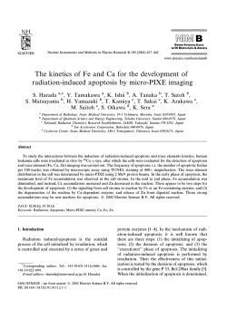
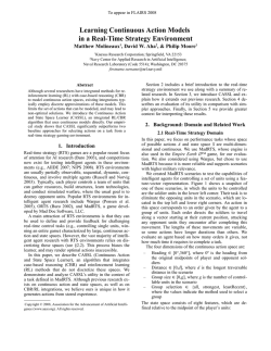
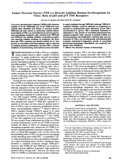
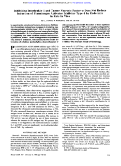
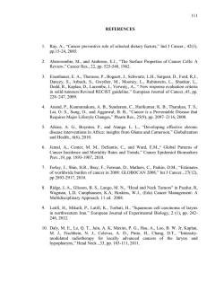
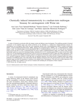
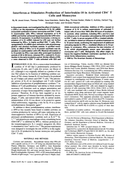
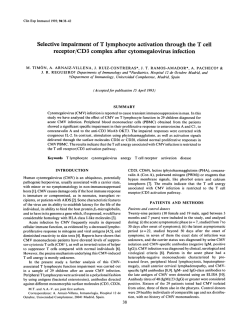
From www.bloodjournal.org by guest on February 6, 2015. For personal use only.
Inhibition of CD4 Cross-Linking-Induced Lymphocytes Apoptosis by
Vesnarinone as a Novel Immunomodulating Agent: Vesnarinone Inhibits Fas
Expression and Apoptosis by Blocking Cytokine Secretion
By Naoki Oyaizu, Thomas W. MCCloskey, Soe Than, and Savita Pahwa
Evidence is accumulatingthat T cellsfrom human immunodeficiency virus type 1 (HIV-l)-infected individuals showacceleratedcell death through apoptosis. We have recently
demonstrated that the cross-linking
of
CD4
molecules
(CD4XL) resultsin death of normal peripheral T cells through
apoptosis and imbalanced cytokine secretion (ie, induction
of tumor necrosis factor-cu [TNF-a1 and interferon-y [IFN-yl
in the absence ofinterleukin-2[IL-21 or IL-4secretion). These
upregulated cytokines (TNF-u/lFN-y) largely contributed to
upregulation of the apoptosis-inducing cell surface molecule, Fas(APO-I/CD95) and apoptosis induction. The present
study investigated the effect of vesnarinone as a novel immunomodulatingagent on CD4XL-induced T-cell apoptosis.
The addition of vesnarinone to peripheral blood mononu-
clearcells(PBMC)
significantly inhibited CD4XL-induced
lymphocyteapoptosis.Thisapoptosis-inhibitoryeffectof
vesnarinone was associated with the blocking ofCD4XLinduced TNF-(U
and IFN-y secretion andof Fas antigen upregulation. However, vesnarinonedid not block effectsof exogenously supplemented TNF-cu/lFN-y on Fas induction. These
data suggestthat vesnarinone inhibits CD4XL-induced TNFcu/lFN-y secretion,thereby blocking subsequentFas upregulation and apoptosis induction. Given
the potent pathogenic
role of imbalanced cytokine secretion observed
in HIV-infection, an agent such as vesnarinone may be of therapeutic
value in slowing disease progression.
0 7996 by The American Society of Hematology.
T
performed in PBMC in vitro, could be one possible explanation for the discordant cytokine upregulation and accelerated
apoptosis observed in HIV-infected individuals. Our findings
suggest that therapies aimed at controlling aberrant cytokine
secretion should prove beneficial in patients with HIV infection.
Here we report that vesnarinone inhibits CD4XL-induced
events, which include ( l ) TNF-a and IFN-y secretion; (2)
Fas upregulation, and (3) apoptosis induction. Vesnarinone
did not block the Fas induction in lymphocytes resulting
from exogenously provided TNF-aIIFN-y. These data suggest that vesnarinone may inhibit apoptosis induction by
suppressing cytokine secretion and subsequent Fas upregulation, which are prerequisite events in T-cell apoptosis induction.
HE LIMITED SUCCESS of nucleotide reverse transcriptase inhibitors in human immunodeficiency virus
type 1 (HIV-l)-infected patients has shifted attention to the
development of drugs that use alternative targets. An oral
inotropic
agent,
vesnarinone
(3,4-dihydro-6-[4-3,4-dimethoxybenzoy1)-l-piperazinyl1-2(1H)-quinolinone),has recently been shown to exert potent antiviral and anticytokine
activities. Vesnarinone has been found to inhibit replication
of HIV-I in vitro without affecting reverse transcriptase or
retroviral proteinase activity.' Vesnarinone was also found
to inhibit lipopolysaccharide (LPS)-induced tumor necrosis
facto-a (TNF-a) and interleukin-6 (IL-6) secretion and phytohemagglutinin (PHA)-induced interferon-y (IFN-y) and
granulocyte/monocyte colony stimulating factor (GM-CSF)
secretion by peripheral blood mononuclear cells (PBMC).'.'
The immunopathogeneic events from the time of initial
infection with HIV until the global deterioration of the immune system, including destruction ofCD4' T cells, are
complex and involve aberrant activation of the immune system and cytokine secretion. A number of laboratories have
reported that T cells from HIV-infected individuals undergo
enhanced apoptotic cell death."7 Considering the fact that
productively infected cells are barely detectable in peripheral
c i r ~ u l a t i o ndirect
, ~ ~ ~ cytopathic effect of HIV cannot account
for the observed substantial degree of apoptosis; rather the
operation of a mechanism for elimination of noninfected
cells is more probable. In this regard, we have recently
shown that the ligation of CD4 molecules and CD4 crosslinking (CD4XL) resulted in the induction of IFN-y and
TNF-a secretion in PBMC, and these induced cytokines contributed largely to the upregulation of Fas (APO-I/CD95),
a cell surface apoptosis-triggering molecule'0,'' and to
apoptosis ind~ction.~.''This discordant cytokine profile induced by CD4XL agrees with that actually observed in HIV
infe~tion.""~
Considering the fact that some level of plasma
viremia is always detectable" and that lymph nodes harbor
a heavy viral l ~ a d , ' ~the
. ' ~ likelihood of CD4XL occurring
in vivo in CD4 antigen bearing cells as a result of highaffinity binding of gp120 to CD4, is extremely high in HIV
infection. Thus, aberrant cytokine induction as a result of
CD4XL occurring in vivo, similar to that seen with CD4XL
Blood, Vol 87,
No 6 (March 15), 1996: pp 2361-2368
MATERIALS AND METHODS
Cells and culture conditions. PBMC were obtained from HIVseronegative healthy donors. PBMC were isolated from heparinized
venous blood by Ficoll-Hypaque density gradient centrifugation.
PHA-induced CD4+ T-cell clones were established and maintained
as described.*' The clones were maintained by the cyclic stimulation
of PHA-P (5 pg/mL, Difco, Detroit, MI) for 3 days followed by 3
days rest in the presence of irradiated (3,000 rads) autologous Epstein-Barr Virus (EBV)-transformed B cells. RPM1 1640 (GIBCO
From the Department of Pediatrics, North Shore University Hospital-Cornell University Medical College, Manhasset, New York,
NY.
Submitted June 12, 1995; accepted October 25, 1995.
Supported by grants from Otsuh Pharmaceutical (Palo Alto, CA)
to S.P. and National Institutes of Health (Bethesda, MD) Grant No.
AI28281.
Address reprint requests to N. Oyaizu, MD, Room 303, Biomedical
Research Building, NSUH-CUMC, 350 Community Drive, Manhasset, New York, NY 11030.
The publication costs of this article were defrayed in part by page
charge payment. This article must therefore be hereby marked
"advenisement" in accordance with 18 U.S.C. section 1734 solely to
indicate this fact.
0 1996 by The American Society of Hematology.
0006-4971/96/8706-0012$3.00/0
2361
From www.bloodjournal.org by guest on February 6, 2015. For personal use only.
2362
Laboratories, Grand Island, NY) supplemented with 10% heat-inactivated FCS (GIBCO), 2 mmoUL L-glutamine (Whittaker Bioproducts, Inc, Walkersville, MD), 100 U/mL penicillin G, and 100 pg/
mL streptomycin was used for all cultures.
Antibodies and reagents. Reagents and sources were as follows:
vesnarinone (OPC-8212, Otsuka Pharmaceutical CO,Tokushima, Japan); cyclosporin A (CsA, Sandoz, East Hanover, NJ); monoclonal
antibody (MoAb) to CD4 (Leu3a; IgGl, Becton Dickinson, Mountain View, CA, OKT4; IgG2b, Ortho Diagnostic System, Inc, Raritan, NJ); MoAb toCD3 (IgG2a, MAb 454,22 a gift from Dr N.
Chiorazzi, North Shore University Hospital, Manhasset, NY); and
goat antimouse immunoglobulin, (GAM; Tago, Inc, Burlingame,
CA). Recombinant human IFN-y (rlFN-y) was obtained from Genzyme (Cambridge, MA); recombinant human TNF-a (50 units/ng
as determined by half-maximal cytotoxicity of L929 cells) was a
kind gift from Dr K.J. Tracey (Picower Institute, Manhasset, NY).
induction of CD4 cross-linking (CD4XL). Induction of CD4XL
were performed as described.’*In brief, cells were treated with Leu3a
or OKT4 at concentrations of 3 pg/2 X IOh cells/mL for 1 hour at
4°C. Cells were then cultured in 24-well Nunc plates (Nunc, Roskible, Denmark) coated with goat antimouse immunoglobulin
(GAM) at 37°C. Various doses of vesnarinone or CsA (400 ng/mL)
were added at the time of culture initiation at 37°C and were present
throughout the culture period. Vesnarinone was initially dissolved
in I N HCI, diluted 1:lO in serum-free RPMI, and the pH was adjusted with IN NaOH to 7.0 just before addition to cell cultures.
CsA was used as a control agent. We confirmed that reagents used
for CD4XL. which include OKT4, Leu3a, and GAM, are free from
endotoxin as determined by the Limulus amebocyte lysate assay (ETOXATE, Sigma),which had an endotoxin detection limit of O. 125
EU/mL.
lmrnunoffuorescence stainingand $ow cytometry. For study of
cell surface expression of Fas, cells were stained with fluorescein
isothiocyanate (F1TC)-conjugated anti-Fas MoAb (clone UB2 IgG 1,
AMAC Inc, Westbrook, ME). FITC-conjugated mouse IgGl antibody (AMAC) was used as a control. In some experiments, antiFas stained cells were subsequently stained with Per-CP-conjugated
anti-CD3 MoAb (Becton Dickinson). All incubations of cells with
antibodies were for 10 minutes at room temperature followed by
washing with Hanks’ Balanced Salt Solution (HBSS) and fixing in
Immunoprep C (Coulter, containing 1% [wtlvol] paraformaldehyde).
The stained cells were analyzed on a flow cytometer (Epics Elite,
Coulter Electronics). A lymphocyte gate was first drawn on a histogram of forward versus right angle light scatter. This gate, which
excluded debris, monocytes, and clumps, was assigned to a histogram of red fluorescence. On this histogram, appropriate gates were
drawn to capture certain populations (CD3’, CD3-l as required.
Fluorescence emissions were collected at 525-nm for FITC (green)
and 675-nm for Per-CP (red), respectively. Single parameter green
histograms representing Fas expression in each gated population
were evaluated by comparing them with the isotypic controls using
Coulter’s cytologic analysis program (Overton’s Cumulative Subtraction Algorithm: OVERSUB) which was implemented to assess
percent of cells positive for Fas expression over control.
Meusurement of upoptosis byftow cytonretry. The percentage of
cells undergoing apoptosis was quantitated by a modification of the
flow cytometric method as described.23Briefly, 2 X IO6 cells were
fixed in 75% ethanol for 1 hour at 4°C. The cells were then washed
and resuspended in 0.5 mL HBSS. To this suspension, 0.5 mL RNase
solution (1 mg/mL HBSS) was added followed by 1 mL propidium
iodide (PI, 100 pg/mL HBSS, Molecular Probes, Eugene, OR). After
gentle mixing, cells were incubated for 72 hours at 4°C in the dark.
The PI fluorescence of individual cells was measured using a flow
cytometer. Cell debris and cell clumps were excluded by gating for
single cells under forward and side light scatter. A distinct cell cycle
OYAIZU ET AL
region of apoptosis (Ao) could be identified below the G,JG, diploid
peak. Percentage of cells in the Ao region were estimated. Cell death
was also evaluated by trypan-blue exclusion.
Determination of cytokineconcentrationsandcytokine
mRNA.
The culture supernatants were collected after 48 hours and cytokine
concentrations were quantitated by using commercial enzyme-linked
immunosorbent assay (ELISA) kits (Genzyme) according to the
manufacturer’s instructions. For the quantitation of cytokine mRNA,
polymerase chain reaction (PCR) assisted mRNA amplification reverse transcriptase (RT-PCR) was employed as described elsewhere.24In brief, cells were collected after a 6-hour culture, and
total cellular RNA was extracted with RNAzolB (Cinna/Biotecx,
Houston, TX) according to the manufacturer’s protocol. Reverse
transcription of RNA to cDNA was performed according to GeneAmp RNA PCR kit protocol (Perkin Elmer Cetus, Nonvalk, CT).
Cytokine specific primer pairs and @-actin were purchased from
Research Genetics (Huntsville, AL). Primer sequences were the same
as those described elsewhere (IFN-y, @-actin,24TNF-a2’). PCR was
performed in a GeneAmp PCR system 9600 (Perkin Elmer Cetus)
for 30 cycles: 1 minute denaturation at 9 4 T , 1.S minutes annealing
at SS”’, and 1 .S minutes extension at 72°C. A final extension period
of 10 minutes followed the 30 cycles. @-actinwas used as an internal
control in all reactions. The reaction products were visualized by
subjecting them to electrophoresis in 1% agarose in 1 X TBE buffer
containing 0.5 pg/mL ethidium bromide, followed by transfer to
nylon membranes, hybridization with ’*P-labeledsense primers, and
exposure to x-ray film.
Stutisticul unalysis. Statistical significance was assessed by Student’s t-test.
RESULTS
Vesnarinone blocks CD4XL-induced lymphocyte apoptosis. As shown in Fig l and Table 1, we confirmedthe
observation that CD4XL resulted in a significant increase of
lymphocyte apoptosis as determined by cellular viability and
by demonstration of the emergence of sub-GdG, peak nuclei
(Ao cells) on flow cytometry. We have previously confirmed
that the majority of cells undergoing apoptosis are composed
of CD3’ T cells.’ Vesnarinone was found to inhibit CD4XLinduced lymphocyte apoptosis in a dose-dependent manner.
Significant inhibition was observed at concentrations of > I O
,ug/mL, and this was associated with a significant increase
of cellular viability. A comparison was made between vesnarinoneand
the well-characterized immunosuppressive
agent, cyclosporine A (CsA), which also inhibited the
CD4XL-induced apoptosis. Vesnarinone, by itself, was not
cytotoxic up to 100 pg/mL (data not shown).
Vesnarinone blocks CD4XL-induced TNF-a and IFN- y
secretion. We have previously demonstrated that CD4XL
results in aberrant cytokine secretion in PBMC, in that cytokines TNF-(Yand IFF-y were induced in the absence of
IL-2 and IL-4.” Moreover, these induced cytokines were
determined to largely contribute to apoptosis-induction in
lymphocytes, which was associated with upregulation of Fas
antigen expression. We therefore examined the effect of
vesnarinone on CD4XL-induced TNF-a and IFN-y secretion. As shown in Table 2, the addition of vesnarinone significantly inhibited CD4XL-induced TNF-a and IFN-7 secretion. The inhibitory effect mediated by vesnarinone was
concentration-dependent and showed a similar dose-response to that required for apoptosis inhibition. CsA was
From www.bloodjournal.org by guest on February 6, 2015. For personal use only.
2363
INHIBITION OFLYMPHOCYTEAPOPTOSISBYVESNARINONE
U
3. CMXL
Fig 1. Vesnarinone
blocks
CD4
cross-linking
(CD4XL)-inducedapoptosis. CD4XL was performed
in normal PBMCs as described in Table 1, and cells
were cultured in the presence of various concentrations [pglmL) of vesnarinone (Ves) indicated for 3
days. Cellswere stained with PI andanalyzedby flow
cytometry. A distinct region of A0 (marked Dt could
be identified below the GolGl peak. Percentage of
cells found in found in the Ao peak l%Aol are indicated.
6
, ;S \&\
L
k
PI fluorescence
also found to block CD4XL-induced TNF-a and IFN-y secretion. We next examined whether vesnarinone affects the
CD4XL-induced TNF-a and IFN-y mRNA accumulation
(Figs 2 and 3). For this purpose, we performed RT-PCR
using mRNAs extracted from CD4 cross-linked PBMC in
the presence or absence of vesnarinone. At concentrations
that significantly blocked cytokine secretion, no discernable
effects were noted on CD4XL-induced TNF-a (Fig 2, lane
3) and IFN-y (Fig 3; Exp. 1, lane 3; Exp. 2 , lane 3) mRNA
accumulation by vesnarinone (cytokine values obtained from
identical samples used for RT-PCR are indicated in inset
tables in Figs 2 and 3). In contrast, CsA blocked both I F N y mRNA accumulation and IFN-7secretion (Fig 3, lane 4).
Table 1. Vesnarinone Blocked CD4 Cross-Linking Induced
LymphocyteCell Death
Treatment Addition (pg/mL)
Medium
Anti-CD4 MoAb alone
GAM alone
CD4XL
+ vesnarinone (3)
+ vesnarinone (10)
+ vesnarinone (30)
+ vesnarinone (60)
CsA
+
% Viability
88.6
80.8
80.4
68.2
68.1
69.8
80.7
82.7
79.7
2 4.2
2
2
-c
2
2
2
2
2
5.0
4.7
5.7
4.9
7.5
4.5*
2.3*
4.1,
Because we performed the CD4XL in unfractioned PBMC
and because CD4 molecules are expressed in both T cells
and monocytes, we examined the cellular source of TNF-a
and IFN-y in response to CD4XL. As shown in inset tables
in Figs 2 and 3, CD4XL of cloned CD4+ T cells in the
absence of any accessory cells resulted in both TNF-a and
IFN-y induction. Soluble anti-CD3 MoAb treatment of
cloned T cells inthe presence of autologous EBV-transformed B cells was used as a positive control. These data
indicate that T cells can be induced by CD4XL to secrete
TNF-a and IFN-y in the absence of accessory cells. We
have confirmed this observation using purified peripheral
T cells and monocytes populations. Upon CD4XL, IFN-y
induction was restricted to T cells, but TNF-a was inducible
both from T cells and, to a lesser degree, from monocytes
(data not shown).
Vesnarinone blocks the CD4XL-induced, but not exoge-
% Ao Cells
15.0
26.1
18.8
41.7
33.5
29.5
22.7
21.3
22.0
2 8.4
2 8.4
-t
8.9
2 3.5
2 5.4
t 8.5*
2 8.8*
2 6.5'
t 4.0*
CD4 cross-linking (CD4XL) was performed in PBMC by treating cells
with anti-CD4 MoAb followed by
culture on goat antimouse lg (GAM)coated 24-well culture plates. Various concentrations of vesnarinone
indicated or cyclosporine A (CsA, 400 ng/mL) were added at the initiation of culture. Following culture for 3 days, cells were harvested
and analyzed for cellular viability by trypan-blue exclusion and for
apoptosis following staining with propidium iodide (PI) by flow cytometry. Values denote the percentage of cells found in Ao peak
(mean t SDI as described in Fig 1. Data represents results of seven
independent experiments.
Values significantly different from CD4XL samples (underscore)
at P i ,001 by the Student's t test are indicated.
Table 2. Vesnarinone BLocks CD4 Cross-Linking InducedTNF-rr
and IFN-y Secretion
Treatment
Addition
(usImL)
Medium
Anti-CD4 MoAb alone
GAM alone
CD4XL
vesnarinone (3)
vesnarinone (10)
+ vesnarinone (30)
vesnarinone (60)
+ vesnarinone (90)
+ CsA
+
+
+
TNFa (pg/rnL)
IFN-.I Ipg/mL)
I%inhibitionl
1% inhibitionl
<l0
19 2 24
56 2 65
1,608 2 399 1-1
1,669 2 347 [ - ]
1,577 t 413 [21
1,002 2 453* [381
431 2 310* I731
504 rt 386* 1691
26 2 19* 1981
<l00
<l00
<l00
1,612 2 894 [-l
1,640 2 741 [-l
1,041 2 701'[551
278 2 316* I831
<loo* [l001
<loo* I1001
1100, [l001
Cytokine levels of 48 to 72 hours PBMC culture supernatants were
measured by ELISA. Levels below which signals were undetectable:
TNF-a 10pglmL; IFN-y 100 pg/mL; respectively. Data represents mean
-L SD of 12 experiments for TNF-a and 5 experiments for IFN-y.
Values significantly different from
CD4 cross-linked PBMC (underscore) at P < .01.
From www.bloodjournal.org by guest on February 6, 2015. For personal use only.
OYAIZU ET AL
2364
$?
&+4
A!
X
+"
&fJ
I
lane # Cells
- pactin
~
l
2
3
TNFa *(pg/ml)
XL
XL+Ves(30)
"
.
517
-
"
TNFa
40
1042
-
PBMC
PBMC
PBMC
C l0
T clone
T clone XL
T+B
aCD3
-
1806
1030
T N F a in 48 h culture supernatant
1
2
3
Fig 2. Effect of vesnarinone on CD4XL-induced TNF-a mRNA induction. Total RNA was extracted from normal PBMCs that were treated
with medium (lane l), anti-CD4 MoAblGAM (XL) in the absence (lane 2) or presence (lane 3) of vesnarinone (30 pg/mL), respectively, for 6
hours. RT-PCR assisted mRNA amplification forTNF-a and p-actin was performed
as described. Top portion shows
agarose gel electrophoresis
stained with ethidium bromide and bottom portion shows autoradiograms
of Southern blotting for TNF-a amplified fragments. Inset table
shows TNF-a values determined by ELISA obtained from a48-hour culture supernatant of identicalsamples used for RT-PCR and from CD4'
T-cell clone that were treated with
medium, Leu3alGAM (XL) in the absence of any accessory cells, or soluble anti-CD3 MoAb (1:lOO) in the
presence of autologous EBV-transformed B cells (T + B), respectively.
Exp. 1
Exp. 2
PBMC
l
2
3
4
5
lane # Cell
~
- INFr
1
2
3
~
6
7
INF -y (pg/rnl)*
~~~
~~
~
PBMC
e100
PBMC X L
904
126
PBMC XL +Ves (90)
PBMC X L +CsA
400
T clone
< l 00
T clone XL
784
T+ B
aCD3
560
* 1NF-y in 48 h culture supernatant
Fig 3. Effect of vesnarinone on CD4XL-induced IFN-y mRNA induction. Exp. l (left): Total RNA was extracted from normal PBMCs that
were treated with medium (lane l),anti-CD4 MoAblGAM (XL) in the absence (lane 2)or presence (lane 31 of vesnarinone (Ves; 30 pglmLI,
respectively, for 6 hours. RT-PCR assisted mRNA amplification for IFN-y and p-actin was performed
as described. Top portion showsagarose
gel electrophoresis stained with ethidium bromide and bottom portion shows autoradiograms of Southern blotting for IFN-y amplified
fragments. Exp. 2 (right): Experimental conditions and IFN-y values determined in a 48-hour culture supernatants by ELSA from identical
samples for RT-PCR are indicated in the inset table. CD4XL were performed in cloned CD4' T cells (T clone) in the absence of any accessory
cells (lane 6);"T + B" denotes that soluble anti-CD3 MoAb treatment (aCD3) of cloned T cells in the presence of EBV-transformed B cells as
positive control (lane 7 ) . Agarose gel electrophoresis stained with ethidium bromide of amplified fragments for p-actin
and IFN-y isshown.
From www.bloodjournal.org by guest on February 6, 2015. For personal use only.
2365
INHIBITION OFLYMPHOCYTEAPOPTOSISBYVESNARINONE
Fas Ag Expression (log fluorescence Intensity)
Fig 4. Vesnarinone blocks CD4XL-induced
Fas upregulation. PBMCs were cultured with medium, Leu3alGAM (XL)
in the absence or presence
of various concentrations of vesnarinone (Ves)indicated [pg/mLI for 48 hours. Far expression was analyzed byflow cytometry. Histograms
obtained from the CD4 cross-linked PBMCs are shaded and superimposed
to the histograms from vesnarinone-treated samples. Mean fluorescence intensities (log scale) are indicated in parentheses. Data represents oneof three independent experiments.
nously supplemented TNF-a- andor IFN- y-induced Fas
expression. To examine whether the apoptosis-blocking
abilities of vesnarinone correlated with Fas antigen expression, Fas expression on lymphocytes was assessed in CD4
cross-linked PBMC in the presence of various concentrations
of vesnarinone. As shown in Fig 4, weconfirmedthat
CD4XL resulted in significant Fas upregulation and found
that vesnarinone inhibited CD4XL-induced Fas upregulation
in a dose-dependent manner. Finally, we examined the effects of vesnarinone on the induction of Fas antigen by exogenously provided TNF-a and IFN--y. As shown in Table 3,
both TNF-a and I F N -y additively augmented Fas expression
in peripheral lymphocytes. However, vesnarinone did not
block this cytokine-mediated Fas upregulation. Further, we
examined these effects in different lymphocyte subpopulations. As shown in Fig 5, addition of TNF-a and IFN-y
resulted in increased Fas expression in both CD3-positive
(A) and CD3-negative lymphocytes subsets (B); addition
of vesnarinone did not reduce the cytokines-induced Fas
upregulation in either subset. These data indicate that vesnarinone does not block TNF-a and IFN-y-med1ated biologic
activities, as assessed by the ability of these cytokines to
upregulate Fas antigen expression.
Panel A
CD3+ cells
Additlon
l.medium
7t7
2 TNFa
+INF-y
3. TNFu
+INF-y
I
tl
+
Table 3. Vesnarinone Does Not Block Exogenous TNF-aandlor
IFN-y-Induced Fas Upregulation
Addition
Medium
TNF-a
TNF-a + vesnarinone
IFN-y
IFN-y + vesnarinone
TNF-n IFN-y
TNF-a + IFN-y + vesnarinone
+
% Fas Positive Cells
67.9
76.6
73.1
76.6
73.8
83.5
80.7
f 6.1
?
6.1
2
7.0
f 3.4
+' 4.2
-C 2.3'
? 2.7*
PBMCs were cultured with recombinant TNF-a (1 pg/rnL) or IFN-y
(1.000 U/mL) in the presence or absence of vesnarinone (50 pg/mL)
for 2 days and analyzed for Fas expression by flowcytometry. Values
denote percent Fas positive cells (mean f SD of three different individuals) over the isotypic control stain cells calculated using the
OVERSUB program.
Values significantly different from medium controls
at P < .01 by
the Student's t-test are indicated.
Fas Ag EXpm3SiOn
(logfluorescence)
Fig 5. Vesnarinone did not blockexogenousTNF-a plus IFN-y
induced Fas upregulation. PBMCs were treated with recombinant
TNF-a (1 pg/mL)/IFN-y (1,OOO UlmL) in theabsence or presence of
vesnarinone (50 pg/mL) for 48 hours. Cells were harvested and analyzed for Fas expression in conjunction with per-CP-labeled CD3
MoAb by flow cytometry. Values denote percent Fas antigen
positive
intensity
cells and values in parentheses represent mean fluorescent
(MFI) on a log scale, respectively. Appropriate gates were drawn
to capture CD3' (left panels) and CD3- (right panels) populations,
respectively, and single parameter green histograms representing
Fas expression in each gatedpopulation was evaluated. Thevertical
line i n d m e s the position of control staining. Values denote percent
Fas positive cells as calculated usingthe OVERSUB program.
From www.bloodjournal.org by guest on February 6, 2015. For personal use only.
2366
OYAIZU ET AL
DISCUSSION
y may play a role not only in upregulating Fas antigen, but
also
for rendering T cells susceptible to Fas-mediated cell
In the present study, we have examined the effects of
death signaling. This notion is supported by thefactthat
vesnarinone on CD4XL-induced lymphocyte apoptosis, cyinduction of sphingomyelin break-down to produce ceratokine secretion, and Fas antigen expression. Here, we report
mide, which plays a role in apoptosis induction, can occur
that vesnarinone can inhibit CD4XL-induced lymphocyte
as
a result of ligation of Fas4' as well as by the cytokines
apoptosis, TNF-a and IFN-y secretion, and Fas upregulation
TNF-cu and I F N - Y . ~Further
~
studies are required to define
in a dose-dependent manner and that significant inhibition
the role of cytokines in regulating sensitivity to Fas-based
was observed at clinically achievable concentrations ( 210
cell death signaling. Nonetheless, our finding that CD4XL
pg/mL). Vesnarinone failed to block effects of exogenously
is sufficient to induce T-cell apoptosis in PBMCand the
supplemented TNF-a and/or IFN-y on Fas upregulation, ininvolvement of Fas antigen in this process is further supdicating that this agent appears not to be inhibitory for cytoported by the recent observations by Wang et a147who have
kine-mediated signaling. Collectively, these data indicate
shownthat (1) administration of anti-CD4 MoAb in vivo
that the primary action of vesnarinone is attributable to inhiresulted in CD4' T-cell depletion through apoptosis in norbition of cytokine secretion, thereby blocking subsequent
mal mice, but not in Fas-defective lpr mice and (2) adminisFas expression and apoptosis induction.
tration of gpl20 and anti-gpl20 antibodies into human CD4HIV infection is characterized not only by global immune
expressing transgenic mice resulted in depletion of human
suppression including progressive decline of CD4+ T cells,
CD4' mouse T-lymphocytes in vivo.4x Thus our present
but also by persistent immune activation as manifested by
findings not onlyprovide an experimental basis for the theraaberrant cytokine secretion. Cytokine/cytokine mRNA deterpeutic benefits of an immunomodulating agent such as vesminationin patients plasma, PBMC, andin lymph node
narinone for the treatment of HIV infection, but also support
mononuclear cells has shown upregulation of TNF-a, IFN-y,
the notion of a major role of aberrantly induced cytokines
XL-6, and IL-10.""' With regard to the observed constitutive
in HIV disease pathogenesis.
cytokine profile in HIV infection, the following findings afVesnarinone is a quinolinone derivative thatwas origiford clues to the mechanism of aberrant cytokine secretion.
nally developed as an oral, positive inotropic agent, and
HIV-I envelope glycoprotein gp120 has been shown to bind
has several complex actions, including possible inhibition of
to its receptor CD4 molecule withhighaffinity."
HTV-I
phosphodiesterase a~tivity.~'
This agent has recentlybeen
gp120 has also been shown to induce a variety of cytokines
confirmed to improve the prognosis of patients with chronic
including TNF-a, TL-6, IL-1, 1L-10, and GM-CSF from
heart failure.'" Subsequently, it was shown that vesnarinone
monocytes/macrophages or T cells through its interaction
possesses immunomodulatory and antiviral activities.'.',"
with CD4 molecule^.*^^^' Further, we have recently shown
Vesnarinone improved mortality in the acute stage of viral
that the CD4XL resulted in the induction of IFN-y and TNFmyocarditis, and this was associated withreduced TNF-a
a in the absence of IL-2 or IL-4 secretion in PBMC obtained
secretion." The principal side effects of vesnarinone include
from HIV seronegative individuals. l 2 This discordant cytoreversible neutropenia, which occurred in 2.5% of patients.'"
kine profile induced by gp120 or CD4XL agrees perfectly
Mechanisms of action of vesnarinone on cytokine secretion
withthat actually observed inHIV infe~tion.'"'~
We and
are not known. It has been reported that increase of intracyothers have also shown that TNF-cu and IFN-y largely contoplasmic CAMPblocks TNF-a secretion at the posttransciptribute to Fas expression and apoptosis i n d u ~ t i o n . ' " ~ ' ~ ~tional
~~~~
~
level.s2,"
One possible explanation for the action of
Although we did not address the issue of Fas-L induction
vesnarinone is that it may block cytokine secretion via its
in this study, it has recently been reported that treatment of
phosphodiesterase inhibitory activity, which results in an
PBMC with anti-CD4 antibodies resulted in Fas-L (APOincrease of intracellular CAMP. Although the precise action
1L) mRNA expression." T cells upregulate Fas antigen and
of vesnarinone on cytokine secretion needs to be elucidated,
itsligand expression within 24hours of T-cell receptor
our results are consistent with this notion. Considering the
(TCR) s t i m ~ l a t i o nbut
, ~ ~they only became sensitive to antipotent role of TNF-a in HIVreplication and apoptosis inducFas-mediated apoptosis several days later.39Freshly isolated
tion, it is important to consider the therapeutic use of other
peripheral T cells are fairly resistant to Fas-based apoptosis
known inhibitors of TNF-a in combination withvesnarialthough they express a substantial amount of Fas antigen.4"
none. The methyl-xanthine derivative pentoxifylline and the
Thus expression of Faspas-L alone is not sufficient to render
agent thalidomide have been shown to inhibit TNF-a secrenormal T cells sensitive to induction of apoptosis. In contion by different mechanisms. Pentoxifylline has been shown
toreduce TNF-a production by inhibiting transcriptional
trast, T cells obtained from HIV-infected patients express
regulation? whereas thalidomide exerts its action by enaugmented F ~ S ~ and
' . ~ 'are extremely sensitive to anti-Fashancing TNF-a mRNA degradati~n.'~Thus vesnarinone,
mediated apopto~is.~'
These observations suggest that Faspentoxifylline, and thalidomide may exert their effects by
mediated apoptosis is regulated not only byits ligand expresdifferent mechanisms operating at distinct points of the cytosion, but also by a permissive signaling pathway downstream
kine biosynthetic pathway, and one can expect synergistic
toFas receptor. In this regard, deletion mapping analysis
effect on inhibition of cytokine production at lower concenhas identified a negative regulatory domain mapped to 15
trations of each drug if used in combination.
amino acids in the C-terminal of Fas that suppresses FasFinally, recent studies have demonstrated that treatment
generated signals leading to cell death." Taken together,
of patients with drugs that directly acton the life cycle
these findings suggest that CD4XL-induced TNF-a and IFN-
From www.bloodjournal.org by guest on February 6, 2015. For personal use only.
INHIBITION OF LYMPHOCYTE APOPTOSIS BY VESNARINONE
2367
by the cDNA for human cell surface antigen fas can mediate
of HIV, including inhibitors for reverse transcriptase and
apoptosis. Cell 66:233, 1991
protenase, resulted in the rapid appearance of drug-resistant
12. Oyaizu N, McCloskey W ,Soe Than, Hu R, Kalyanaraman
m ~ t a n t s . It
~ ~appears
. ~ ~ that administration of these drugs
VS, Pahwa S: Crosslinking of CD4 molecules up-regulates Fas antialone is insufficient in controlling disease progression and
gen expression in lymphocytes by inducing interferon y and tumor
that combination therapies including drugs that indirectly
necrosis factor-cy secretion. Blood 84:2622, 1994
regulate HIV replication may be desirable. Given the clear13. Lahdevirta J, Maury CPJ, Teppo A-M, Rep0 H: Elevated
cut role of T N F - a m - y in inducing HIV r e p l i ~ a t i o n ~ ~ levels
. ~ ~ of circulating cahectidtumor necrosis factor in patients with
and in promoting T-cell apoptosis i n d u ~ t i o n , ' ~drugs
, ~ ~ .that
~~
acquired immunodeficiency syndrome. Am J Med 85:289, 1988
are able to suppress imbalanced cytokine secretion in HIV
14. Fuchs D, Hausen A, Reibnegger G , Werner ER, Wernerinfection should be considered. Thus, vesnarinone has the
Felmayer G , Dierich MP, Wachter H: Interferon-? concentration are
increased in sera from individuals infected with human immunodepotential to slow HIV disease progression by two mechaficiency virus type 1. J Acquire Immune Defic Syndr 2:158, 1989
nisms, an anti-apoptosis ability and suppression of HIV repli15. Emille D, Permutter M, Malliot MC, Crevon MC, Brousse
cation.
ACKNOWLEDGMENT
The authors thank Dr Rong Hu for providing T-cell clones, Dr
Xue Ping Wang for cytokine measurement by ELISA, Dr Nicholas
Chiorazzi for MoAb 454; and Dr K.J. Tracy for providing recombinant TNF-a.
REFERENCES
1. Maruyama I, Maruyama Y, Nakajima T, Kitajima I, Osame
M, Zhao J-Q, Chen ISY, Nakai S, Ikeda M, Yabu-uchi Y, Adachi M:
Vesnarinone inhibits production of HIV-1 incultured cells. Biochem
Biophys Res Commun 195:1264, 1993
2. Busch F W , Tillmann A, Becker EW, Owsianowski M, Berg
PA: The inhibitory effects of a positive quinolinone derivative, 3,4dihydro-6-[4-(3,4-dimethoxybenzoyl)-l-piperazinyl]-2(1H)quinoline (OPC-8212), on bone-marrow progenitor cells and peripheral lymphocytes. Eur J Pharamacol 42:629, 1992
3. Groux H, Torpier G, Monte D, Mouton Y, Carpon A, Ameisen
JC: Activation-induced death by apoptosis in CD4' T cells from
human immunodeficiency virus-infected asymptomatic individuals.
J Exp Med 175:331, 1992
4. Meyaard L, Otto SA, Jonker RR, Mijnster MJ, Keet RPM,
Miedema F: Programmed cell death of T cells in HIV-I infection.
Science 257:217, 1992
S . Oyaizu N, McCloskey TW, Coronesi M, Chirmule N, Kalyanaraman VS, Pahwa S: Accelerated apoptosis in peripheral blood
mononuclear cells (PBMC) from human immunodeficiency virus
type-l infected patients and in CD4 cross-linked PBMCs from normal individuals. Blood 82:3392, 1993
6. Lewis DE, Ng-Tang DS, Adu-Oppong A, Schober W, Rodger
JR: Anergy and apoptosis in CD8' T cells from HIV-infected persons. J Immunol 153:412, 1994
7. Carbonari M, Cibati M, Cherchi M, Sbarigia D, Pesce AM,
Dell' Anna L, Modica A, Fiorilli M: Detection and characterization of
apoptotic peripheral blood lymphocytes in human immunodeficiency
virus infection and cancer chemotheraphy by an novel flowimmunocytometric method. Blood 83:1268, 1994
8. Patterson BK, Till M, Otto P, Goolsby C, Furtado MR,
McBride LJ, Wolindky SM: Detection of HIV-1 DNA and messenger RNA in individual cells by PCR-driven in situ hybridization and
flow cytometry. Science 260976, 1993
9. Saksela K, Stevens C, Rubinstein P, Baltimore D: Human immunodeficiency virus type 1 mRNA expression in peripheral blood
cells predicts disease progression independently of the numbers of
CD4+ lymphocytes. Proc Natl Acad Sci USA 91:1104, 1994
IO. Yonehara S, Ishii A, Yonehara M: A cell-killing monoclonal
antibody (anti-Fas) to a cell surface antigen co-downregulated with
the receptor of tumor necrosis factor. J Exp Med 169:1747, 1989
11. Itoh N, Yonehara S, Ishii A, Yonehara M, Mizushima S,
Sameshima M, Hase A, Seto Y, Nagata S: The polypeptide encoded
N, Delfraissy JF, Dormont J, Galanaud P Production of interleukins
in human immunodeficiency virus-l-replicating lymph nodes. J Clin
Invest 86:148, 1990
16. Fan J, Bass H Z , Fahey JL: Elevated IFN-7and decreased IL2 gene expression are associated with HIV infection. J Immunol
151:5031, 1993
17. Graziosi C, Pantaleo G, Gantt K R , Fortin J-P, Demarest JF,
Cohen OJ, Sekaly RP, Fauci AS: Lack of evidence for the dichotomy
of Thl and Th2 predominance in HIV-infected individuals. Science
265:248, 1994
18. Piatak M, Saag MS, Yang LC, Clark SJ, Kappes JC, Luk
K-C, Hahn BH, Shaw GM, Lifson JD: High levels of HIV-I in
plasma during all stages of infection determined by competitive
PCR. Science 259:1749, 1993
19. Pantaleo G, Graziosi C, Demarest JF, Butini L, Montroni M,
Fox CH, Orenstein JM, Kotler DP, Fauci AS: HIV infection is active
and progressive in lymphoid tissue during the clinically latent stage
of disease. Nature 362:355, 1993
20. Embretson J, Zupancic M, Ribas JL, Burke A, Racz P, Tenner-Racz K, Haase AT: Massive covert infection of helper T lymphocytes and macrophages by HIV during the incubation period of
AIDS. Nature 362:359, 1993
21. Hu R, Oyaizu N. Kalyanaraman VS, Pahwa S: HIV-I gp160
as a modifier of TH1 and TH2 cytokine response: gp160 suppresses
INF-7 and IL-2 production concomitantly with enhanced IL-4 production in vitro. J Immunol Immunopathol 73:245, 1994
22. Stohl W, Posnett DN, Chiorazzi N: Induction of T cell-dependent B cell differentiation by anti-CD3 monoclonal antbodies. J
Immunol 138:1667, 1987
23. McCloskey TW, Oyaizu N, Pahwa S: Use of flow cytometric
assay to quantitate apoptosis in human lymphocytes. Clin Immunol
Immunopathol 7 1:14, 1994
24. Ehlers S, Smith KA: Differentiation of T cell lymphokine
gene expression: The in vitro acquisition of T cell memory. Proc
Natl Acad Sci USA 173:25, 1991
25. Yamamura M, Uemura K, Deans RJ, Weinberg K, Rea TH,
Bloom BR, Modlin RL: Defining protective responses to pathogens:
Cytokine profiles in leprosy lesions. Science 254:277, 1991
26. Landau NR, Warton M, Littman D R The envelope glycoprotein of human immunodeficiency virus binds to the immunoglobulinlike domain of CD4. Nature 334:159, 1988
27. Clouse KA, Cosentino LM, Weih KA, Pyle SW, Robbins PB,
Hochstein HD, Natarajan V, Farrar WL: The HIV-l gp120 envelope
protein has the intrinsic capacity to stimulate monokine secretion. J
Immunol 147:2892,1991
28. Soe Than, Oyaizu N, Kalyanaraman VS, Pahwa S: Effect of
HIV-I envelope perotein gp160 on cytokine production from cord
blood T cells. Blood 84:184, 1994
29. Oyaizu N, Chirmule N, Ohnishi Y, Kalyanaraman VS, Pahwa
S: Human immunodeficiency virustype 1 envelope glycoprotein
From www.bloodjournal.org by guest on February 6, 2015. For personal use only.
2368
gp120 and gp160 induce interleukin-6 production in CD4+ T-cell
clones. J Virol 65:6277, 1991
30. Wahl LM, Corcoran ML, Pyle SW, Arthur LO, Harel-Bellan
A, Farrar WL: Human immunodeficiency virus glycoprotein (gp120)
induction of monocyte arachidonic acid metabolites and interleukin
I. Proc Natl Acad Sci USA 86:621, 1989
31. Borghi P, Fantuzzi L, Varano B, Gessani S, Puddu P, Conti
L, Capobianchi MR, Ameglio F, Belardelli F: Induction of interleukin-l0 by human immunodeficiency virus type 1 and its gp120 protein in human monocytedmacrophages. J Virol 69:1284, 1995
32. Liu Y, Janeway, Jr CA: Interferon y plays a critical role in
induced cell death of effector T cell: A possible third mechanism
of self-tolerance. J Exp Med 172:1735, 1990
33. Groux H, Monte D, Plouvier B, Capon D, Ameisen J-C: CD3mediated apoptosis of human medullary thymocytes and activated
peripheral T cells: respective roles of IL-1, IL-2, interferone-y and
accessory cells. Eur J Immunol 23:1623, 1993
34. Wong GHW, Goeddel DV: Fas antigen and p55 TNF receptor
signal apoptosis through distinct pathways. J Immunol 152:1751,
I994
35. Wang J, Stohlman SA, Dennert G: TCR cross-linking induces
CTL death via internal action of TNF. J Immunol 152:3824, 1994
36. Clement M-V, Stamenkovic I: Fas and tumor nectrosis factormediated cell death: Similarities and distinction. J Exp Med 180557,
1994
37. Westendorp MO, Frank R, Ochsenbauer C, Stricker K, Dhein
J, Walczak H, Debatin K-M, Krammer PH: Sensitization of T cells
to CD95-mediated apoptosis by HIV-1 tatand
gp120. Nature
375:495, 1995
38. Dhein J, Walczac H, Baumler C, Debatin K-M, Krammer PH:
Autocrine T-cell suicide mediated by APO-l(FasKD95). Nature
373:438, 1995
39. Klas C, Debatin K M , Jonker RR, Krammer PH. Activation
interferences with APO-I pathway in mature human T cells. Int
Immunol 5625, 1993
40. Owen-Schaub LB, Yonehara S, CrumpIII WL, Grimm EA:
DNA fragmentation and cell death is selectively triggerd in activated
human lymphocytes by Fas antigen expression. Cell Immunol
140:197, 1992
41. Debatin K-M, Enenke-Stoogt S, Benner A, Krammer PH:
High expression of Apo-l (CD95) on T lymphocytes from human
immunodeficiency virus-l-infected children. Blood 83:3101, 1994
42. McCloskey TW, Oyaizu N, Kaplan M, Pahwa S: Expression
of the Fas antigen in patients infected with human immunodeficiency
virus. Cytometry 22: 11I , 1995
43. Katsikis PD, Wunderlich ES, Smith CA, Hersenberg LA, Herzenberg LA: Fas antigen stimulation induces marked apoptosis of T
lymphocytes in human immunodeficiency virus-infected individuals.
J Exp Med 181:2029, 1995
44. Itoh N, Nagata S: A novel protein domain required for
apoptosis. Mutation analysis ofhuman Fas antigen. J Biol Chem
268:10932, 1993
45. Gulbins E, Bissonnette R, Mahboubi A, Martins S, Nishioka
W, Brunner T, Baier G, Baier-Bitterlich G, Byrd C, Lang F, Kolensnick R, Altman A, Green D: Fas-induced apoptosis is mediated via
a ceramide-initiated Ras signaling pathway. Immunity 2:341, 1995
OYAIZU ET AL
46. Kim K-Y, Linarhc C, Obeid L, Hannun Y: Identification of
sphingomyelin turnover as an effector mechanism for the action of
tumor necrosis factor a and y-interferon. J BiolChem 266:484,
1991
47. Wang 2, Dudhane A, Orlikowsky T, Clarke K, Li X, Darzynkiewicz Z, Hoffmann MK: CD4 engagement induces Fas antigendependent apoptosis in vivo. Eur J Immunol 24:1549, 1994
48. Wang Z, Orlikowsky T, Dudhane R, Mittler R, BIum M, Lacy
E, Riethmuller G, Hoffmann MK: Deletion of T lymphocytes in
human CD4 transgenic mice induced by HIV-gpl20 and gp120specific antibodies from AIDS patients. Eur J Immunol 24:1553,
1994
49. Endoh M, Yanagisawa T, Taira N, Brinks JR: Effects of
new inotropic agents on cyclic nucleotide metabolism and calcium
transients in canine venticular muscle. Circulation 73 (3F’t2):III I 17,
1986
50. Felman AM, Bristow MR, Parmley WW, Carson PE, Pepine
CJ, Gilbert EM, Strobeck JE, Hendrix GH, Powers ER, Bain BP,
White BG, for the Vesnarinone Study Group: Effects of vesnarinone
on morbidity and mortality in patients with heart failure. N Engl J
Med 329: 149, 1993
51. Matsui S, Matsumoto A, Mataba Y, Uchida A, Sasayama S :
Treatment of virus-induced myocardial injury with a novel immunomodulating agent, vesnarinone. J Clin Invest 94:1212, l994
52. Renz H, Gong J-H, Schmidt A, Nain M, Gemsa D: Release
of tumor necrosis factor-a from macrophages. Enhancement and
suppression are dose-dependently regulated by prostaglandin E2 and
cyclic nucleotides. J Immunol 141:2388, 1988
53. Severn A, Rapson NT, Hunter CA, Liew FY: Regulation of
tumor necrosis factor production by adrenaline and P-adrenergic
agonists. J Immunol 148:3441, 1992
54. Doherty GM, Jensen JC, Alexander HR, Buresh CM, Norton
JA: Pentoxifylline suppression of tumor necrosis factor gene transcription. Sugery 110:192, 1991
55. Moreira AL, Sampaio EP, Zmuidzinas A,FrindtP,
Smith
KA,Kaplan G: Thalidomide exerts its inhibitory action on tumor
necrosis factor a by enhancing mRNA degradation. J Exp Med
177:1675, 1993
56. Wei P, Ghosh SK, Taylor ME, Johnson VA,EminiEA,
Deutsch P, Lifson JD, Bonhoeffer S, Nowak MA, Hahn BH, Saag
MS, Shaw GM: Viral dynamics in human immunodeficiency virus
type I infection. Nature 373:117, 1995
57. Ho DD, Neumann AU, Perelson AS, Chen W, Leonard JM,
Markowitz M: Rapid turnover of plasma virions and CD4 lymphocytes in HIV-1 infection. Nature 373:123, 1995
58. Duh EJ, Maury WJ, Folks TM, Fauci AS, Rabson AB: Tumor
necrosis factor-alpha activates human immunodeficiency virus type
I through induction of nuclear factor binding to the NF-KB sites in
the long terminal repeat. Proc Natl Acad Sci USA 865974, 1989
59. Biswas P, Poli G, Kinter AL, Kinter AL, Justement JS, Stanley SK, Maury WJ, Bressler P, Orenstein JM, Fauci AS: Interferongamma induces the expression of human immunodeficiency virus
in persistently infected promonocytic cells (UI) and redirects the
production of virions to intracytoplasmic vacuoles in phorbol myristate acetate-differentiated U1 cells. J Exp Med 176:739, 1992
From www.bloodjournal.org by guest on February 6, 2015. For personal use only.
1996 87: 2361-2368
Inhibition of CD4 cross-linking-induced lymphocytes apoptosis by
vesnarinone as a novel immunomodulating agent: vesnarinone
inhibits Fas expression and apoptosis by blocking cytokine secretion
N Oyaizu, TW McCloskey, S Than and S Pahwa
Updated information and services can be found at:
http://www.bloodjournal.org/content/87/6/2361.full.html
Articles on similar topics can be found in the following Blood collections
Information about reproducing this article in parts or in its entirety may be found online at:
http://www.bloodjournal.org/site/misc/rights.xhtml#repub_requests
Information about ordering reprints may be found online at:
http://www.bloodjournal.org/site/misc/rights.xhtml#reprints
Information about subscriptions and ASH membership may be found online at:
http://www.bloodjournal.org/site/subscriptions/index.xhtml
Blood (print ISSN 0006-4971, online ISSN 1528-0020), is published weekly by the American
Society of Hematology, 2021 L St, NW, Suite 900, Washington DC 20036.
Copyright 2011 by The American Society of Hematology; all rights reserved.


© Copyright 2026