Selective impairment of T lymphocyte activation
Clin Exp Immunol 1993; 94:38-42
Selective impairment of T lymphocyte activation through the T cell
receptor/CD3 complex after cytomegalovirus infection
M. TIMON, A. ARNAIZ-VILLENA, J. RUIZ-CONTRERAS*, J. T. RAMOS-AMADOR*, A. PACHECOt &
J. R. REGUEIROt Departments of Immunology and *Paediatrics, Hospital 12 de Octubre Madrid, and
tDepartment of Immunology, Universidad Complutense, Madrid, Spain
(Acceptedfor publication 15 April 1993)
SUMMARY
Cytomegalovirus (CMV) infection is reported to cause transient immunosuppression in man. In this
study we have analysed the effect of CMV on T lymphocyte function in 29 children diagnosed for
acute CMV infection. Peripheral blood mononuclear cells (PBMC) obtained from the patients
showed a significant specific impairment in their proliferative response to enterotoxins A and Cl, to
concanavalin A and to the anti-CD3 MoAb OKT3. The impaired responses were corrected with
exogenous IL-2. In contrast, stimulation using phytohaemagglutinin, as well as activation signals
delivered through the surface molecules CD26 or CD28, elicited normal proliferative responses in
CMV PBMC. The results indicate that the T cell anergy associated with CMV infection is restricted to
the T cell receptor/CD3 activation pathway.
Keywords T lymphocyte cytomegalovirus
anergy
T cell receptor activation disease
CD28, CD69), lectins (phytohaemagglutinin (PHA), concanavalin A (Con A), pokeweed mitogen (PWM)) or reagents that
bypass membrane signals, like phorbol esters and calcium
ionophores [7]. The results indicate that the T cell anergy
associated with CMV infection is restricted to the T cell
receptor/CD3 activation pathway.
INTRODUCTION
Human cytomegalovirus (CMV) is an ubiquitous, potentially
pathogenic herpesvirus, usually associated with a carrier state,
with minor or no symptomatology in non-immunosuppressed
hosts [1]. CMV causes damage only if the host immune response
is immature or compromised, as in neonates, transplant recipients, or patients with AIDS [2]. Some characteristic features
of the virus are its ability to establish latency for the life of the
individual, its ability to bind the host protein f2-microglobulin,
and to have in its genome a gene which, if expressed, would have
considerable homology with HLA class I-like molecules [3].
Acute infection by CMV frequently results in depressed
cellular immune function, as evidenced by a decreased lymphoproliferative response to mitogens and viral antigens [4,5], and
diminished reactivity to skin tests [4]. Reports have shown that
CMV mononucleosis patients have elevated levels of suppressor-cytotoxic T cells (CD8 +), as well as inverted ratios of helper
to suppressor T cells compared with normal individuals [6].
However, the precise mechanism underlying this CMV-induced
T cell anergy is mostly unknown.
In the present study a further analysis of this CMVassociated T lymphocyte function impairment was carried out
PATIENTS AND METHODS
Patients and control donors
Twenty-nine patients (10 female and 19 male, aged between 3
months and 7 years) were included in the study, and analysed
during: (i) the acute symptomatic phase (n = 7, studied from 7 to
30 days after onset of symptoms); (ii) the latent asymptomatic
period (n = 22, studied beyond 50 days after the onset of
symptoms; in seven of them the exact date of infection was
unknown, and the carrier status was diagnosed by urine CMV
isolation and CMV-specific antibodies (negative IgM, positive
IgG)). CMV infection was diagnosed by clinical, serological and
virological criteria [8]. Patients in the acute phase had a
heterophile-negative mononucleosis characterized by protracted fever, peripheral blood lymphocytosis, hepatosplenomegaly, small anterior cervical lymphadenopathy, and CMVspecific IgM antibodies [8,9]. IgM- and IgG-class antibodies to
the late antigen of CMV were detected using an ELISA [10].
Antibody titres of 40 (IgM)/23 (IgG) or greater were considered
positive. Sixteen of the 29 patients tested had CMV isolated
from urine, three of them also in the pharynx. Control donors
were 29 healthy individuals of comparable age and sex distribution, with no history of CMV mononucleosis.
in a sample of 29 children after an acute CMV infection.
Peripheral T lymphocytes were activated in a polyclonal fashion
by using antigens (bacterial enterotoxins), antibodies directed
against different monomorphic surface molecules (CD3, CD26,
M.T. and A.A.-V. are joint first authors.
Correspondence: A. Arnaiz-Villena, Inmunologia, Hospital 12 de
Octubre, Universidad Complutense. 28041 Madrid. Spain.
38
T lymphocyte anergy in CMV infection
39
Table 1. Peripheral blood mononuclear cell phenotype of cytomegalovirus-infected patients
Patients (n = 29)
Monoclonal antibody (subset)
Lymphocytes
Total lymphocytes/pl
OKTl1 (total Tcells+NK cells)
OKT3 (mature T cells)
Leu-4 (mature T cells)
OKT4 (helper T cells)
OKT8 (cytotoxic/suppressor T cells)
DR (mature B cells + activated T cells)
OKB7 (B cells)
Leu-7 (NK cells)
Monocytes
Total monocytes/ilOKM14
OKT4
DR
Controls (n = 29)
CD
equivalent
Per cent of
total cells
Absolute
no. per il
Per cent of
total cells
Absolute
no. per /l
CD2
CD3
CD3
CD4
CD8
CD21
CD57
81 +2
64+ 1**
69+2
34+2***
26+ 1
20+ 1
15+ 1**
10+ 1**
5606+435***
4552+382***
3575+ 298**
3874+318***
2001 + 182
1400 + 117**
1153 + 136**
834+92***
570+92
84+2
72 + 2
73+2
44+2
28 + 2
18 +0
10+ 1
15+ 1
3394+ 150
3002+ 143
2421 + 115
2447+ 105
1518+98
916 + 70
639 +43
360+35
526+58
CD14
CD4
-
86 + I***
24+4***
82+3***
683 + 71
577 + 65**
202+39***
596+83
97+0
53+5
94+ 1
848 + 56
823 + 54
471 +56
779+57
Results are expressed as mean + s.e.m.
*P<0-05 compared with normal controls.
**P<0-01 compared with normal controls.
***P < 0-001 compared with normal controls.
NK, Natural killer.
Immunofluorescence staining andflow cytometry
Whole blood samples were stained by direct immunofluorescence with different MoAbs, their erythrocytes lysed and the
remaining cells fixed for flow cytofluorometry analysis by
standard techniques (Q-Prep; Coulter, Hialeah, FL) [11]. The
results were recorded as the percentage of positive cells for each
MoAb within the lymphocyte population (electronically gated
by size/complexity criteria). A logarithmic scale of fluorescence
intensity was used, and cells whose intensity exceeded the upper
limit of values for the negative control were considered positive.
The cells were stained with the following MoAbs: OK series
from Ortho Pharmaceuticals (Raritan, NJ), and Leu-7 from
Becton Dickinson (Sunnyvale, CA). Anti-CD25 (IL-2-Ra)
MoAb was purchased from Coulter, and anti-CD28 (IOT28)
from Immunotech. The expression level of the different molecules was simultaneously recorded in arbitrary units for
comparative purposes only.
Isolation of lymphocytes andfunctional tests
Peripheral blood mononuclear cells (PBMC) were isolated from
heparinized peripheral blood by density gradient centrifugation
and Ficoll-Hypaque (Lymphoprep, Nycomed AS, Oslo, Norway) [12] and resuspended in RPMI 1640 (Flow, Flow Labs,
UK) supplemented with 10% fetal calf serum (FCS; Flow), 1%
L-glutamine (200 mM) (Flow). PBMC (1 x 10⁵ cells/well) were
cultured in triplicate in complete medium in 96-well roundbottomed microwell plates (Nunc, Roskilde, Denmark). The
following reagents or their combinations were used: anti-CD3
(OKT3) 12-5 ng/ml, anti-CD28 (Kolt-2) 50 ng/ml, Con A
(Calbiochem, La Jolla, CA) 10 pg/ml, PHA (Difco, Detroit, MI)
10 jug/ml, phorbol myristate acetate (PMA; Sigma, St Louis,
MO) 1-10 ng/ml, ionomycin (Calbiochem, La Jolla, CA) 1 yzM,
rIL-2 (Genzyme, Boston, MA) 100 U/ml, PWM (Difco) 1% v/v,
anti-CD26 (TAI, Coulter) 0-2 yg/ml, anti-CD69 (Leu-23,
Becton Dickinson) 0-25 pg/ml. The plates were incubated at
37 C for 90 h in a humidified atmosphere containing 5% CO2 in
air. Cell proliferation was measured by 3H-thymidine (3H-TdR)
incorporation (1 pCi/well; sp. act. 25 Ci/mmol; Amersham, UK)
during the last 15-18 h of culture. The samples were washed and
harvested on glass fibre filters, resuspended in a liquid scintillation mixture, and the incorporated radioactivity determined in
a liquid scintillation counter (LKB Betaplate). Results are
expressed as ct/min x 10-3 3H-TdR. For cytokine determination, supernatants of 1 x 10⁵ PBMC/well from patients and
controls were collected at day 5 after stimulation and assayed
for IL-2 activity. Cytokine concentrations were measured in
each supernatant using ELISA kits, according to the manufacturer's technical guidelines (R & D Systems, Minneapolis, MN).
Statistical analysis
Mann-Whitney U-test (Sigma Horus Hardware, New York,
NY) was used for all comparisons between patients and normal
controls. Only P values below 0 05 were considered significant.
Data are presented as mean + s.e.m. The s.e.m. of the mean was
calculated as s.d./I/n.
RESULTS
Peripheral blood mononuclear cell phenotype (Table 1)
The main significant differences between CMV patients and
controls were as expected and already described, increased
numbers of total and mature T cells (CD2, CD3), mostly due to
the CD8 subset, as well as of total B cells (CD2 1), and decreased
monocytes (CD14). The percentage of CD4 T cells was, as a
consequence, significantly reduced. In addition, a higher expression of CD8 molecules on the surface of CMV patients' T
M. Timon et al.
40
Table 2. Stimulation of cytomegalovirus (CMV) peripheral blood
lymphocytes with antigens and lectins
Table 3. Stimulation of cytomegalovirus (CMV) peripheral blood
lymphocytes with antibodies against surface molecules
3H-TdR incorporation
(ct/min x 10 - 3)
3H-TdR incorporation
(ct/min x 10 - 3)
Stimulus
Medium
rI L-2
PMA
Enterotoxin A
Enterotoxin C I
PHA
PHA + rIL-2
PHA + PMA
Con A
Con A+ rl L-2
Con A+PMA
PWM
PWM + rlL-2
PWM+PMA
lonomycin + PMA
Patients
(n = 29)
Controls
(n = 29)
significance
1+0
13+2
5+1
36 +4
11+3
113+15
174 + 18
177+ 19
56 +9
159 + 18
116+ 17
15 + 2
14+2
40 + 5
148 + 30
2+0
11+1
7+0
70 + 7
30 +4
141 + 19
228 + 15
189 + 18
110+ 13
166 + 16
161 +27
29 + 3
32 +4
71+6
193 + 27
NS
NS
P<0.05
P<0 001
P<0 001
NS
P<0.05
NS
P<0.001
NS
NS
P<0-001
P<000I
P<0.001
NS
Statistical
Stimulus
Medium
rlL-2
PMA
aCD3
iCD3 +rlL-2
YCD3+PMA
XCD3 +iCD28
zCD28 + rIL-2
iCD28 + PMA
xCD26 + PMA
oCD69+PMA
lonomycin + PMA
PBMC were cultured in the presence of the indicated stimuli and
pulsed for 18 h with I pCi/well 3H-TdR. The mean ct/min of triplicate
samples was determined by liquid scintillation on day 3. Results
Patients
(n = 29)
1 +0
13+2
5+ 1
6+0
52 + 6
74+ 13
16 +4
6+3
96 + 25
119+28
55+ 13
148 + 30
Controls
(n = 29)
Statistical
significance
2+0
NS
NS
P<0 05
P<0 001
NS
P<0 001
P<0 001
NS
NS
NS*
11+1
7+0
30+5
67 + 5
151 + 15
73 + 10
11 + I
147 +28
152 + 35
174+49
193 + 27
P<0.05*
NS
*n = 8 for these stimuli.
Peripheral blood mononuclear cells (PBMC) were cultured in the
presence of the indicated stimuli and pulsed for 18 h with I pCi/well 3HTdR. The mean ct/min of triplicate samples was determined by liquid
scintillation on day 3. Results represent mean+ s.e.m.
PMA, Phorbol myristate acetate; NS, not significant.
represent mean + s.e.m.
PHA, Phytohaemagglutinin; PMA, phorbol myristate acetate;
PWM, pokeweed mitogen; Con A, concanavalin A; NS, not significant.
£
IL-2
3r
IL-2
E600
I
(b)
~~~~~~~IL-?
)T
00
2500F
rLE[~~~~~U
2500i
0Medium
A
PMA
Fig.
1.
PMAl
after
synthesis
IL-2
+
stimulation
of cytomegalovirus
(CMV)
peripheral blood lymphocytes with lectins (a) or anti-CD3 antibodies
(b). A small sample of six patients and nine controls was analysed.
Controls;
bol
*U,
CMV patients. PHA, Phytohaemagglutinin ; PM
myristate
acetate;
Con
A.
concanavalin
PWM,
A;
A,
fl,
phor-
pokeweed
mitogen.
cells was recorded
normals,
(148±+
P<005).
Our
1 1 arbitrary units
groups
of acute
versus 1 16 +10 in
and
latent
CMV
patients did not show significant differences for these phenotypical (or functional, see below) parameters, except for a higher
P<0001). This was not due to a general proliferation impairment of CMV PBMC, because stimulation of the same cells with
PHA induced normal proliferation. In contrast, CMV PBMC
showed a decreased (around two-fold) proliferative response to
two other lectins, PWM and Con A. Interestingly, the impaired
proliferation of CMV PBMC in response to Con A, but not
PWM, was recovered by addition of exogenous rIL-2 or PMA (a
protein kinase C activator). These results indicate that T cells
from individuals infected with CMV show a defined pattern of
unresponsiveness to mitogens, probably due to insufficient
signalling to protein kinase C (PKC) moieties and, as a
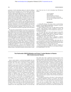
consequence, lower induction of IL-2. Direct assessment of
cytokine induction in a small sample of patients (n =6) and
controls (n=9) confirmed the existence of an IL-2 synthesis
defect after stimulation of CMV PBMC with PHA (around twofold reduction), Con A (three-fold) and PWM (three-fold) (Fig.
la). Thus, PWM and Con A stimulation, which caused a
significantly decreased proliferation in CMV PBMC (Table 2),
induced an average two-fold lower IL-2 production than PHA
in the patients; these PHA-induced IL-2 levels may have been
sufficient to allow a normal proliferation in response to PHA, as
shown in Table 2. Lastly, addition of PMA completely normalized the lectin-induced synthesis of IL-2.
number of CD8 cells in acute patients. For practical reasons,
they were pooled for the analysis, but the conclusions hold if the
two separate
Stimulation
sets of patients are maintained (data not shown).
with antigens and lectins (Table 2)
Upon stimulation
recognize
the
V/I
of CMV
region
PBMC
of the
T
with enterotoxins which
cell
receptor
(TCR),
a
significant decrease in T cell proliferation was observed (compared with control cells) both with enterotoxin A (around twofold,
P<0001)
and
with
enterotoxin
Cl
(almost
four-fold,
Stimulation with antibodies against CD3, CD28, CD26 and CD69
(Table 3)
Further analysis of T cell function was performed using specific
MoAbs. The proliferative response to anti-CD3 was markedly
lower in CMV than in control PBMC samples (around five-fold,
P<0 001). Co-stimulation of CMV PBMC with anti-CD3 and
antibodies directed against any other signal-transducing molecules, like CD28, also resulted in impaired proliferative
responses (around five-fold, P<0001) compared with control
T lymphocyte anergy in CMV infection
cells. This could not be due to a CD28 signalling defect in CMV
PBMC, because a specific antibody was capable of inducing
significant responses in the presence of PMA. The impaired
response to anti-CD3 is corrected with exogenous rIL-2, but
only in part with PMA, which directly activates PKC. Direct
assessment of IL-2 induction as above confirmed the existence
of an IL-2 synthesis defect after stimulation of CMV PBMC
with anti-CD3 (five-fold reduction) or anti-CD3 plus PMA (10fold reduction) (Fig. lb). Two other T cell surface molecules,
CD26 and CD69, which, like CD28, are capable of inducing T
cell proliferation in the presence of PMA, were triggered in a
small number of patients (n = 8). The results showed that CD26,
but not CD69, induced normal proliferation in CMV PBMC.
Lastly, the distal cytoplasmic signal-transducing biochemistry
of CMV PBMC seemed to be adequate, since their proliferative
response to the transmembrane activators PMA (which directly
activates PKC) plus ionomycin (which increases cytosolic Ca2+)
was comparable to that of normal controls. This suggests that
the proliferation defects observed in CMV PBMC using surface
stimuli are probably due to TCR/CD3 membrane-proximal
defects.
DISCUSSION
Secondary immunodeficiencies are a heterogeneous group of
disorders of the immune system that occur together with other
pathological conditions [13]. Due to the transient nature of the
alterations, the molecular basis of secondary immunodeficiencies is mostly unknown, precluding the development of rational
therapies. In this study, an attempt has been made to characterize a common herpesvirus-induced secondary immunodeficiency due to CMV infection.
Our results show that CMV infection, both acute and latent,
causes a selective anergy of the TCR/CD3 activation pathway.
This is supported by the observed low proliferative response of
PBMC obtained from CMV patients to enterotoxins, certain
lectins (Con A) and anti-CD3 MoAbs, whereas stimulation
using other lectins (PHA) or anti-CD28 or -CD26 antibodies
elicited normal proliferations. The impaired responses to antiCD3 antibodies and Con A were corrected with exogenous IL-2,
suggesting the existence of a TCR/CD3-associated signalling
defect to certain response genes like that of IL-2. Direct
assessment of IL-2 synthesis confirmed that it was more severely
reduced in response to Con A and anti-CD3. The defect was
probably proximal to the TCR/CD3 complex, because direct
activation of PKC using phorbol ester PMA corrected the
impaired proliferative responses to anti-CD3 only partially.
Although tested in only a small number of patients, the low
responses elicited through CD69 compared with CD26 (or
CD28) may suggest that CD69 activation is more CD3dependent than CD26 or CD28 [14], at least in this model.
A range of secondary immunodeficiencies is associated with
defects in T cell activation [15]. Secondary defects in CD3mediated, but not CD2-mediated (or CD28-mediated [15]) T
cell activation have been reported in certain pathological
situations like uraemia [16], HIV infection [17], Epstein-Barr
virus (EBV) infection [18]. Conversely, defects in the CD2 but
not in the CD3 activation pathway are believed to be secondary
to several disease states, like Sjogren's syndrome [19], lupus [20],
atopy [21], alcoholic liver disease [22] and Hodgkin's disease
[23]. In certain patients both the CD2 and CD3 activation
41
pathways show quantitative impairments, but a normal transmembrane activation biochemistry is preserved, as in Down's
syndrome [24], healthy aged individuals [25] and bone marrow
recipients [26]. More recently, biochemical defects in certain
TCR-associated proteins have been observed in tumour-bearing
mice and humans [27].
The molecular basis in each case is undoubtedly heterogeneous. However, the association of at least three viral infections
(HIV, EBV and CMV) with selective CD3-associated activation
defects may suggest the existence of a common evolutive
immunosuppression mechanism. The ability of these viruses to
encode for cell surface protein homologues of immunological
significance (CD4 ligands, CD21 ligands and HLA molecules,
respectively) may underlie the defect. Other interpretations are
possible, however. First, intracellular interferences like the
induction of apoptosis rather than proliferation, as shown for
HIV [28], may be involved in the observed CMV-associated T
cell anergy. Second, the observed increased levels of peripheral
blood CD8+ T cells in CMV patients may cause the selective
defect of T cell activation, as CD8 + cells are thought to contain
suppressor as well as cytotoxic lymphocytes [29]. Third, as we
have not assessed which subsets of T cells actually proliferate in
controls versus patients, a strong expansion of a set of anergic T
cells (in terms of proliferation to certain stimuli ex vivo) after
CMV infection could explain our data. In the last two cases,
immune response to CMV, rather than the virus itself, would
mediate the T cell anergy. Further immunological and virological work is required to elucidate the molecular basis of the
interaction of CMV with the human immune system. This, in
turn, will allow rational therapies to be developed for this (and
other) common secondary immunodeficiencies.
ACKNOWLEDGMENTS
We thank Ana Garcia for her help. This work was supported in part by
grants from FIS (91/324) and DGICYT (92/270).
REFERENCES
I Weller TH. The cytomegalovirus, ubiquitous agents with protean
clinical manifestations. N Engl J Med 1971; 285:203-67.
2 Rinaldo CRJ, De Bianco RL. Alteration of immunoregulatory
mechanism during cytomegalovirus mononucleosis: effect of in vitro
culture of lymphocyte blastogenesis to viral antigens. Clin Immunol
Immunopathol 1983; 28:46.
3 Browne H, Smith G, Beck S. Menson T. A complex between the
MHC class I homologue encoded by human cytomegalovirus and ,B2
microglobulin. Nature 1990; 347:770.
4 Hirsh MS, Felsenstein D. Cytomegalovirus induced immunosuppression. Ann NY Acad Sci 1984; 437:8.
5 Schrier RD, Rice GPA, Oldstone MBA. Suppression of natural
killer cell activity and T cell proliferation by fresh isolates of human
cytomegalovirus. J Infect Dis 1986; 153: 1084.
6 Carney WP, Rubin RH, Hoffman RA, Hansen WP, Healey K,
Hirsch MS. Analysis of T lymphocyte subsets in cytomegalovirus
mononucleosis. J Immunol 1981; 126:2114.
7 Moretta A, Ciccone E, Pantaleo G et al. Surface molecules involved
in the activation and regulation of T or natural killer lymphocytes in
humans. Immunol Rev 1989; 111:145-75.
8 Britt WJ, Pass RF, Stagno S, Alford CA. Pediatric cytomegalovirus
infection. Transplant Proc 1991; 23:115-7.
9 Carney WP, Tacoviello V, Hirsch MS. Functional properties of T
lymphocytes and their subsets in cytomegalovirus mononucleosis.
J Immunol 1983; 130:390-5.
42
M. Timon et al.
10 Middeldorp JM, Jongsma J, Terhoor A, Schirm J, The TH.
Conditions for the detection of IgM and IgG antibodies against
CMV early (CMV-EA) and late (CMV-LA) antigens by ELISA.
J Clin Microbiol 1984; 20:763.
11 Caldwell CW, Taylor HM. A rapid, no-wash method for immunophenotypic analysis by flow cytometry. Am J Clin Path 1986;
86:600-7.
12 Regueiro JR, Lopez-Botet M, Landazuri MO et al. An in vivo
functional immune system lacking polyclonal T cell surface expression of the CD3/Ti (WT3 1) complex. Scand J Immunol 1987;
26:699-707.
13 Amman AJ. Immunodeficiency diseases. In: Stites DP, Stobo JD,
Wells JV, eds. Basic and clinical immunology. East Norwalk:
Appleton and Lange, 1987:347-53.
14 Rinc6n M, Cebridn M, Sanchez-Madrid F, Lopez-Botet M. Induction of T cell function via the gp 33/27 activation inducer molecule
(AIM) requires co-expression of the CD3/TCR complex. Eur J
Immunol 1989; 19:959-62.
15 Arnaiz-Villena A, Tim6n M, Rodriguez-Gallego C, Perez-Blas M,
Corell A, Martin-Villa M, Regueiro JR. Human T-cell activation
deficiencies. Immunol Today 1992; 13:259-65.
16 Meuer SC, Hauer M, Kurz P, Meyer zum Buschenfelde KH, Kohler
H. Selective blockade of the antigen-receptor-mediated pathway of
T cell activation in patients with impaired primary immune responses. J Clin Invest 1987; 80:743-9.
17 Linette GP, Hatzman RJ, Ledbetter JA, June CH. HIV- I -infected T
cells show a selective signaling defect after perturbation of CD3/
antigen receptor. Science 1988; 241:573-6.
18 Perez-Blas M, Regueiro JR, Ruiz-Contreras J, Arnaiz-Villena A. T
lymphocyte anergy during acute infectious mononucleosis is restricted to the clonotypic receptor activation pathway. Clin Exp
Immunol 1992; 89:83-88.
19 Dauphinee MJ, Tovar Z, Ballester A, Talal N. The expression and
function of CD3 and CD5 in patients with primary Sjogren's
syndrome. Arthritis Rheum 1989; 32:420-9.
20 Fox DA, Millard JA, Treisman J et al. Defective CD2 pathway T cell
activation in systemic lupus erythematosus. Arthritis Rheum 1991;
34:561-71.
21 Romano MF, Valerio G, Turco MC et al. Defect of CD2- and CD3mediated activation pathways in T cells of atopic patients: role of
interleukin 2. Cell Immunol 1992; 139:91-97.
22 Spinozzi F, Bertotto A, Rondoni F et al. T-lymphocyte activation
pathways in alcoholic liver disease. Immunology 1991; 73:140-6.
23 Meuer SC, Roux MM, Schraven B. The alternative pathway of T cell
activation: biology, pathophysiology, and perspectives for immunopharmacology. Clin Immunol Immunopathol 1989; 50:133-9.
24 Bertotto A, Crupi S. Arcangeli C et al. T-cell response to phorbol
ester PMA and calcium ionophore A23187 in Down's syndrome.
Scand J Immunol 1989; 30:583-6.
25 Beckman I, Dimopoulos K, Xu X, Ahern M, Bradley J. Age-related
changes in the activation requirements of human CD4+ T-cell
subsets. Cell Immunol 1991; 132:17-25.
26 Yamagami M, McFadden PW, Koethe SM, Ratanatharathorn V,
Lum LG. Failure of T cell receptor-anti-CD3 monoclonal antibody
interaction in T cells from marrow recipients to induce increases in
intracellular ionized calcium. J Clin Invest 1990; 86:1347-51.
27 Mizoguchi H, O'Shea JJ, Longo DL, Loeffler CM, McVicar DW,
Ochoa AC. Alterations in signal transduction molecules in T
lymphocytes from tumor-bearing mice. Science 1992; 258:1795-98.
28 Groux H, Tarpier G, Maute D, Montan Y, Capron A, Ameisen JC.
Activation-induced death by apoptosis in CD4 + T cells from human
immunodeficiency virus-infected asymtomatic individuals. J Exp
Med 1992; 175:331-40.
29 Takata M, Maiti PK, Kubo RT, Chen YH, Holford-Stevens V,
Rector ES, Sehon AH. Cloned suppressor T cells derived from mice
tolerized with conjugates of antigen and monomethoxypolyethylene
glycol. Relationship between monoclonal T suppressor factor and
the T cell receptor. J Immunol 1990; 145:2846-53.
© Copyright 2026
