
Aversion and Attraction through Olfaction
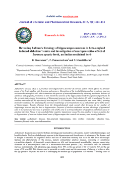
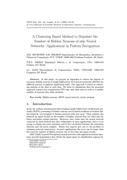
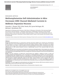
Current Biology 25, R120–R129, February 2, 2015 ª2015 Elsevier Ltd All rights reserved
http://dx.doi.org/10.1016/j.cub.2014.11.044
Aversion and Attraction through Olfaction
Qian Li and Stephen D. Liberles*
Sensory cues that predict reward or punishment are
fundamental drivers of animal behavior. For example,
attractive odors of palatable food or a potential mate predict reward, while aversive odors of pathogen-laced food
or a predator predict punishment. Aversive and attractive
odors can be detected by intermingled sensory neurons
that express highly related olfactory receptors and display
similar central projections. These findings raise basic
questions of how innate odor valence is extracted from
olfactory circuits, how such circuits are developmentally
endowed and modulated by state, and how innate and
learned odor responses are related. Here, we review
odors, receptors and neural circuits associated with stimulus valence, discussing salient principles derived from
studies on nematodes, insects and vertebrates. Understanding the organization of neural circuitry that mediates
odor aversion and attraction will provide key insights into
how the brain functions.
Introduction
Our five basic external senses — touch, taste, vision, hearing, and smell — and our internal sensory systems that regulate bodily homeostasis are strong drivers of behavior. For
example, mice exhibit fear responses to the smell of a cat,
the sight of a looming hawk or the sound of an unknown
animal rustling in nearby leaves. Anatomically distinct brain
regions initially process smell, sight and sound, yet responding neural pathways can converge on similar control centers
to execute common behaviors, such as fear responses.
Conversely, other sensory stimuli, such as the smells of
mates, food or offspring, will evoke different behaviors
related to reproduction, feeding, or parental care. So, within
a sensory system, neural pathways that are anatomically
quite similar can diverge centrally for execution of specific
responses. Understanding how input from different sensory
systems might converge, whereas inputs within a sensory
system might diverge, presents an important challenge for
study. In general, the routing logic at the interface of sensory
systems and descending motor programs that enables
execution of stimulus-appropriate behaviors remains poorly
defined.
Here, we discuss recent advances in understanding the
molecular basis of odor attraction and aversion behavior as
a model for deciphering how a sensory system can evoke
divergent responses. Olfaction is a powerful model system
for unraveling the molecular basis of behavior — many species rely on their sense of smell for survival, and olfactory
circuits are highly streamlined, using a small number of
synaptic connections to convert sensory inputs into behavioral outputs [1,2]. Our hope is that principles gleaned from
understanding odor aversion and attraction will shed light
on how the olfactory system can drive other divergent behaviors, such as responses to pheromones that evoke or
Department of Cell Biology, Harvard Medical School, Boston, MA
02115, USA.
*E-mail: [email protected]
Review
inhibit sexual behavior, aggression and parental care [3].
We take an integrative analysis, describing odors, receptors
and neural circuits associated with aversion and attraction
across species, with a focused discussion of model organisms where advances have been numerous.
Odor Valence in the Nematode Caenorhabditis elegans
Attractive and Aversive Odors for C. elegans
The nematode C. elegans, with its simple nervous system,
provides a powerful model for mechanistic dissection of
odor aversion and attraction behavior. Like other animals,
C. elegans uses its olfactory system for social behavior,
foraging and pathogen avoidance [4]. Many attractants are
essential nutrients, minerals and food-associated odors
that signal the presence of nearby bacterial prey [5]. Chemotaxis towards water-soluble attractants involves a characteristic movement pattern similar to the bacterial random walk,
where animals pirouette and change direction at a frequency
inversely related to changes in attractant concentration [6].
In addition, social cues, such as pheromones, can be attractive [7]. C. elegans releases and detects pheromone blends
containing structurally related glycolipids called ‘ascarosides’ [8–10]. Ascarosides released by C. elegans hermaphrodites attract males, and depending on concentration and
the social nature of the strain, cause aggregation or dispersal
behavior in other hermaphrodites [11,12]. C. elegans also
displays long-range chemotaxis behavior towards chemically diverse airborne stimuli [13], including the odor diacetyl
(2,3-butanedione) for which the first nematode chemosensory receptor was identified [14].
C. elegans displays stereotyped avoidance behaviors to
carbon dioxide and many volatile odors, including long
chain alcohols and ketones [13,15,16]. Detection of pathogen-derived cues, such as the lipopeptide serrawettin
W2 produced during Serratia marcescens swarming
behavior or biofilm-promoting metabolites from Pseudomonas aeruginosa, enables avoidance of pathogen-infested bacterial lawns [17,18]. Odors of other pathogenic
bacteria are not innately aversive, but associated illness
causes learned odor avoidance through a serotonin-mediated mechanism [19]. Other stimuli also evoke olfactory
learning in C. elegans, such as nutrient abundance or scarcity [4,20], with distinct neural circuits mediating innate and
acquired responses [21].
C. elegans Olfactory Receptors and Sensory Neurons
C. elegans contains three pairs of chemosensory neurons
that play a major role in odor detection, the AWA, AWB and
AWC neurons (Figure 1A), as well as other sensory neurons
that detect water-soluble chemicals, pheromones, oxygen,
carbon dioxide and nociceptive stimuli [4]. AWA, AWB, and
AWC sensory neurons detect odors using hundreds of coexpressed chemosensory G protein-coupled receptors
(GPCRs) [4]. Each sensory neuron expresses a large and
unique receptor repertoire, suggesting an ability to detect
chemically diverse stimuli [4]. Activation of any chemosensory GPCR within a particular neuron likely has the capacity
to trigger common signaling pathways, neural circuits and
behavioral responses. Laser microbeam-induced ablation
of AWA and AWC neurons eliminated several odor attraction responses, while ablation of AWB neurons eliminated
Review
R121
Figure 1. Aversion and attraction in
C. elegans.
(A) Location of olfactory sensory neurons
(AWA, AWB, AWC) in the worm head. Each
AWA, AWB, and AWC neuron pair extends
sensory cilia that are embedded in the sheath
near the amphid pore. Image adapted from
[128] and WormAtlas. (B) AWC neurons are
inhibited by attractive odors, and control
turning rate by gating the activity of AIB and
AIY interneurons through glutamate release.
A
several odor aversion responses
[13,22]. These and other neurons have
also been genetically linked to attracB
tion and aversion behaviors by their
response properties and cell-type-speChemotaxis
cific rescue of key sensory signaling
molecules [23,24].
One C. elegans chemosensory
receptor, Odr-10, mediates attraction
to the volatile odor diacetyl [14], as
mutant animals lacking Odr-10 have a
specific chemotaxis deficit to diacetyl.
Local search
Odr-10 is expressed in AWA neurons
that mediate attraction responses, but
interestingly, misexpression of Odr-10
in AWB neurons instead of AWA neurons reverses the valence of the odor
response, causing worms to avoid
diacetyl [22]. These findings elegantly demonstrate that the
valence of a C. elegans odor response is guided by an
intrinsic property of the responding neuron — such as its
connectivity or synaptic release properties — that is independent of the responding receptor.
Higher-Order Processing of Odor Valence in C. elegans
The complete wiring diagram of the C. elegans nervous
system has revealed potential avenues for information flow
during odor aversion and attraction behavior. With only a
few synaptic connections — from sensory neuron to sensory
interneuron to command neuron to motor neuron — an odor
can drive a behavioral response [25]. For example, AWC
neurons mediate either attractive chemotaxis behavior
or local search behavior by gating the relative activity of
different interneurons (Figure 1B) [23]. AWC neurons detect
several attractive odors and respond by hyperpolarizing
and decreasing neurotransmitter release [23]. AWC neurons
form excitatory glutamatergic synapses with AIB interneurons, which promote turning behavior, and inhibitory glutamatergic synapses with AIY interneurons, which restrict
turning behavior [23]. Thus, an attractive odor will restrict
turning behavior by removing sensory neuron-mediated
inhibition of AIY interneurons. Conversely, abrupt removal
of an attractive food-odor source activates AWC neurons
and AIB interneurons, causing the opposite response: a
dramatic increase in turning frequency characteristic of local
search behavior.
Despite the apparent simplicity of the neural wiring diagram in C. elegans, the flow of information can be strikingly
complex and dynamic, with neurons changing roles and
synapses changing strength, depending on experience and
state [21,24,26]. Olfactory information can be controllably
AWB aversion
AWA
attraction
AWC
AIB
Attractive
odors
AWC
Decrease turning
AIY
AIB
Attractive
odors
Increase turning
AWC
AIY
Current Biology
routed by modulating the response or release properties of
neurons throughout the circuit [19,26–29], and new neurons
can be recruited to the circuit during odor learning [21].
Considering sensory neurons alone, output can be changed
by odor adaptation, odor sensitization, feedback from neuromodulators, such as biogenic amines and insulin-like peptides, and gap junctions that function as part of a hub-andspoke circuit (see below) [12,20,26–28]. Neuromodulators
can impact sensory neuron responses, synaptic strength,
and perhaps even the basic logic gate (or shift in behavioral
salience) of a synapse [24,27,29]. Dynamic modulation of
competing sensory neuron inputs to a particular interneuron
can create a so-called ‘push–pull’ circuitry motif, with the
relative synaptic strengths instructing behavioral outcome
[24]. The same neuromodulator can also be used in different
contexts; for example, serotonin released from different
neurons can either signal the rewarding presence of food
or the aversive effects of pathogen-induced illness [19,20].
Furthermore, the function of a single sensory neuron type
can be flipped. Mutations in a presynaptic signaling pathway
involving guanylate cyclase and diacylglycerol reverse the
valence of the AWC neuron-mediated behavioral response
from specifying attraction to aversion behavior, and perhaps
toggling of this pathway underlies natural state-dependent
changes in odor responses [29].
Electrical coupling, as occurs in the hub-and-spoke circuit
motif, provides another potential mechanism for dynamic
control of sensory information [12]. The RMG neuron is
an interneuron at the center of a hub-and-spoke circuit
that controls the choice between pheromone-evoked
attraction (social aggregation) or aversion (dispersal) [12].
The RMG neuron is electrically coupled to multiple sensory
neurons that receive input about hermaphrodite population
Current Biology Vol 25 No 3
R122
density — for example, through detection of ascarosides or
oxygen. Activity in the RMG neuron is broadly transmitted
through these sensory neurons, helping to amplify and coordinate weak responses. Social C. elegans strains have mutations in a receptor (Npr-1) for an inhibitory neuromodulator,
causing a boost in hub-and-spoke output [12,30]. Npr-1
signaling possibly restricts RMG function by dampening
basal activity or by reducing the extent of electrical coupling.
Signaling through hub-and-spoke circuitry can be integrated
with other neuromodulator effects, enabling complex sexand state-specific changes in neuron output [24]. The many
opportunities for dynamic circuit modulation in C. elegans
odor responses indicate that a full understanding of nervous system structure through a connectivity map provides
only a starting point for understanding how the flow of sensory information through neural circuits can be flexibly
routed to evoke behaviors that vary with state and experience [31].
Odor Valence in Fruit Flies and Mosquitoes
Attractive and Aversive Odors for Drosophila and Aedes
Odors that attract or repel insects provide tools to thwart
agricultural pests and potentially disease vectors [32,33].
Here, we focus on insect model systems that have enabled
genetic access to the olfactory system: the fruit fly,
Drosophila melanogaster [34–37], and more recently the
mosquito Aedes aegypti [38,39].
Drosophila display innate attraction to food-related odors,
including farnesol in citrus fruit peels, amines associated
with protein breakdown, as well as complex odor mixtures
emitted from apple cider vinegar, yeast and natural blends
of spoiled and unspoiled fruit [35,40–42]. It should be noted
that the valence of odor responses can be concentrationdependent — increasing odor levels can recruit additional
olfactory receptors, change central odor representations
and even alter perceived valence [35]. Attraction to food
odors is also state-dependent, with behavioral responses
enhanced by food deprivation [43,44]. Mosquitoes and flies
display different feeding preferences; mosquitoes generally
feed on nectar, with females about to lay eggs also feeding
on blood. Female mosquitoes locate human targets using
multiple sensory inputs, including sweat and skin odors,
carbon dioxide in breath, and non-olfactory stimuli such as
heat and visual cues [33,38,39]. Whereas mosquitoes are attracted to carbon dioxide (0.1% above ambient), Drosophila
displays a different and complex response. Walking flies
avoid elevated carbon dioxide, but flies in flight track a
carbon dioxide plume, suggesting state-dependent responses [45]. Carbon dioxide may function as an alarm
signal released by stressed flies and/or indicate fruit palatability [36,46]. Flies also avoid geosmin, an odor produced
by harmful microbes, high concentrations of acids and
various other volatiles [34,47,48]. Both mosquitoes and
flies innately avoid the widely used insect repellant DEET
(N,N-diethyl-3-methylbenzamide), and other chemicals that
target DEET-activated sensory neurons [33].
Attractive sex pheromones have been identified from
thousands of insect species, including agricultural pests
that are effectively controlled by luring to pheromone-laced
traps [32]. The first pheromone identified in any species
was bombykol, a fatty acid derivative produced by the
female silkworm which is a powerful long-range attractant
for conspecific males [49]. Similar far-acting attractants
have not been identified in the fly, but the male sex
pheromone 11-cis-vaccenyl acetate (cVA) controls courtship
and male aggression, providing a powerful tool for study of
sexually dimorphic neural circuitry [2,50–53].
Drosophila Olfactory Receptors and Sensory Neurons
Drosophila has w2,600 olfactory sensory neurons located in
sensilla of the antenna and maxillary palp (Figure 2A; for a
recent review of Drosophila olfaction, see [54]). Drosophila
sensory neurons are bipolar neurons containing both sensory dendrites that detect odors and long axons that transmit information to the antennal lobe of the brain [54]. In the
antennal lobe, sensory neurons communicate with secondorder projection neurons at specialized structures termed
‘glomeruli’ [54]. Olfactory sensory neurons detect odors
using large families of chemosensory receptors: odorant
receptors (ORs), ionotropic receptors (IRs) and gustatory
receptors (GRs) [37,55–58]. Drosophila ORs, IRs and GRs
are not GPCRs, but instead are heteromeric ligand-gated
ion channels [55,59–61]. Sensory neurons typically express
one or a few IRs, ORs or GRs, and ORs are usually expressed
together with an obligate co-receptor (Or83b or ORCO) [37].
Sensory neurons containing the same receptor target dedicated glomeruli in the fly brain, with the small size of the
Drosophila olfactory system enabling a nearly comprehensive assignment of antennal lobe glomeruli and the sensory
neurons that innervate them [62,63].
Several insect ORs and IRs are required for aversion and
attraction responses to particular odors. Fly attraction to
apple cider vinegar involves Or42b and Or92a, while high vinegar concentrations recruit an additional low affinity receptor,
Or85a, which mediates dominant aversion responses [35].
Specific fly olfactory receptors also mediate attraction to
amines (IR92a) [42], attraction to farnesol (Or83c) [41], aversion to acids (IR64a) [34,47] and aversion to the microbeassociated odor geosmin (Or56a) [48]. Fruit-associated
odors influence egg-laying and male courtship through specific olfactory receptors (OR19a, IR84a), presumably to
promote offspring deposition on preferred energy-rich
substrates [64,65]. In fly larvae, two different olfactory receptors (Or42a, Or42b) mediate behavioral attraction to low and
high concentrations of ethyl acetate [66]. Flies and mosquitoes display different behavioral responses to carbon dioxide
(0.1–1.0% above ambient), but use orthologous receptors for
detection, suggesting species-specific differences in receptor-associated neural circuitry [36,39]. The repellant DEET activates multiple chemosensory receptors, including an IR, an
OR and a GR, and may act as a ‘confusant’ that distorts other
receptor-ligand interactions [33,38]. Aversive and attractive
odor responses are mediated by intermingled and often adjacent glomeruli in the antennal lobe (Figure 2B), although some
spatial organization of odor valence has been proposed [67].
Together, these findings indicate that neurons with related
sensory receptors and similar projections to the antennal
lobe can generate opposing behaviors in the fly.
Higher-Order Processing of Odor Valence in Drosophila
Each of the w50 antennal lobe glomeruli in Drosophila is
innervated by, on average, four projection neurons, with
most projection neurons innervating a single glomerulus
[68,69]. Projection neurons generally respond to the same
odors as their connected sensory neurons, with response
gain and kinetics modulated by local interneuron circuits of
the antennal lobe [54]. Projection neurons transmit olfactory
information to two higher order olfactory centers, the lateral
Review
R123
Innate
A
Learned
LH
MB
AL
AL
Antenna
Maxillary palp
B
DA2
DM5
DC3
VA2
DM1
DC4
1
VM
horn and the mushroom body, which are thought to mediate
innate and learned odor responses, respectively.
Roles for the lateral horn in innate behaviors — including
attraction, aversion, and pheromone responses — have
largely been inferred by observing the residual olfactory behaviors that persist after mushroom body ablation [70,71].
The lateral horn receives input from antennal lobe projection
neurons, and this input has been systematically mapped
using single neuron genetic approaches [68,69,72,73]. The
axons of projection neurons are highly stereotyped and
regionalized within the lateral horn, with those of a given
glomerulus typically clustering spatially. (There are exceptions, such as the divergently innervating projection neurons
from the CO2-responsive glomerulus [42].) In contrast, input
from different glomeruli is more variably distributed, with
projection neurons from neighboring glomeruli sometimes
targeting quite distant locations [68,69]. The lateral horn is
proposed to contain multiple input zones, including dedicated zones for processing pheromones and attractive
food odors [73]. Projections from individual glomeruli linked
to odor attraction (amines) and aversion (acids and CO2)
target topographically distinct regions of the lateral horn,
suggesting that valence is spatially encoded in the lateral
horn [42]. However, lateral horn inputs are not mapped
solely based on ethological salience, as for instance
projection neurons responsive to the food-associated
odor farnesol innervate the putative pheromone-response
zone [41].
Third-order lateral horn neurons can be classified based
on their anatomy and breadth of response properties
[72,74]. For example, one group of lateral horn neurons responds broadly to odors that activate several invariant
glomeruli, perhaps allowing for channeling of ecologically
related odors, such as food odors, into a common hardwired response [74]. In contrast, a second group of lateral
horn neurons receives input from one glomerulus but responds more narrowly due to stereotyped inhibitory input
from other coactivated glomeruli [74]. Thus, third-order neurons of the lateral horn can have either broader or narrower
response fields than their connected projection neurons.
How do lateral horn neurons evoke particular innate behaviors? One possibility is that different classes of lateral horn
neurons are connected to different descending motor outputs. Experiments that traced an entire pheromone-responsive circuit from input to output revealed a class of
highly tuned pheromone-responsive lateral horn neurons
with sexually dimorphic projections, including to male-specific descending neurons that enter the ventral nerve cord
[2]. Identifying other classes of lateral horn output neurons
that evoke either innate aversion or attraction would provide
a foundation for understanding how these behaviors are extracted from particular sensory inputs.
The mushroom body is the major site of associative olfactory learning in insects [71,75,76]. The fly mushroom body
contains w2,500 intrinsic third-order neurons called ‘Kenyon
cells’ that together receive sensory input from w200 projection neurons. (Other insects can have far more Kenyon cells;
for example, the honeybee has 170,000 [76].) Projection neurons send diffuse collaterals across the mushroom body
calyx, with extensive intermingling of projection neurons
from different glomeruli [68,69,72]. Each Kenyon cell forms
synapses with incoming projection neurons at the calyx,
with Kenyon cell dendrites displaying a characteristic clawlike morphology (termed a ‘dendritic claw’) that envelops
V
D
Aversion glomeruli
L
Attraction glomeruli
Current Biology
Figure 2. Anatomical organization of fly olfactory circuits.
(A) Distinct olfactory circuits mediate innate (blue) and learned (red)
odor responses. Image adapted from [75]. (B) Glomeruli in the fly
antennal lobe that mediate aversion (red) and attraction (blue) are intermingled and can be adjacent. Depicted glomeruli respond to vinegar
(low threshold: VA2, DM1; high threshold: DM5), farnesol (DC3), amines
(VM1), geosmin (DA2), acids (DC4), and CO2 (V). Anterior to posterior
antennal lobe representations are depicted clockwise from the top
left. Image adapted from [62,63].
the axon of an incoming projection neuron [77]. Each Kenyon
cell has, on average, 7 dendritic claws [78], with each claw
connecting to a different projection neuron and responding
Current Biology Vol 25 No 3
R124
Box 1
A model for olfactory learning in the Drosophila mushroom body.
What are the mechanisms of associative attraction and aversion learning in the mushroom body? One compelling model is that learning is
regulated at the synapse between Kenyon cells that report odor identity and mushroom body output neurons that report behavioral
significance [75,76]. Synapses between Kenyon cells and output neurons would be strengthened by learning, via neuromodulators that
provide signals of unconditioned reward or punishment and whose release coincides with Kenyon cell activity. The same neurotransmitter,
dopamine, provides an unconditioned signal for both reward (sugar) and punishment (electric shock) [130–134]. Dopamine neurons that
mediate attraction and aversion learning are largely, but not exclusively, located in different neuron clusters extrinsic to the mushroom body
and innervate spatially segregated axonal compartments of different Kenyon cell classes [133,134]. It is possible that a single Kenyon cell
couples to different readouts through different synapses. Alternatively, recent evidence indicates that aversion and attraction may be driven
by non-overlapping Kenyon cell populations [135]. In the latter model, it is important that odors are represented across each subtype of
Kenyon cell. Either way, output neurons must be in tight opposition to particular dopamine neurons to ensure appropriate responses- this is
appreciated for one class of mushroom body output neuron (MB-V2 neurons) that mediates aversion but not appetitive memory performance
[136]. Interestingly, MB-V2 neurons send projections to the lateral horn, raising the possibility that execution of learned odor responses
involves recruitment of circuitry components underlying innate odor responses [136]. A comprehensive understanding of the diversity of
output neurons and associated neuromodulatory neurons available to Kenyon cells may inform on the repertoire of learnable fly behaviors.
to different odors [77]. Different dendritic claws of the same
Kenyon cell receive input from random projection neurons,
with little or no assignment bias due to the location, response
properties or ethological salience of projection neurons, or
due to the class of the responding Kenyon cell [78]. Kenyon
cells require activation of multiple claws to fire, with convergent input from multiple claws summed to drive Kenyon cells
to spike threshold [77]. This anatomical organization and
requirement for coincidence detection is consistent with
the sparse and distributed pattern of odor-evoked activity
observed across the Kenyon cell population [79,80]. These
findings are consistent with a primary role for the mushroom
body in associative learning; it is difficult to imagine how
innate responses could be specifically encoded through a
random wiring architecture of unsupervised design (Box 1).
Innate and learned odor responses can be further modulated
by internal state and sex to ensure situation-appropriate
display of attraction or aversion. For example, hungry flies
display increased attraction to food odors, an effect due at
least in part to neuropeptide modulation of neurons throughout
olfactory circuits [43,44]. In sensory neurons, starvationinduced decreases in insulin promote expression of the short
Neuropeptide F (sNPF) receptor, through which sNPF induces
presynaptic facilitation, potentiation of glomerular responses,
and enhanced food-seeking behavior [44]. A related neuropeptide, Drosophila Neuropeptide F, gates activity of mushroom
body-innervating dopamine neurons important for hunger
state-dependent memory retrieval [43]. Olfactory circuits also
differentially route pheromone input to evoke sex-specific behaviors [2]. Sexually dimorphic neural circuits develop under
control of the male-specific isoform of the transcription factor
fruitless (FruM), which is expressed in and/or shapes the architecture of first-order sensory neurons, second-order projection neurons, third-order lateral horn neurons and fourthorder descending neurons of a pheromone-response pathway
[2]. It seems that in Drosophila, as in C. elegans, state- and sexdependent behaviors are produced through multi-tiered control of neurons at multiple levels in the circuit.
Odor Valence in Mice
Attractive and Aversive Odors for Mice
Mice, like the other species discussed, display attraction
to food and mates as well as aversion to predators and
pathogens. Aversion and attraction responses in mice, as
in insects and nematodes, are strongly guided by learning.
Mice display learned attraction to odors associated with
energy-rich meals, through integration with taste or internal
sensory pathways, and learned aversion to odors conditioned by post-ingestive illness. Mice even locate their first
meal, milk, by chemotaxis towards maternal odors learned
in utero and during parturition [81]. Mice also use olfactory
cues for social behavior, displaying robust attraction to
conspecific scent marks, pheromone-rich urine deposits
used for territorial marking and mate attraction. Many odors
present in mouse urine are attractive, including trimethylamine, methylthio-methylthiol (MTMT), (Z)-5-tetradecen-1ol, dehydro-exo-brevicomin, farnesenes, and a urinary
protein darcin [82–86]. Neonatal odors can be attractive to
parents but neutral (mice) or aversive (rat) to sexually naive
animals, with associated neural circuitry modulated by pregnancy hormones and experience [87]. Predator odors are
aversive to mice upon first exposure, suggesting a hardwired response. Predator odors avoided by mice include lipocalin proteins, the carnivore odor 2-phenylethylamine and
the fox odor 2,5-dihydro-2,4,5-trimethylthiazoline (TMT)
[88–90]. Conversely, certain prey odors such as skunk odor
thiols and the mouse odor trimethylamine repel potential
predators [83], and mice reportedly release alarm pheromones to warn nearby conspecifics of danger [91]. Mice
also avoid carbon dioxide, spoiled food odors such as isoamylamine, aliphatic acids and aliphatic aldehydes, as well as
the carrion odor cadaverine [83,89,92–94].
Each of these innate aversion responses can be flexible
between species: for example, the carnivore odor 2-phenylethylamine is aversive to mice but is proposed to function as
a sex pheromone in tigers [88,95]. Carrion odors provide
aversive pathogen-related danger signals to some animals,
but attractive food-associated cues to scavengers like vultures and burying beetles [94,96]. Finally, trimethylamine is
an attractive scent odor constituent in mice but repels rats
(who eat mice) and humans [83]. Such species-specific behaviors suggest that the olfactory system can morph during
evolution to change the valence of specific odor responses.
Aversive and attractive odors provide valuable tools for understanding neural pathways that encode stimulus valence
(see below).
Review
R125
Figure 3. Olfactory pathways in the mouse.
(A) The main olfactory system involves different brain nuclei that may mediate innate
(blue) and learned (red) responses. Signals
from the main olfactory epithelium (MOE) are
transmitted to the main olfactory bulb (MOB)
and then to several brain regions including
the anterior olfactory nucleus (AON), piriform
cortex (PC), olfactory tubercle (OT), posterolateral cortical amygdaloid nucleus (PLCN),
and anterior cortical nucleus (ACN). In the
accessory olfactory pathway (green), signals
from the VNO are sent to the accessory olfactory bulb (AOB) and then to the medial amygdala (MeA) and posteromedial cortical amygdalaoid nucleus (PMCN). Image adapted from
[129]. (B) The adjacent projections of TAAR4
(green) and TAAR5 (red) sensory neurons
were revealed by antibody staining [109].
A
AOB
MOE
MOB
AON
ACN
MeA
PC
VNO
B
Mouse Olfactory Receptors and
Sensory Neurons
Mice have about five million olfactory
sensory neurons located in the main
olfactory epithelium, with additional
chemosensory neurons located in the
vomeronasal organ (VNO), Grueneberg
ganglion and septal organ [3]. As in
Drosophila, mouse olfactory sensory
neurons are bipolar neurons, containing both sensory dendrites and long
axons that innervate glomeruli in the
olfactory bulb. Sensory neurons detect
odors using >1,500 receptors from five
evolutionarily distinct GPCR families:
odorant receptors (ORs) and trace
amine-associated receptors (TAARs)
in the main olfactory epithelium, and
vomeronasal receptors (V1Rs, V2Rs)
and formyl peptide receptors (FPRs) in the vomeronasal organ [97–104]. In addition, rare sensory neurons detect odors
using a membrane-associated guanylate cyclase [105].
Large-scale ablation of >100 glomeruli throughout the dorsal olfactory bulb (DD mutants) produced striking deficits
in aversion responses to leopard urine, the fox odor TMT,
2-methylbutyric acid, isoamylamine and other odors [89].
Interestingly, despite the absence of innate aversion to
TMT and 2-methylbutyric acid, DD mutant animals could still
detect these cues and learn to like or dislike them using other
glomeruli. These findings indicate that TMT and 2-methylbutyric acid can activate multiple glomeruli with only specific
glomeruli mediating innate responses.
Which olfactory sensory neurons mediate innate responses
and how do they develop appropriate circuit connectivity?
Only a few mouse olfactory receptors have been linked to aversion and attraction behaviors. Certain receptors detect attractants, including trimethylamine (TAAR5), (Z)-5-tetradecen-1-ol
(Olfr288), and MTMT (Olfr1509), and other receptors detect
repellants, including the carnivore odor 2-phenylethylamine
(TAAR4), hexanal (many ORs), hexanoic acid (many ORs),
N-methylpiperidine (TAAR8c, TAAR9) and isoamylamine
(TAAR3) [83,86,88,106–108]. Knockout of mouse Taar4 eliminates 2-phenylethylamine aversion while knockout of mouse
Taar5 eliminates trimethylamine attraction [83,92]. TAAR4
PLCN
OT
PMCN
Attraction
Aversion
TAAR4
TAAR5
Current Biology
and TAAR5 are encoded by adjacent genes and neurons that
express these receptors project axons to adjacent glomeruli
in the brain (Figure 3B) [109]; nevertheless, these receptors
mediate behavioral responses of opposing valence in mouse.
Interestingly, neurons initially committed to express a Taar5
knockout allele can switch receptor choice, with some neurons secondarily expressing Taar4 [109]. This observation
suggests that the commitment of a sensory neuron to couple
to aversion or attraction circuitry may arise relatively late in
development, seemingly after receptor choice has stabilized.
Alternatively, it is possible that TAAR5-mediated attraction
involves a learned override of a developmentally established
aversion circuit.
Higher-Order Processing of Odor Valence in Mice
Olfactory information in mice, as in flies, is processed in parallel by multiple brain regions. Sensory neurons from the
main olfactory epithelium and vomeronasal organ target
the main and accessory olfactory bulbs (MOB and AOB)
respectively, which then couple to strikingly divergent
pathways deeper in the brain (Figure 3A) [1]. MOB projection
neurons (termed ‘mitral’ or ‘tufted’ cells) provide dense
innervation of several cortical and limbic areas including
the piriform cortex, cortical amygdala and olfactory tubercle.
In contrast, AOB mitral cells directly target limbic system
Current Biology Vol 25 No 3
R126
nuclei, including the vomeronasal amygdala and bed nucleus of the stria terminalis (BNST). Despite these anatomical
differences, the main and accessory olfactory systems
can both mediate aversion and attraction behaviors
[83,85,88,90].
Responses to predator odors are arguably the best studied innate odor-driven behaviors in mice [110]. Different
classes of predator odors (volatile vs non-volatile) are detected by the main olfactory epithelium (TMT, 2-phenylethylamine, crude predator urine) and vomeronasal organ
(lipocalins, crude cat dander or collar odors) [88–90,110].
These different predator odor classes activate non-overlapping neural pathways, as determined by focal lesion analysis and patterns of immediate early gene expression.
Aversion to TMT requires several limbic system nuclei,
including the medial BNST and the lateral septum, while
the spoiled food odor 2-methylbutyric acid activates neurons in the adjacent lateral BNST [89,111]. VNO-activating
predator odors activate a different neural network termed
the medial hypothalamic defensive system, composed of
the medial amygdala, the dorsomedial region of the ventromedial hypothalamus, the premamillary nucleus, periaqueductal grey and other brain regions [112]. Both main and
accessory olfactory pathways — as well as predatorderived visual and auditory inputs — ultimately engage
common effectors in the limbic system, such as the hypothalamic-pituitary-adrenal axis, which coordinates systemic
stress responses.
The routing logic between sensory inputs and brain centers that control innate responses remains poorly defined
in the mouse. Dense, stereotyped projections from the
MOB arise in the cortical amygdala, which functions in innate
odor responses [113]. Projections from the AOB densely
innervate the nearby vomeronasal amygdala, which may
serve a similar organizing role. The odors of males, females,
juveniles and predators activate neuron subsets in the
medial amygdala [114–117], suggesting that the vomeronasal system contains parallel processing streams relevant
for different behaviors. One model is that the medial amygdala acts as a hub, receiving inputs from a large number of
different vomeronasal receptors, and funneling information
selectively to evoke appropriate behavioral outcomes. It
will be exciting to understand the diversity of medial amygdala neuron subtypes, their roles in different innate behaviors and whether (and how) they display stereotyped
connections with specific classes of projection neurons.
Hormone signaling can sculpt olfactory circuits to
generate sex- and state-dependent behaviors. In the rat,
neonatal odors evoke different responses in mothers and
sexually naive females [87]. A change in neonatal odor
valence from aversion to attraction has been attributed to
the ordered release of pregnancy hormones, which in turn
cause a re-routing of amygdala outputs. Sex-hormone
signaling also shapes the architecture and function of olfactory circuits during a perinatal critical period and at puberty
[118]. Testosterone produced during a perinatal surge is
converted locally in the brain into estradiol, which perhaps
counterintuitively functions as an organizing signal to
masculinize neural circuits. Higher-order olfactory nuclei,
including the medial amygdala, ventromedial hypothalamus
and other areas, display sexual dimorphism in neuron number and projections, spine density, neurotransmitter usage
and gene-expression programs. It will be interesting to understand how hormone-responsive signaling pathways
function at the molecular and cellular levels to mold olfactory
neural circuits and change behaviors.
Finally, odor aversion and attraction responses can be
shaped in the mouse by experience. Associative olfactory
learning can involve the piriform cortex, a large third-order
olfactory nucleus that shares many similarities with the insect mushroom body. The piriform cortex is a three-layered
paleocortex that receives dense and direct MOB input [119–
122]. The axons of incoming mitral and tufted cells innervate
the superficial layer (layer 1), where they form synapses with
third-order pyramidal neurons whose cell bodies are predominantly located in layer 2. Inputs from individual
glomeruli are broadly dispersed across the piriform cortex,
with little or no bias based on glomerulus position in the olfactory bulb [119–122]. Individual piriform pyramidal cells
are coincidence detectors, contacting mitral cells from
several spatially uncorrelated glomeruli and requiring multiple active inputs to fire [119]. Individual odors activate 3–
15% of piriform pyramidal cells [119,123], suggesting that
odor identity is encoded by neural activity patterns across
cells rather than by ultra-rare neurons that uniquely define
odor identity (so-called ‘grandmother cells’) [124]. Interestingly, activation of a stochastic ensemble of w500 piriform
pyramidal cells can elicit either attraction or aversion behavior following training [125]. Both aversion and attraction
could be similarly entrained by ensembles in different locations of the piriform cortex, suggesting that valence
might not be topographically encoded. How can an arbitrary
neuron ensemble be entrained to evoke divergent behaviors? One model is that each ensemble contains different
classes of piriform pyramidal cells relevant for aversion or
attraction; alternatively, all piriform pyramidal cells may
display equal and diverse synaptic contacts to output neurons involved in aversion, attraction and presumably other
behaviors, with the relative strengths of different output synapses gated by experience to allow for flexible information
flow. The latter model immediately poses the question of
how much functional diversity exists among fourth-order
neurons. Higher order brain regions that receive input from
the piriform cortex, including the ventral striatum, olfactory
tubercle, and amygdala, can assign odor valence through
dopamine-dependent learning [126,127]. It is intriguing to
consider that olfactory learning in both the fly and mouse
may involve dopamine-dependent strengthening of the third
synapse in the circuit.
Conclusions
Recent work has identified odors and receptors that evoke
aversion and attraction behavior in several model organisms,
providing an important framework for understanding how
associated neural circuits are organized and modulated by
internal state, sex and experience. The olfactory systems
of nematodes, insects and vertebrates are charged with
similar tasks — to find food and mates and avoid predators
and pathogens. Unique olfactory system features arose in
each lineage: for example, vertebrates and nematodes predominantly sense odors with GPCRs, while insects use ion
channels. Also, in nematodes, individual olfactory sensory
neurons express large repertoires of chemosensory receptors while in insects and vertebrates, individual olfactory
sensory neurons express one or a small number of receptors. Nevertheless, many common principles have emerged.
In each model organism, receptors and sensory neurons
have been identified that specify innate aversion or attraction
Review
R127
behavior. Furthermore, in flies and mice, olfactory sensory
neurons form synapses at analogous brain structures,
termed ‘glomeruli’, and odor inputs are routed to different
higher-order olfactory centers involved in innate and learned
responses. Third-order neurons within learning centers (insect mushroom body and rodent piriform cortex) display
sparse odor responses, a distributed connectivity to projection neurons, and sensitivity to neuromodulators such as
dopamine. A comparative analysis of olfactory physiology
across model systems has revealed general principles of
neural coding, with advances in each model system synergistically guiding progress in others.
However, many basic questions remain about how neural
circuits differentially process aversive and attractive odors.
These opposing behaviors can be generated by intermingled
sensory neurons with related olfactory receptors and similar
central projections, pushing the question of how valence is
encoded to higher-order synapses in the circuit. We will be
required to understand the functional diversity of second-order projection neurons, third-order neurons in the lateral
horn, mushroom body, cortical amygdala and piriform cortex, neuromodulatory neurons in learning centers and
fourth-order neurons that export olfactory information from
brain nuclei that process innate and learned odor responses.
At each level of the circuit, we will need to understand the diversity of neuron projections, responses, and behavioral
roles. Do specific higher order neurons specify aversion,
attraction, or even more complicated behaviors such as
aggression, mating or fear? If so, where in the circuit does
behavioral specification occur, and what are the rules that
govern connectivity to appropriate sensory neurons? Understanding the structure of odor aversion and attraction
circuitry, and how information may flow through it with
state-dependent dynamics, will provide insights into how a
sensory system can evoke divergent behaviors, and into nervous system function more generally.
Acknowledgments
We thank Zecai Liang, David Strochlic and Erika Williams for
comments on the manuscript, and an NIH grant (RO1DC013289 to SDL)
for support.
11.
Simon, J.M., and Sternberg, P.W. (2002). Evidence of a mate-finding cue in
the hermaphrodite nematode Caenorhabditis elegans. Proc. Natl. Acad.
Sci. USA 99, 1598–1603.
12.
Macosko, E.Z., Pokala, N., Feinberg, E.H., Chalasani, S.H., Butcher, R.A.,
Clardy, J., and Bargmann, C.I. (2009). A hub-and-spoke circuit drives pheromone attraction and social behaviour in C. elegans. Nature 458, 1171–
1175.
13.
Bargmann, C.I., Hartwieg, E., and Horvitz, H.R. (1993). Odorant-selective
genes and neurons mediate olfaction in C. elegans. Cell 74, 515–527.
14.
Sengupta, P., Chou, J.H., and Bargmann, C.I. (1996). odr-10 encodes a
seven transmembrane domain olfactory receptor required for responses
to the odorant diacetyl. Cell 84, 899–909.
15.
Hallem, E.A., and Sternberg, P.W. (2008). Acute carbon dioxide avoidance
in Caenorhabditis elegans. Proc. Natl. Acad. Sci. USA 105, 8038–8043.
16.
Bretscher, A.J., Busch, K.E., and de Bono, M. (2008). A carbon dioxide
avoidance behavior is integrated with responses to ambient oxygen and
food in Caenorhabditis elegans. Proc. Natl. Acad. Sci. USA 105, 8044–8049.
17.
Meisel, J.D., Panda, O., Mahanti, P., Schroeder, F.C., and Kim, D.H. (2014).
Chemosensation of bacterial secondary metabolites modulates neuroendocrine signaling and behavior of C. elegans. Cell 159, 267–280.
18.
Pradel, E., Zhang, Y., Pujol, N., Matsuyama, T., Bargmann, C.I., and Ewbank, J.J. (2007). Detection and avoidance of a natural product from the
pathogenic bacterium Serratia marcescens by Caenorhabditis elegans.
Proc. Natl. Acad. Sci. USA 104, 2295–2300.
19.
Zhang, Y., Lu, H., and Bargmann, C.I. (2005). Pathogenic bacteria induce
aversive olfactory learning in Caenorhabditis elegans. Nature 438, 179–184.
20.
Chao, M.Y., Komatsu, H., Fukuto, H.S., Dionne, H.M., and Hart, A.C. (2004).
Feeding status and serotonin rapidly and reversibly modulate a Caenorhabditis elegans chemosensory circuit. Proc. Natl. Acad. Sci. USA 101,
15512–15517.
21.
Ha, H.I., Hendricks, M., Shen, Y., Gabel, C.V., Fang-Yen, C., Qin, Y., ColonRamos, D., Shen, K., Samuel, A.D., and Zhang, Y. (2010). Functional
organization of a neural network for aversive olfactory learning in Caenorhabditis elegans. Neuron 68, 1173–1186.
22.
Troemel, E.R., Kimmel, B.E., and Bargmann, C.I. (1997). Reprogramming
chemotaxis responses: sensory neurons define olfactory preferences in
C. elegans. Cell 91, 161–169.
23.
Chalasani, S.H., Chronis, N., Tsunozaki, M., Gray, J.M., Ramot, D.,
Goodman, M.B., and Bargmann, C.I. (2007). Dissecting a circuit for olfactory behaviour in Caenorhabditis elegans. Nature 450, 63–70.
24.
Jang, H., Kim, K., Neal, S.J., Macosko, E., Kim, D., Butcher, R.A., Zeiger,
D.M., Bargmann, C.I., and Sengupta, P. (2012). Neuromodulatory state
and sex specify alternative behaviors through antagonistic synaptic pathways in C. elegans. Neuron 75, 585–592.
25.
Faumont, S., Lindsay, T.H., and Lockery, S.R. (2012). Neuronal microcircuits for decision making in C. elegans. Curr. Opin. Neurobiol. 22, 580–591.
26.
Chen, Z., Hendricks, M., Cornils, A., Maier, W., Alcedo, J., and Zhang, Y.
(2013). Two insulin-like peptides antagonistically regulate aversive olfactory learning in C. elegans. Neuron 77, 572–585.
27.
Chalasani, S.H., Kato, S., Albrecht, D.R., Nakagawa, T., Abbott, L.F., and
Bargmann, C.I. (2010). Neuropeptide feedback modifies odor-evoked dynamics in Caenorhabditis elegans olfactory neurons. Nat. Neurosci. 13,
615–621.
28.
Harris, G.P., Hapiak, V.M., Wragg, R.T., Miller, S.B., Hughes, L.J., Hobson,
R.J., Steven, R., Bamber, B., and Komuniecki, R.W. (2009). Three distinct
amine receptors operating at different levels within the locomotory circuit
are each essential for the serotonergic modulation of chemosensation in
Caenorhabditis elegans. J. Neurosci. 29, 1446–1456.
29.
Tsunozaki, M., Chalasani, S.H., and Bargmann, C.I. (2008). A behavioral
switch: cGMP and PKC signaling in olfactory neurons reverses odor preference in C. elegans. Neuron 59, 959–971.
30.
de Bono, M., and Bargmann, C.I. (1998). Natural variation in a neuropeptide
Y receptor homolog modifies social behavior and food response in
C. elegans. Cell 94, 679–689.
31.
Bargmann, C.I. (2012). Beyond the connectome: how neuromodulators
shape neural circuits. Bioessays 34, 458–465.
32.
Jurenka, R. (2004). Insect pheromone biosynthesis. Top Curr. Chem. 239,
97–132.
33.
Kain, P., Boyle, S.M., Tharadra, S.K., Guda, T., Pham, C., Dahanukar, A.,
and Ray, A. (2013). Odour receptors and neurons for DEET and new insect
repellents. Nature 502, 507–512.
34.
Ai, M., Min, S., Grosjean, Y., Leblanc, C., Bell, R., Benton, R., and Suh,
G.S. (2010). Acid sensing by the Drosophila olfactory system. Nature
468, 691–695.
35.
Semmelhack, J.L., and Wang, J.W. (2009). Select Drosophila glomeruli
mediate innate olfactory attraction and aversion. Nature 459, 218–223.
36.
Suh, G.S., Wong, A.M., Hergarden, A.C., Wang, J.W., Simon, A.F., Benzer,
S., Axel, R., and Anderson, D.J. (2004). A single population of olfactory
sensory neurons mediates an innate avoidance behaviour in Drosophila.
Nature 431, 854–859.
References
1.
Dulac, C., and Wagner, S. (2006). Genetic analysis of brain circuits underlying pheromone signaling. Annu. Rev. Genet. 40, 449–467.
2.
Ruta, V., Datta, S.R., Vasconcelos, M.L., Freeland, J., Looger, L.L., and
Axel, R. (2010). A dimorphic pheromone circuit in Drosophila from sensory
input to descending output. Nature 468, 686–690.
3.
Liberles, S.D. (2014). Mammalian pheromones. Annu. Rev. Physiol. 76,
151–175.
4.
Bargmann, C.I. (2006). Chemosensation in C. elegans. WormBook, 1–29.
5.
Bargmann, C.I., and Horvitz, H.R. (1991). Chemosensory neurons with overlapping functions direct chemotaxis to multiple chemicals in C. elegans.
Neuron 7, 729–742.
6.
Pierce-Shimomura, J.T., Dores, M., and Lockery, S.R. (2005). Analysis of
the effects of turning bias on chemotaxis in C. elegans. J. Exp. Biol. 208,
4727–4733.
7.
Edison, A.S. (2009). Caenorhabditis elegans pheromones regulate multiple
complex behaviors. Curr. Opin. Neurobiol. 19, 378–388.
8.
Jeong, P.Y., Jung, M., Yim, Y.H., Kim, H., Park, M., Hong, E., Lee, W., Kim,
Y.H., Kim, K., and Paik, Y.K. (2005). Chemical structure and biological activity of the Caenorhabditis elegans dauer-inducing pheromone. Nature 433,
541–545.
9.
10.
Srinivasan, J., Kaplan, F., Ajredini, R., Zachariah, C., Alborn, H.T., Teal, P.E.,
Malik, R.U., Edison, A.S., Sternberg, P.W., and Schroeder, F.C. (2008). A
blend of small molecules regulates both mating and development in Caenorhabditis elegans. Nature 454, 1115–1118.
Butcher, R.A., Fujita, M., Schroeder, F.C., and Clardy, J. (2007). Small-molecule pheromones that control dauer development in Caenorhabditis elegans. Nat. Chem. Biol. 3, 420–422.
Current Biology Vol 25 No 3
R128
37.
Vosshall, L.B., Amrein, H., Morozov, P.S., Rzhetsky, A., and Axel, R. (1999).
A spatial map of olfactory receptor expression in the Drosophila antenna.
Cell 96, 725–736.
64.
38.
DeGennaro, M., McBride, C.S., Seeholzer, L., Nakagawa, T., Dennis, E.J.,
Goldman, C., Jasinskiene, N., James, A.A., and Vosshall, L.B. (2013). orco
mutant mosquitoes lose strong preference for humans and are not repelled
by volatile DEET. Nature 498, 487–491.
65.
39.
McMeniman, C.J., Corfas, R.A., Matthews, B.J., Ritchie, S.A., and Vosshall,
L.B. (2014). Multimodal integration of carbon dioxide and other sensory
cues drives mosquito attraction to humans. Cell 156, 1060–1071.
67.
40.
Ruebenbauer, A., Schlyter, F., Hansson, B.S., Lofstedt, C., and Larsson,
M.C. (2008). Genetic variability and robustness of host odor preference in
Drosophila melanogaster. Curr. Biol. 18, 1438–1443.
68.
41.
Ronderos, D.S., Lin, C.C., Potter, C.J., and Smith, D.P. (2014). Farnesol-detecting olfactory neurons in Drosophila. J. Neurosci. 34, 3959–3968.
42.
Min, S., Ai, M., Shin, S.A., and Suh, G.S. (2013). Dedicated olfactory neurons
mediating attraction behavior to ammonia and amines in Drosophila. Proc.
Natl. Acad. Sci. USA 110, E1321–1329.
43.
Krashes, M.J., DasGupta, S., Vreede, A., White, B., Armstrong, J.D., and
Waddell, S. (2009). A neural circuit mechanism integrating motivational
state with memory expression in Drosophila. Cell 139, 416–427.
44.
Root, C.M., Ko, K.I., Jafari, A., and Wang, J.W. (2011). Presynaptic facilitation by neuropeptide signaling mediates odor-driven food search. Cell 145,
133–144.
71.
45.
Wasserman, S., Salomon, A., and Frye, M.A. (2013). Drosophila tracks carbon dioxide in flight. Curr. Biol. 23, 301–306.
72.
46.
Faucher, C., Forstreuter, M., Hilker, M., and de Bruyne, M. (2006). Behavioral responses of Drosophila to biogenic levels of carbon dioxide depend
on life-stage, sex and olfactory context. J. Exp. Biol. 209, 2739–2748.
73.
47.
Silbering, A.F., Rytz, R., Grosjean, Y., Abuin, L., Ramdya, P., Jefferis, G.S.,
and Benton, R. (2011). Complementary function and integrated wiring of the
evolutionarily distinct Drosophila olfactory subsystems. J. Neurosci. 31,
13357–13375.
74.
48.
49.
Stensmyr, M.C., Dweck, H.K., Farhan, A., Ibba, I., Strutz, A., Mukunda, L.,
Linz, J., Grabe, V., Steck, K., Lavista-Llanos, S., et al. (2012). A conserved
dedicated olfactory circuit for detecting harmful microbes in Drosophila.
Cell 151, 1345–1357.
Butenandt, A., Beckmann, R., Stamm, D., and Hecker, E. (1959). U¨ber den
Sexuallockstoff des Seidenspinners Bombyx mori. Reindarstellung und
Konstitution. Z. Naturforsch. 14b, 283–284.
66.
69.
70.
75.
76.
77.
50.
Datta, S.R., Vasconcelos, M.L., Ruta, V., Luo, S., Wong, A., Demir, E.,
Flores, J., Balonze, K., Dickson, B.J., and Axel, R. (2008). The Drosophila
pheromone cVA activates a sexually dimorphic neural circuit. Nature 452,
473–477.
51.
Kohl, J., Ostrovsky, A.D., Frechter, S., and Jefferis, G.S. (2013). A bidirectional circuit switch reroutes pheromone signals in male and female brains.
Cell 155, 1610–1623.
79.
52.
Kurtovic, A., Widmer, A., and Dickson, B.J. (2007). A single class of olfactory neurons mediates behavioural responses to a Drosophila sex pheromone. Nature 446, 542–546.
80.
53.
Wang, L., and Anderson, D.J. (2010). Identification of an aggression-promoting pheromone and its receptor neurons in Drosophila. Nature 463,
227–231.
54.
Wilson, R.I. (2013). Early olfactory processing in Drosophila: mechanisms
and principles. Annu. Rev. Neurosci. 36, 217–241.
55.
Benton, R., Vannice, K.S., Gomez-Diaz, C., and Vosshall, L.B. (2009).
Variant ionotropic glutamate receptors as chemosensory receptors in
Drosophila. Cell 136, 149–162.
56.
Clyne, P.J., Warr, C.G., Freeman, M.R., Lessing, D., Kim, J., and Carlson,
J.R. (1999). A novel family of divergent seven-transmembrane proteins:
candidate odorant receptors in Drosophila. Neuron 22, 327–338.
57.
Gao, Q., and Chess, A. (1999). Identification of candidate Drosophila olfactory receptors from genomic DNA sequence. Genomics 60, 31–39.
58.
Scott, K., Brady, R., Jr., Cravchik, A., Morozov, P., Rzhetsky, A., Zuker, C.,
and Axel, R. (2001). A chemosensory gene family encoding candidate gustatory and olfactory receptors in Drosophila. Cell 104, 661–673.
59.
Sato, K., Pellegrino, M., Nakagawa, T., Vosshall, L.B., and Touhara, K.
(2008). Insect olfactory receptors are heteromeric ligand-gated ion channels. Nature 452, 1002–1006.
60.
Sato, K., Tanaka, K., and Touhara, K. (2011). Sugar-regulated cation channel formed by an insect gustatory receptor. Proc. Natl. Acad. Sci. USA 108,
11680–11685.
61.
Wicher, D., Schafer, R., Bauernfeind, R., Stensmyr, M.C., Heller, R., Heinemann, S.H., and Hansson, B.S. (2008). Drosophila odorant receptors are
both ligand-gated and cyclic-nucleotide-activated cation channels. Nature
452, 1007–1011.
62.
Couto, A., Alenius, M., and Dickson, B.J. (2005). Molecular, anatomical, and
functional organization of the Drosophila olfactory system. Curr. Biol. 15,
1535–1547.
63.
Fishilevich, E., and Vosshall, L.B. (2005). Genetic and functional subdivision
of the Drosophila antennal lobe. Curr. Biol. 15, 1548–1553.
78.
81.
82.
83.
84.
85.
86.
87.
88.
89.
90.
Grosjean, Y., Rytz, R., Farine, J.P., Abuin, L., Cortot, J., Jefferis, G.S., and
Benton, R. (2011). An olfactory receptor for food-derived odours promotes
male courtship in Drosophila. Nature 478, 236–240.
Dweck, H.K., Ebrahim, S.A., Kromann, S., Bown, D., Hillbur, Y., Sachse, S.,
Hansson, B.S., and Stensmyr, M.C. (2013). Olfactory preference for egg
laying on citrus substrates in Drosophila. Curr. Biol. 23, 2472–2480.
Kreher, S.A., Mathew, D., Kim, J., and Carlson, J.R. (2008). Translation of
sensory input into behavioral output via an olfactory system. Neuron 59,
110–124.
Knaden, M., Strutz, A., Ahsan, J., Sachse, S., and Hansson, B.S. (2012).
Spatial representation of odorant valence in an insect brain. Cell Rep. 1,
392–399.
Marin, E.C., Jefferis, G.S., Komiyama, T., Zhu, H., and Luo, L. (2002). Representation of the glomerular olfactory map in the Drosophila brain. Cell
109, 243–255.
Wong, A.M., Wang, J.W., and Axel, R. (2002). Spatial representation of the
glomerular map in the Drosophila protocerebrum. Cell 109, 229–241.
Heimbeck, G., Bugnon, V., Gendre, N., Keller, A., and Stocker, R.F. (2001). A
central neural circuit for experience-independent olfactory and courtship
behavior in Drosophila melanogaster. Proc. Natl. Acad. Sci. USA 98,
15336–15341.
de Belle, J.S., and Heisenberg, M. (1994). Associative odor learning in
Drosophila abolished by chemical ablation of mushroom bodies. Science
263, 692–695.
Tanaka, N.K., Awasaki, T., Shimada, T., and Ito, K. (2004). Integration of
chemosensory pathways in the Drosophila second-order olfactory centers.
Curr. Biol. 14, 449–457.
Jefferis, G.S., Potter, C.J., Chan, A.M., Marin, E.C., Rohlfing, T., Maurer,
C.R., Jr., and Luo, L. (2007). Comprehensive maps of Drosophila higher olfactory centers: spatially segregated fruit and pheromone representation.
Cell 128, 1187–1203.
Fisek, M., and Wilson, R.I. (2014). Stereotyped connectivity and computations in higher-order olfactory neurons. Nat. Neurosci. 17, 280–288.
Perisse, E., Burke, C., Huetteroth, W., and Waddell, S. (2013). Shocking
revelations and saccharin sweetness in the study of Drosophila olfactory
memory. Curr. Biol. 23, R752–763.
Heisenberg, M. (2003). Mushroom body memoir: from maps to models. Nat.
Rev. Neurosci. 4, 266–275.
Gruntman, E., and Turner, G.C. (2013). Integration of the olfactory code
across dendritic claws of single mushroom body neurons. Nat. Neurosci.
16, 1821–1829.
Caron, S.J., Ruta, V., Abbott, L.F., and Axel, R. (2013). Random convergence of olfactory inputs in the Drosophila mushroom body. Nature 497,
113–117.
Honegger, K.S., Campbell, R.A., and Turner, G.C. (2011). Cellular-resolution
population imaging reveals robust sparse coding in the Drosophila mushroom body. J. Neurosci. 31, 11772–11785.
Murthy, M., Fiete, I., and Laurent, G. (2008). Testing odor response stereotypy in the Drosophila mushroom body. Neuron 59, 1009–1023.
Logan, D.W., Brunet, L.J., Webb, W.R., Cutforth, T., Ngai, J., and Stowers,
L. (2012). Learned recognition of maternal signature odors mediates the
first suckling episode in mice. Curr. Biol. 22, 1998–2007.
Jemiolo, B., Xie, T.M., and Novotny, M. (1991). Socio-sexual olfactory preference in female mice: attractiveness of synthetic chemosignals. Physiol.
Behav. 50, 1119–1122.
Li, Q., Korzan, W.J., Ferrero, D.M., Chang, R.B., Roy, D.S., Buchi, M.,
Lemon, J.K., Kaur, A.W., Stowers, L., Fendt, M., et al. (2013). Synchronous
evolution of an odor biosynthesis pathway and behavioral response. Curr.
Biol. 23, 11–20.
Lin, D.Y., Zhang, S.Z., Block, E., and Katz, L.C. (2005). Encoding social signals in the mouse main olfactory bulb. Nature 434, 470–477.
Roberts, S.A., Simpson, D.M., Armstrong, S.D., Davidson, A.J., Robertson,
D.H., McLean, L., Beynon, R.J., and Hurst, J.L. (2010). Darcin: a male pheromone that stimulates female memory and sexual attraction to an individual male’s odour. BMC Biol. 8, 75.
Yoshikawa, K., Nakagawa, H., Mori, N., Watanabe, H., and Touhara, K.
(2013). An unsaturated aliphatic alcohol as a natural ligand for a mouse
odorant receptor. Nat. Chem. Biol. 9, 160–162.
Numan, M., Fleming, A.S., and Levy, F. (2006). Maternal behavior. In Knobil
and Neill’s Physiology of Reproduction, Volume 2, 3 Edition, Neill, J.D., ed.
(St. Louis, MO: Elsevier Academic Press), pp. 1921–1993.
Ferrero, D.M., Lemon, J.K., Fluegge, D., Pashkovski, S.L., Korzan, W.J.,
Datta, S.R., Spehr, M., Fendt, M., and Liberles, S.D. (2011). Detection and
avoidance of a carnivore odor by prey. Proc. Natl. Acad. Sci. USA 108,
11235–11240.
Kobayakawa, K., Kobayakawa, R., Matsumoto, H., Oka, Y., Imai, T.,
Ikawa, M., Okabe, M., Ikeda, T., Itohara, S., Kikusui, T., et al. (2007). Innate
versus learned odour processing in the mouse olfactory bulb. Nature 450,
503–508.
Papes, F., Logan, D.W., and Stowers, L. (2010). The vomeronasal organ mediates interspecies defensive behaviors through detection of protein pheromone homologs. Cell 141, 692–703.
Review
R129
91.
Brechbuhl, J., Moine, F., Klaey, M., Nenniger-Tosato, M., Hurni, N., Sporkert, F., Giroud, C., and Broillet, M.C. (2013). Mouse alarm pheromone
shares structural similarity with predator scents. Proc. Natl. Acad. Sci.
USA 110, 4762–4767.
117.
92.
Dewan, A., Pacifico, R., Zhan, R., Rinberg, D., and Bozza, T. (2013). Nonredundant coding of aversive odours in the main olfactory pathway. Nature
497, 486–489.
118.
93.
Hu, J., Zhong, C., Ding, C., Chi, Q., Walz, A., Mombaerts, P., Matsunami, H.,
and Luo, M. (2007). Detection of near-atmospheric concentrations of CO2
by an olfactory subsystem in the mouse. Science 317, 953–957.
94.
Hussain, A., Saraiva, L.R., Ferrero, D.M., Ahuja, G., Krishna, V.S., Liberles,
S.D., and Korsching, S.I. (2013). High-affinity olfactory receptor for the
death-associated odor cadaverine. Proc. Natl. Acad. Sci. USA 110,
19579–19584.
95.
Brahmachary, R.L., and Dutta, J. (1979). Phenylethylamine as a biochemical marker of tiger. Z Naturforsch C 34, 632–633.
96.
DeVault, T.L., Rhodes, O.E., and Shivik, J.A. (2003). Scavenging by vertebrates: behavioral, ecological, and evolutionary perspectives on an
important energy transfer pathway in terrestial ecosystems. Oikos 102,
225–234.
119.
120.
121.
122.
123.
97.
Buck, L., and Axel, R. (1991). A novel multigene family may encode odorant
receptors: a molecular basis for odor recognition. Cell 65, 175–187.
124.
98.
Dulac, C., and Axel, R. (1995). A novel family of genes encoding putative
pheromone receptors in mammals. Cell 83, 195–206.
125.
99.
Herrada, G., and Dulac, C. (1997). A novel family of putative pheromone receptors in mammals with a topographically organized and sexually dimorphic distribution. Cell 90, 763–773.
126.
100.
Liberles, S.D., and Buck, L.B. (2006). A second class of chemosensory receptors in the olfactory epithelium. Nature 442, 645–650.
127.
101.
Liberles, S.D., Horowitz, L.F., Kuang, D., Contos, J.J., Wilson, K.L., SiltbergLiberles, J., Liberles, D.A., and Buck, L.B. (2009). Formyl peptide receptors
are candidate chemosensory receptors in the vomeronasal organ. Proc.
Natl. Acad. Sci. USA 106, 9842–9847.
128.
129.
102.
Matsunami, H., and Buck, L.B. (1997). A multigene family encoding a
diverse array of putative pheromone receptors in mammals. Cell 90,
775–784.
103.
Riviere, S., Challet, L., Fluegge, D., Spehr, M., and Rodriguez, I. (2009).
Formyl peptide receptor-like proteins are a novel family of vomeronasal
chemosensors. Nature 459, 574–577.
104.
Ryba, N.J., and Tirindelli, R. (1997). A new multigene family of putative pheromone receptors. Neuron 19, 371–379.
105.
Fulle, H.J., Vassar, R., Foster, D.C., Yang, R.B., Axel, R., and Garbers, D.L.
(1995). A receptor guanylyl cyclase expressed specifically in olfactory sensory neurons. Proc. Natl. Acad. Sci. USA 92, 3571–3575.
132.
106.
Duan, X., Block, E., Li, Z., Connelly, T., Zhang, J., Huang, Z., Su, X., Pan,
Y., Wu, L., Chi, Q., et al. (2012). Crucial role of copper in detection of
metal-coordinating odorants. Proc. Natl. Acad. Sci. USA 109, 3492–
3497.
133.
107.
Ferrero, D.M., Wacker, D., Roque, M.A., Baldwin, M.W., Stevens, R.C., and
Liberles, S.D. (2012). Agonists for 13 trace amine-associated receptors provide insight into the molecular basis of odor selectivity. ACS Chem. Biol. 7,
1184–1189.
134.
108.
Saito, H., Chi, Q., Zhuang, H., Matsunami, H., and Mainland, J.D. (2009).
Odor coding by a mammalian receptor repertoire. Sci. Signal 2, ra9.
109.
Johnson, M.A., Tsai, L., Roy, D.S., Valenzuela, D.H., Mosley, C., Magklara,
A., Lomvardas, S., Liberles, S.D., and Barnea, G. (2012). Neurons expressing trace amine-associated receptors project to discrete glomeruli and
constitute an olfactory subsystem. Proc. Natl. Acad. Sci. USA 109,
13410–13415.
110.
Apfelbach, R., Blanchard, C.D., Blanchard, R.J., Hayes, R.A., and McGregor, I.S. (2005). The effects of predator odors in mammalian prey species:
a review of field and laboratory studies. Neurosci. Biobehav. Rev. 29,
1123–1144.
111.
Fendt, M., and Endres, T. (2008). 2,3,5-Trimethyl-3-thiazoline (TMT), a
component of fox odor - just repugnant or really fear-inducing? Neurosci.
Biobehav. Rev. 32, 1259–1266.
112.
Canteras, N.S. (2002). The medial hypothalamic defensive system: hodological organization and functional implications. Pharmacol. Biochem. Behav. 71, 481–491.
113.
Root, C.M., Denny, C.A., Hen, R., and Axel, R. (2014). The participation of
cortical amygdala in innate, odour-driven behaviour. Nature 515, 269–273.
114.
Bergan, J.F., Ben-Shaul, Y., and Dulac, C. (2014). Sex-specific processing
of social cues in the medial amygdala. Elife 3, e02743.
115.
Choi, G.B., Dong, H.W., Murphy, A.J., Valenzuela, D.M., Yancopoulos, G.D.,
Swanson, L.W., and Anderson, D.J. (2005). Lhx6 delineates a pathway
mediating innate reproductive behaviors from the amygdala to the hypothalamus. Neuron 46, 647–660.
116.
Ferrero, D.M., Moeller, L.M., Osakada, T., Horio, N., Li, Q., Roy, D.S., Cichy,
A., Spehr, M., Touhara, K., and Liberles, S.D. (2013). A juvenile mouse pheromone inhibits sexual behaviour through the vomeronasal system. Nature
502, 368–371.
130.
131.
135.
136.
Haga, S., Hattori, T., Sato, T., Sato, K., Matsuda, S., Kobayakawa, R., Sakano, H., Yoshihara, Y., Kikusui, T., and Touhara, K. (2010). The male mouse
pheromone ESP1 enhances female sexual receptive behaviour through a
specific vomeronasal receptor. Nature 466, 118–122.
Manoli, D.S., Fan, P., Fraser, E.J., and Shah, N.M. (2013). Neural control of
sexually dimorphic behaviors. Curr. Opin. Neurobiol. 23, 330–338.
Davison, I.G., and Ehlers, M.D. (2011). Neural circuit mechanisms for
pattern detection and feature combination in olfactory cortex. Neuron 70,
82–94.
Ghosh, S., Larson, S.D., Hefzi, H., Marnoy, Z., Cutforth, T., Dokka, K., and
Baldwin, K.K. (2011). Sensory maps in the olfactory cortex defined by
long-range viral tracing of single neurons. Nature 472, 217–220.
Miyamichi, K., Amat, F., Moussavi, F., Wang, C., Wickersham, I., Wall, N.R.,
Taniguchi, H., Tasic, B., Huang, Z.J., He, Z., et al. (2011). Cortical representations of olfactory input by trans-synaptic tracing. Nature 472, 191–196.
Sosulski, D.L., Bloom, M.L., Cutforth, T., Axel, R., and Datta, S.R. (2011).
Distinct representations of olfactory information in different cortical centres. Nature 472, 213–216.
Stettler, D.D., and Axel, R. (2009). Representations of odor in the piriform
cortex. Neuron 63, 854–864.
Gross, C.G. (2002). Genealogy of the ‘‘grandmother cell’’. Neurosci. 8,
512–518.
Choi, G.B., Stettler, D.D., Kallman, B.R., Bhaskar, S.T., Fleischmann, A., and
Axel, R. (2011). Driving opposing behaviors with ensembles of piriform neurons. Cell 146, 1004–1015.
Rosenkranz, J.A., and Grace, A.A. (2002). Dopamine-mediated modulation
of odour-evoked amygdala potentials during pavlovian conditioning. Nature 417, 282–287.
Setlow, B., Schoenbaum, G., and Gallagher, M. (2003). Neural encoding
in ventral striatum during olfactory discrimination learning. Neuron 38,
625–636.
Ludewig, A.H., and Schroeder, F.C. (2013). Ascaroside signaling in C. elegans. WormBook, 1–22.
Ferrero, D.M., and Liberles, S.D. (2010). The secret codes of mammalian
scents. Wiley Interdiscip Rev. Syst. Biol. Med. 2, 23–33.
Aso, Y., Siwanowicz, I., Bracker, L., Ito, K., Kitamoto, T., and Tanimoto, H.
(2010). Specific dopaminergic neurons for the formation of labile aversive
memory. Curr. Biol. 20, 1445–1451.
Burke, C.J., Huetteroth, W., Owald, D., Perisse, E., Krashes, M.J., Das, G.,
Gohl, D., Silies, M., Certel, S., and Waddell, S. (2012). Layered reward signalling through octopamine and dopamine in Drosophila. Nature 492,
433–437.
Claridge-Chang, A., Roorda, R.D., Vrontou, E., Sjulson, L., Li, H., Hirsh, J.,
and Miesenbock, G. (2009). Writing memories with light-addressable reinforcement circuitry. Cell 139, 405–415.
Liu, C., Placais, P.Y., Yamagata, N., Pfeiffer, B.D., Aso, Y., Friedrich, A.B.,
Siwanowicz, I., Rubin, G.M., Preat, T., and Tanimoto, H. (2012). A subset
of dopamine neurons signals reward for odour memory in Drosophila. Nature 488, 512–516.
Waddell, S. (2013). Reinforcement signalling in Drosophila; dopamine does
it all after all. Curr. Opin. Neurobiol. 23, 324–329.
Perisse, E., Yin, Y., Lin, A.C., Lin, S., Huetteroth, W., and Waddell, S. (2013).
Different kenyon cell populations drive learned approach and avoidance in
Drosophila. Neuron 79, 945–956.
Sejourne, J., Placais, P.Y., Aso, Y., Siwanowicz, I., Trannoy, S., Thoma, V.,
Tedjakumala, S.R., Rubin, G.M., Tchenio, P., Ito, K., et al. (2011). Mushroom
body efferent neurons responsible for aversive olfactory memory retrieval
in Drosophila. Nat. Neurosci. 14, 903–910.
© Copyright 2026