
Revealing hallmark histology of hippocampus neurons in beta
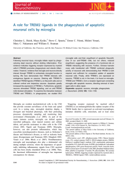
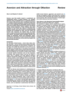
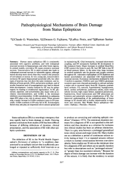
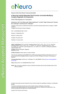
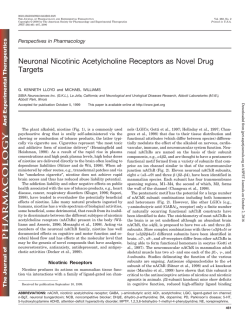
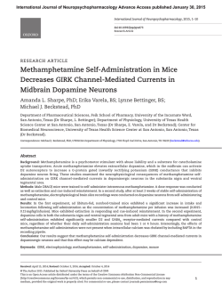
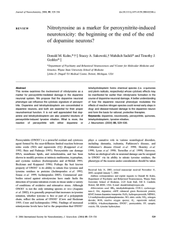
Available online www.jocpr.com
Journal of Chemical and Pharmaceutical Research, 2015, 7(1):424-434
Research Article
ISSN : 0975-7384
CODEN(USA) : JCPRC5
Revealing hallmark histology of hippocampus neurons in beta-amyloid
induced alzheimer’s mice and investigation of neuroprotective effect of
Ipomoea aquatic forsk, an Indian medicinal herb
D. Sivaraman*1, P. Panneerselvam2 and P. Muralidharan3
1
Centre for Laboratory Animal Technology and Research, Sathyabama University, Jeppiaar Nagar, Rajiv Gandhi
Salai, Chennai, Tamil Nadu, India
2
Department of Pharmaceutical Chemistry, C. L. Baid Metha College of Pharmacy, Jyothi Nagar, Rajiv Gandhi
Salai, Thoraipakkam, Chennai,Tamil Nadu, India
3
Department of Pharmacology and Toxicology, C. L. Baid Metha College of Pharmacy, Jyothi Nagar, Rajiv Gandhi
Salai, Thoraipakkam, Chennai, Tamil Nadu, India
_________________________________________________________________________________________
ABSTRACT
Alzheimer’s disease (AD) is a potential neurodegenerative disorder of nervous system which affects the primary
areas of the brain dealing with learning and memory. Deposition of the insolublebeta-amyloid protein on neurons
activates the microglial cells which stimulates the process of neuroinflammation by releasing mediators. Release of
cytokines and apoptotic promoters in turn shrink the neurons of the hippocampus leads to cognitive impairment. In
the present study change in histology of hippocampi neurons in mice brain during AD will be evaluated by intra
cerebro ventricular (ICV) injection of beta-amyloid 25-35.Toluidine blue, haematoxylin and eosin (H&E) staining
methodswereutilized for analyzing the neuronal morphology of Cornuammonis (CA) and dentate gyrus (DG) zones
of hippocampus. Results obtained from the histopathological study reveals that decrease in the number of
hippocampi neurons may be due to degeneration. Presence of darken condensed nucleus, shrinkage of pyramidal
cells in CA zones and decrease in granular cells of DG zone is a strong indication of apoptosis and stress cause by
amyloid injection. Hence it was concluded that cognitive impairment caused by beta-amyloid in mice is mainly due
to degeneration of neurons in functional zones of hippocampus that controls the memory and learning behavior.
Key words: Alzheimer’s disease, beta-amyloid, hippocampus, intra cerebro ventricular, toluidine blue,
haematoxylin and eosin, neuroinflammation.
_________________________________________________________________________________________
INTRODUCTION
Alzheimer's disease is associated with brain shrinkage and localised loss of neurons, mainly in the hippocampus and
basal forebrain. The loss of cholinergic neurons in the hippocampus and frontal cortex is a feature of the disease, and
is thought to underlie the cognitive deficit and loss of short-term memory that occur in AD. Two microscopic
features are characteristic of the disease, namely extracellular amyloid plaques, consisting of amorphous
extracellular deposits of β-amyloid protein (known as Aβ), and intraneuronal neurofibrillary tangles, comprising
filaments of a phosphorylated form of a microtubule-associated protein (Tau). Prevalence rates for dementia
increase exponentially with advancing age, ranging from 10% in the age group of 60-65 years to 36% in the age
group of 90 years[1]. This disease may be presenile or senile onset and the occurrence of disease is before or after
the age group of 60 years, but now it is challenging the median age of population. AD was the 7th leading cause of
424
D. Sivaraman et al
J. Chem. Pharm. Res., 2015, 7(1):424-434
______________________________________________________________________________
death in 2004 with 65,829 numbers of deaths [2]. It has been estimated that nearly 24million people with dementia
worldwide [3].
Aβ protein is a potent neurotoxin both in vitro and in vivo. It has been substantiated that the 11 amino acid sequence
(25-35) of beta amyloid is neurotoxic for primary neurons [4]. Aβ is secreted by normal cells in cultures and are
detected as circulating peptide in the plasma and the cerebrospinal fluid of healthy humans and other
mammals[5].Formation of Aβ involves cleavage at two different points, including one in the transmembrane domain
of APP by β and γ-secretases. Gamma secretase is a clumsy enzyme that lacks precision and cuts APP at different
points in the same victinity, generating Aβ fragments of different lengths including Aβ 40 and Aβ 42, mutations
favour the formation of Aβ42.Amyloid hypothesis points to cytotoxicity of mature aggregated amyloid fibrils, which
are believed to be the toxic form of protein responsible for disturbing the cells calcium ion homeostasis and thus
including apoptosis [6]. The calcium permeable channels in the membranes resulting in excessive calcium influx
and cause the induction of neurotoxic cascades [7].
In brain the hippocampus involved in controlling the complex process of forming, organizing, and storing memories.
The hippocampi and their connections to the dorsal medial nuclei of the thalamus and the mamillary nuclei of the
hypothalamus constitute a limbic system network crucial for learning and processing of events for long-term
storage. The basic information required by the memory is registered by the prominent areas of the brain such as
primary somatosensory, auditory, or visual cortex. The hippocampus may mediate the representation of spatial and
temporal memories. The key aspect of these kinds of memories is that they are relational, flexible, and permit
creative behavior. The hippocampus is critical for encoding information about an animal's environment and the
episodes that occur within those environments. In so doing, it forms and stores representations of spaces and spatial
contexts; these are called cognitive maps. Experimental support for these ideas has come from many different
approaches to the study of hippocampal function [8, 9] .The cognitive map theory of hippocampal function suggests
that discrete and selective lesions of the hippocampus will produce a deficit in place learning or exploration of a
novel environment The kinds of memories that the hippocampus stores have also been referred to as relational,
that’s information on how specific objects or events, existing in some spatial or temporal arrangement, are related to
each other [10].
Inside the hippocampi nerve impulse and memory related message passes through distinct regions like
Cornuammonis (CA) and dentate gyrus (DG). CA further spread into three main zones which are CA1, CA2 and
CA3 which constitute the trisynaptic loop of the hippocampus. The disease of central nervous system in particular
with Alzheimer’s, Epilepsy, schizophrenia and some severe depressions may affects the hippocampi neurons which
may leads to cognitive impairments that affects the social life of the individual. Apart from this functional ability of
the hippocampus may declines with aging. It was estimated that approximately 20 percentage of the neurons will
lose their ability but this doesn’t applies to all aged persons.
Neuroinflammation is now recognized as a fundamental response of the CNS not only to acute injury, but also to
chronic neurodegenerative disease. This is perhaps no better demonstrated than in AD, where the severity of the
neuroinflammatory response parallels the disease course.
Plants have formed the basis of sophisticated traditional medicine systems that have been existence for thousands of
years and continue to provide mankind with new remedies. Although some of the therapeutic properties attributed to
plants have proven to be enormous, medicinal plant therapy is based on the empirical findings of hundreds and
thousands of years.
Ipomoea aquatic Forsk is a tender, trailing, vascular semiaquatic or floating perennial plant, found on moist soil
along the margin of fresh water, ditches, marches and wet rice fields. It is commercially cultivated as an edible green
leafy vegetable in Hong kong, Taiwan, China and also in rural part of India [11]. It is runner type plant with
numerous small flowers. Only a very few scientific studies have been conducted on its medicinal aspects. These
include oral hypoglycemic [12] and anti-oxidant activity [13] .Treatment on liver diseases [14], constipation[15].
Ipomoea aquatic is considered a tonic and it is used to treat gastric and intestinal disorders [16, 17].
Recently several biological indicators are available for predicting the severity of the Alzheimer’s disease in animals
one such potential tool is histopathological analysis of the brain tissue. Staining tissues with dyes is quite old
technique but often provided some valuable information about the cytoplasm of neurons and glial cells, though it is
425
D. Sivaraman et al
J. Chem. Pharm. Res., 2015, 7(1):424-434
______________________________________________________________________________
possible to recognize several cell-types on the basis of their stained nuclei. Hematoxylin and eosin is the most
widely used stain for diagnostic histopathology of neurodegenerative diseases like AD. Toluidine blue is a
metachromatic stain that stains nucleus blue and cytoplasm light blue. It is very often used to squash preparation of
brain parenchymal tissue.
The aim of the current research work is to determine the neuronal histology of various regions of hippocampus
insulted by intra cerebro ventricular injection of beta-amyloid 25-35.Further pathological change in neuronal
architecture is evaluated by Toluidine blue, haematoxylin and eosin staining. This work reveals valuable information
about the structural changes attributes to the neurons of the hippocampus under stress full condition which may be
valuable for the researcher in the field of neuropharmacology.
EXPERIMENTAL SECTION
Plant material.
The fresh leaves of Ipomoea aquatica (IA) were collected from (Perambur region of chennai, Tamilnadu, India).The
plant was identified and authenticated by two botanist one Dr. Sasikala Ethirajulu. Captain srinivasamurthy research
Foundation, Chennai, Tamil nadu, India. and Dr. A. Aravind, Assistant Professor ,Department of Medicinal Botany
,National institute of Siddha, Chennai 600047,Tamilnadu, India. Reg no: NIS/MB/73/2012, The specimen voucher
was deposited in the Department of Pharmacology and toxicology, C. L. Baid Metha College of Pharmacy, Chennai,
Tamil nadu, India.
Preparation of the Plant Extract.
The fresh leaf of IA was collected and washed with running water. It was shade dried at room temperature and 1kg
of the dried leaf was made in to coarse powder. The powder was passed through a 60 No mesh sieve. Air dried
Powdered drug was extracted with mixture of Ethanol: water (6:4) (hydro-alcoholic extract) by using soxhlet
extraction. Hydroalcoholic extract of Ipomoea Aquatica (HAEIA) was filtered, concentrated by rotary vacuum pump
to get the solid mass.
Experimental Animal
Healthy Swiss albino male mice weighing 20–25 gm, obtained from C. L. Baid Metha College of pharmacy was
used for the pharmacological studies. The animals were kept under standard conditions maintained at 25±2oC, 12 hr
light/dark cycle and given standard pellet diet (Hindustan lever, Bangalore) and water provided ad libitum. The
animals were acclimatized to the laboratory conditions for a week prior to the experimentation .All the animals were
housed in polypropylene cages using paddy husk bedding. Principles of animal handling were strictly adhered to and
the handling of animals was made under the supervision of animal ethics committee of the institute. The
experimental protocol was approved by Institutional animal ethics committee (IAEC) of CPCSEA (Committee for
the Purpose of Control and Supervision of Experiments on Animals). C. L. Baid Metha College of Pharmacy,
Chennai, Tamil nadu, India.
Intra Cerebro Ventricular injection of Aβ peptide
The administration of a Aβ 25-35 was performed by identifying the bregma pointing the skull, each mouse was
injected at bregma with a 50µl Hamilton micro syringe fitted with a 26-gauge needle that was inserted to a depth of
2.4 mm. in brief, the needle was inserted unilaterally 1 mm to the right of the middle point equidistant from each eye
slightly angled towards 450 1 mm to the right of the middle point equidistant from the eyes perpendicular to the
plane of the skull. Mice exhibited normal behavior within 1 min after injection [18].
Experimental Design
Animals were divided into five groups
GROUP I
GROUP II
GROUP III
mg/kg (p.o.)
GROUP IV
mg/ kg (p.o.)
GROUP V
: Animals injected with phosphate buffered saline. (10µl)
: Animals injected with Aβ peptide (10µl) by I.C.V
: Animals injected by Aβ peptide (10µl) by I.C.V and treated with HAEIA 200
: Animals injected with Aβ peptide (10µl) by I.C.V and treated with HAEIA 400
: Animals injected with Aβ peptide (10µl) by I.C.V and treated Donepezil 5mg/kg (p.o.)
426
D. Sivaraman et al
J. Chem. Pharm. Res., 2015, 7(1):424-434
______________________________________________________________________________
Amnesia was induced by intra cerebro ventricular injection (I.C.V.) of Aβ peptide (25-35) preparation to II, III, IV
and V groups on the 21st day after pretreatment with HAEIA and continues for the period of 07 days. Control
animals were injected with only phosphate buffered saline.
Histopathology
At the end of the study animals were sacrificed by cervical dislocation and whole brain of animals was removed
quickly and fixed with 10% formalin. Later the brain tissues were processes for histopathological study. The
pathological changes were viewed under light microscope after staining with haematoxylin and eosin.
Hematoxylin and Eosin Staining
Principle
A transverse section of tissue with approximately 0.5 cm thickness is taken from the mice brain. Tissue pieces were
fixed in 10 per cent buffered neutral formalin and trimmed to a thickness of about 3 mm. The tissues were
dehydrated, cleared and paraffin embedded in a routine manual processing. Tissues were cut at 3 to 5 µm thickness,
stained with haematoxylin and eosin and mounted with DPX for histopathological examinations [19].
Reagents
1. Neutral buffered formalin: To 900 ml distilled water, 4g of sodium dihydrogen phosphate, 6.5g of disodium
hydrogen phosphate anhydrous and 100 ml of 40% formalin was added and the pH was adjusted to 7.4.
2. Harris’s haematoxylin: 1 gm of haematoxylin stain was dissolved in 10 ml of absolute alcohol and mixed with a
solution containing 20 gm of potassium alum which was previously dissolved in hot distilled water. After bringing
this mixture to boiling point, 0.5 gm of mercuric oxide was added. This mixture was cooled rapidly under cold
water. This was then filtered and stored in an amber colored bottle.
3. Eosin stain (0.5%): 500 mg of eosin yellow powder was dissolved in 100 ml of distilled water, filtered and
stored.
Tissue processing procedure
After fixation the tissue sample were washed thoroughly in running water overnight. It was dehydrated by ascending
rates of alcohol (50, 60, 70, 80, 90 and 100% I and 100% II) each for 60 minutes. Then the tissues were cleared with
two changes of xylene each with 30 minutes. Embedding was done with two changes of molten paraffin for 30 min
to remove xylene. Mounted in L blocks with molten paraffin and solidified. The solidified blocks were trimmed to
small size and sectioned using microtome (Spencer U.S.A) 3 to 5 micron thickness.
H&E staining procedure
The sections were deparaffinised by xylene I and II each with 5-10 minutes ,xylene was removed by descending
grades of alcohol each with 5 minutes and then washed in tap water and stained with harris’s haematoxylin for 3-4
minute again washed in tap water followed by this differentiated with acid alcohol (15-30 seconds) .The sections
were kept in tap water 5-10 mins and counterstained with eosin 0.5% until section appeared light pink (15 to 30
seconds) ,blotted dehydrated in ascending grades of alcohol each with 1 minute further cleared with xylene I and II
each for 5 minutes finally mounted in DPX Mountant and cover slipped and viewed under light microscope.
Toluidine blue Staining
Toluidine blue is a metachromatic stain that stains nucleus which appears blue in color. The isolated mice brain was
fixed and processed as per the earlier procedure, and then brain tissue slices were prepared for toluidine blue
staining. The slice was incubated with a 0.5% (w/v) solution of toluidine blue at 56°C for 20 minutes. After being
dehydrated, cleared and mounted, brain slices were examined under a light microscope. Morphological changes in
hippocampi neurons in CA3 area of mice hippocampal slices were observed [20].
RESULTS AND DISCUSSION
Effect of HAEIA on histopathological analysis of CA1 zone of mice brain hippocampus
Sample belongs to group I control animal shows five to six layer of compactly aranged small pyramidal cells of CA1
region, most with vesicular nuclei. Two to three layer of shrinked pyramidal cells of CA1 region with irregular
arrangment and most of the nucleus apperas pale in colour was observed in group II injected with beta amyloid.
427
D. Sivaraman et al
J. Chem. Pharm. Res., 2015, 7(1):424-434
______________________________________________________________________________
Increased layer of pyramidal cells with dark prominent nuclues appers in CA1 region.Arragngement of neurons still
improper in group III animals treated with HAEIA 200 mg/kg where as four to five layers of pyramidal cells with
regular arrangment at deepre layer was observed group IV animals treated with HAEIA 400 mg/kg.
GROUP I (H&E X 400)
GROUP II (H&E X 400)
GROUP III (H&E X 400)
GROUP IV (H&E X 400)
GROUP V (H&E X 400)
Figure 1: Histopathology of mice brain hippocampus CA1 zone
428
D. Sivaraman et al
J. Chem. Pharm. Res., 2015, 7(1):424-434
______________________________________________________________________________
Five to six layers of regularly arranged pyramidal cells with prominent nucleus appearing in CA1 region of sample
belongs to group V treated with standard Donepezil 5mg/kg. As shown in Figure 1.
Effect of HAEIA on histopathological analysis of CA3 zone of mice brain hippocampus
Compactly arranged organized layer of larger pyramidal cells with prominent nucleus was observed in CA3 zone of
control group. Completely disorganized with shrunken pattern of pyramidal cells was observed in CA3 region of
negative control group. Increase in the number of larger pyramidal cells with few shrunken cells was observed in
CA3 zone of group III animals treated with HAEIA 200 mg/kg .Regular arrangement pyramidal cells was observed
in more deeper layer when compare to outer layer which projects scattered pattern of cells was observed in group III
animals.
Neuronal cells are well organized with more number of larger pyramidal cells appearing in CA3 region. Cells shows
regular arrangement in deeper layer but outer layer is still markedly scattered in group IV treated with HAEIA 400
mg/kg. Cells are completely organized with prominent dark colored nucleus .Both the deeper and outer layer of cells
shows regular arrangement in group V treated with standard Donepezil 5mg/kg as shown in Figure 2.
Effect of HAEIA on histopathological analysis of hippocampus arch.
In control group sample the hippocampus proper is formed of Cornu Ammonis CA1, CA2, CA3, CA4 and Dentate
gyrus DG regions. CA1 and CA2 zones comprises of small pyramidal cells .CA3 and CA4 zones of hippocampi
comprises large pyramidal cells. DG region composed on granular cells. Areas in between compact zones of cells
comprise the molecular layer which consists of neuronal processes (axons and dendrites), glial cells, and scattered
nerve cells.
GROUP I (H&E X 400)
GROUP II (H&E X 400)
GROUP III (H&E X 400)
GROUP IV (H&E X 400)
429
D. Sivaraman et al
J. Chem. Pharm. Res., 2015, 7(1):424-434
______________________________________________________________________________
GROUP V (H&E X 400)
Figure 2: Histopathology of mice brain hippocampusCA3 zone
GROUP I (H&E X 100)
GROUP II (H&E X 100)
GROUP III (H&E X 100)
GROUP IV (H&E X 100)
430
D. Sivaraman et al
J. Chem. Pharm. Res., 2015, 7(1):424-434
______________________________________________________________________________
GROUP V (H&E X 100)
Figure 3: Histopathology of hippocampi arch
Minimal layer of Cells observed in CA1 and CA2 regions. Total shrinkage of zonal arc was observed in this group.
Disorganization in cell arrangement was observed in CA 3 and CA4 zones. Loss of cells in DG region was observed
in amyloid injected group. Increased layer of Cells observed in CA1 and CA2 regions. Restoration in cell
arrangement was observed in CA 3 and CA4 zones. Increased number of granular cells appeared in DG region of
group III animals treated with HAEIA 200 mg/kg as shwon in Figure 3.
Arrangement of Cells observed in CA1, CA2 .CA3 and CA4 zones are almost normal further with thick bundles of
pyramidal cells. Increased mass of granular cells appeared in DG region of group IV animals treated with HAEIA
400 mg/kg. Intact arrangement of pyramidal cells was observed in CA1, CA2 .CA3 and CA4 zones. Increase in
thickness of DG layer projected with numerous mass of granular cells was observed in group V treated with
standard Donepezil 5mg/kg.
Effect of HAEIA on histopathological analysis of Zone of Apoptosis.
Control group shows regular arrangement of cells with no signs of apoptosis and further no cells with darken
condensed nucleus. Samples belongs to negative control group projects disorganized layer of larger pyramidal cells
in CA3 and CA4 zones with increased apoptotic cells marked with arrow (↑).Neurons are markedly organized with
minimized appearance of apoptotic cells was observed in animals treated with HAEIA 200 mg/kg. Neuronal cells
are almost organized but with very few projection apoptotic cells were observed in animals treated with HAEIA 400
mg/kg. Neuronal organization and arrangement are almost restored back to normal with rare appearance of apoptotic
cells in group V treated with standard Donepezil 5mg/kg as shown in Figure 4.
GROUP I (H&E X 400)
GROUP II (H&E X 400)
431
D. Sivaraman et al
J. Chem. Pharm. Res., 2015, 7(1):424-434
______________________________________________________________________________
GROUP III (H&E X 400)
GROUP IV (H&E X 400)
GROUP V (H&E X 400)
Figure 4: Histopathology of zone of apoptosis
Effect of HAEIA on histopathological analysis of toluidine blue staining of hippocampi neuronal architecture
Toluidine blue staining of neurons in CA3 area of mice hippocampal slices reveals the following observation.
Neurons in the control group exhibited a very normal size, shape and a regularly ordered arrangement. With dark
blue colored nucleus. Neuronal morphology project larger cell bodies with intact arrangement. No evidence of
apoptosis and nuclear condensation was observed.
Group treated with beta amyloid shows very less neuronal count when compare to control group. Architecture of the
neuronal cell surface project more cell shrinkage which may be a sign of apoptosis Sample belongs to group III shos
marginal increase in the size of nerve cell bodies was observed in the low dose treated group when compare to beta
amyloid treated group. Some neurons with condensed chromosomes were clearly detected in between the neurons
with larger cell bodies. Further this group shows less cell shrinkage and less condensed neurons. Mixed pattern of
larger and shrunken neurons was observed
Neurons of animal belongs to group IV shows larger cell bodies with less condensed nuclei when compare to beta
amyloid treated group. Neurons have almost normal morphology with large cell body and also with regular
arrangement, very minimal evidence of condensed neurons. Further increased neuronal count was observed in this
group V animal when compare to amyloid beta treated group as shown in Figure 5.
432
D. Sivaraman et al
J. Chem. Pharm. Res., 2015, 7(1):424-434
______________________________________________________________________________
GROUP I (Toluidine blue X 400)
GRPOUP II (Toluidine blue X 400)
GROUP III (Toluidine blue X 400)
GROUP IV (Toluidine blue X 400)
GROUP V (Toluidine blue X 400)
Figure 5: Histopathology of Toluidine blue staining of hippocampi neuronal architecture
CONCLUSION
Apoptosis is a Programmed cell death it’s a form of cellular demise mediated by an intracellular program [21].
Unlike necrosis, which is a form of cell death that results from acute tissue injury, and provokes an inflammatory
response.
433
D. Sivaraman et al
J. Chem. Pharm. Res., 2015, 7(1):424-434
______________________________________________________________________________
Apoptosis involves a series of biochemical events leading to characteristic changes in cell morphology and death.
These changes include blebbing, loss of membrane symmetry and attachment, cell shrinkage, nuclear fragmentation,
chromatin condensation and chromosomal DNA fragmentation [22]. Beta amyloid induced oxidative stress results in
the generation of reactive oxygen species that can damage various cellular components such as cell membrane
proteins, mitochondrial DNA, lipids, and cytoplasmic proteins. In the brains of Alzheimer's patients these
components have been shown to exhibit elevated levels of oxidative damage [23].
Observation of the current study clearly reveals that injection of Aβinduces the condition of apoptosis and neuronal
shrinkage might be the reason for cognitive impairment in mice as observed from beta-amyloid injected group. It is
evident that control group animals has increased neuronal count and well projected neuronal architecture with
multiple layers of larger nucleated pyramidal cells when compare to the negative control group.
Increased zone of apoptosis with condensed and darkened nucleus were observed in group II animals further
decreased neuronal count with complete destruction of pyramidal structure in CA zones of hippocampus evident the
induction of apoptosis by Aβ protein.
Restoration of neuronal structure and increased neuronal count observed in group III, IV and V projects the
ameliorating potential of HAEIA against the stress induced by Aβ injection. Treatment with HAEIA protects
neurons from the oxidative and apoptotic damage induced by amyloid injection. Hence from the observation it was
concluded that ICV injection of beta amyloid in mice model of AD may undergo neuronal degeneration mainly
because of apoptotic cell death in the marginal and prioritized zones of hippocampus and further research needs to
be carried out in order to find out the exact molecular mechanism underlying the neuronal damage due to amyloid
insult.
Acknowledgement
The authors are grateful to Dr. Grace Rathnam (Principal, C. L. Baid Metha college of Pharmacy) for her technical
assistance and also acknowledge Dr. N. Mohana, Veterinary pathologist for her analytical support in sample
interpretation.
REFERENCES
[1] A Sharma ;V Parikh; M Singh,IJEB., 1997,35,1146-115.
[2] P Sloane ;S Zimmerman ; C Suchindran ; P Reed ,Annu Rev Pub Health.,2002, 23:13-31.
[3] LE Hebert;PA Scherr; JL Bienias,Arch. Neurol.,2003, 60,1119–1122.
[4] C Pereira;MS Santos; C Oliveira,Neurobiol. Dis .,1999,6, 209-219.
[5] P Seubert,Nature., 1992, 359,325–327.
[6] HP Rang; M Dale ,Science., 1990, 250, 279-282.
[7] RJ Mark; JW Geddes; K Uchida; MPMattson,J. Neurosci., 1997, 17, 1046-1054.
[8] L Nadel,Frontiers in Neuroscience.1996, 2(74), 305-317.
[9] T Otto ;NJ Cohen; HEichenbaum ,Behav. Neural Biol.,1992, 57, 2-36.
[10] H Eichenbaum ; T Otto; NJ Cohen ,Behav. Brain Sci.,1994, 17, 449-518.
[11] HH Edie; BWC Ho,Econ Bot.,1969, 23, 32-36.
[12] N MitalManvar; TR Desai,Ind J. med sci.,2013, 67(3),49-60.
[13] DJ Haung; HJ Chen; CDLin;YH Lin ,Bot Bull AcaSinica (Taipei)., 2005,46(2),99.
[14] SM Badruzzaman; W Husain,Fitoterapia.,1992, 63(3), 245-247.
[15] G Samuelsson; MH Farah ;P Claeson;MHagos,J. Ethnopharmacol., 1992, 37, 47-70.
[16] G Watt G,Dehra Dun.,1972,47- 52.
[17] E Westphal,Number Vegetables.,1993,181-184.
[18] E Susan;JK Belknap,J Pharmacol Methods., 1986, 16,355-357.
[19] SC Landis;G Guidry,Auto Neuro Bas and Clinical., 2000, 82, 97-108.
[20] L Jianyu ; QYuting; L Hui; C Ying ,Bas Res In Neural Reg.,2013,8(31), 2932-2941.
[21] HS Engelberg,PLOS Genet., 2006, 2(10), 135.
[22] DS Kerr; LWCampbell,Science., 1989, 245(4925), 1505‐9.
[23] P Mecocci; UMacGarvey U,Ann Neurol .,1994, 36(5), 747‐51.
434
© Copyright 2026