
Morphological and histological identification of
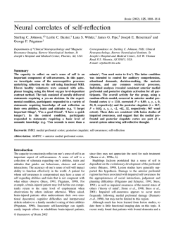
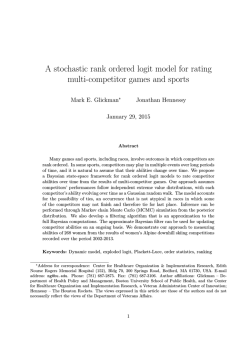
Veterinary World, EISSN: 2231-0916
Available at www.veterinaryworld.org/Vol.8/January-2015/24.pdf
RESEARCH ARTICLE
Open Access
Morphological and histological identification of Paramphistomum cervi
(Trematoda: Paramiphistoma) in the rumen of infected sheep
Vijayata Choudhary1, J. J. Hasnani1, Mukesh K. Khyalia2, Sunanda Pandey2, Vandip D. Chauhan1,
Suchit S. Pandya1 and P. V. Patel1
1. Department of Veterinary Parasitology, College of Veterinary Science & Animal Husbandry, Anand Agricultural
University, Anand - 388 001, Gujarat, India; 2. Department of Veterinary Pathology, College of Veterinary Science &
Animal Husbandry, Anand Agricultural University, Anand - 388 001, Gujarat, India.
Corresponding author: Vijayata Choudhary, e-mail: [email protected],
JJH: [email protected], MKK: [email protected], SP: [email protected],
VDC: [email protected], SSP: [email protected], PVP: [email protected]
Received: 03-11-2014, Revised: 19-12-2014, Accepted: 25-12-2014, Published online: 30-01-2015
doi: 10.14202/vetworld.2015.125-129. How to cite this article: Choudhary V, Hasnani JJ, Khyalia MK, Pandey S,
Chauhan VD, Pandya SS, Patel PV (2015) Morphological and histological identification of Paramphistomum cervi (Trematoda:
Paramiphistoma) in rumen of infected sheep, Veterinary World, 8(1): 125-129.
Abstract
Aim: This study was undertaken to identify Paramphistomum cervi on the basis of its morphology and histology to be
the common cause of paramphistomosis in infected sheep and its differentiation from other similar Paramphistomes in
Gujarat.
Materials and Methods: Adult rumen flukes were recovered from the rumen of naturally infected sheep slaughtered in
various abattoirs in Gujarat. Some adult flukes were flattened and stained in Borax carmine, and some were sectioned in the
median sagittal plane and histological slides of the flukes were prepared for detailed morphological and histological studies.
Result: Microscopic pictures of the parasite used in identification define the similarity in the morphology and histology of
the anterior sucker, pharynx, esophagus, genital atrium, posterior sucker (acetabulum) and testes to the P. cervi.
Conclusion: It can be concluded that the most common species found in sheep infected with Paramphistomosis is P. cervi
on the basis of its histo-morphological appearance in Gujarat.
Keywords: abattoirs, histology, morphology, paramphistomosis, posterior sucker, rumen fluke, sheep.
Introduction
Paramphistomum spp. are Platyhelminth (flatworm) parasites (Platyhelminthes: Trematoda:
Digenea) responsible for Paramphistomosis i.e. gastrointestinal parasitic disease in domesticated animals, which causes heavy economic losses [1] to the
livestock industry to the tune of several thousand
crores of rupees annually [2]. It has been a neglected
trematode infectious disease in ruminants, but has
recently come out as a significant cause of productivity loss [3-7]. Distribution of Paramphistomosis
is worldwide [8], but the highest prevalence has
been accounted in tropical and subtropical regions,
particularly in Africa, Asia, Australia, and Eastern
Europe [1,9].
It is caused by specific species of the parasite
depending on the regions [3]. The common rumen
fluke, Paramphistomum cervi is considered to be one
of the most important species of Paramphistomes [2].
Adult flukes are located in the rumen of ruminants
and immature flukes in the small intestine mainly in
the duodenum [1,10]. P. cervi has di-heteroxenous life
cycle, which involve snail (Bulinus spp., Glyptanisus
gilberti, Indoplanorbis exustus, Planorbis planorbis
Copyright: The authors. This article is an open access article licensed
under the terms of the Creative Commons Attributin License (http://
creative commons.org/licenses/by/2.0) which permits unrestricted
use, distribution and reproduction in any medium, provided the
work is properly cited.
Veterinary World, EISSN: 2231-0916
and Lymnaea bulimoides) as intermediate host and
mammalian as definitive host [11,12].
P. cervi are plug feeders [9] and cause serious
disease by burying themselves into the sub mucosa
of the duodenum and feeding on the epithelial cells
of the Brunner’s gland resulting in anorexia, profuse
fetid diarrhea, drop in plasma protein concentration
and anemia, which weaken the host [13]. Mature
Paramphistomes are also responsible for rumenitis,
irregular rumination, un-thriftiness, lower nutrition
conversion and loss of body condition, resulting in
considerable economic loss [9].
The species identification is still neglected as the
various species of the family Paramphistomatidae are
difficult to be detected through a systematic point of
view, and most of the reports do not quote the main
one [14].
The rumen fluke, P. cervi has economic importance in ruminants but presently no reliable methods
are available to identify and differentiate this parasite
from other Paramphistomes. The present study represents the morphological and histological identification of P. cervi using Borax carmine and H and E
staining methods, in order to provide a base for future
molecular studies.
The present study on morphological and histological identification of P. cervi was designed to fulfill
the need for molecular understanding of this economically important parasite.
125
Available at www.veterinaryworld.org/Vol.8/January-2015/24.pdf
Materials and Methods
Collection of adult parasites
The rumen of 350 sheep between 1 and 2 years
of age was inspected for the presence of rumen
flukes during slaughter at various local abattoirs of
Anand, Ahmedabad and Vadodara District in Gujarat
state, of which 17 sheep were found positive for
Paramphistomes. Infected part of the rumen was
brought to the laboratory of Department of Veterinary
Parasitology, where it was washed thoroughly. After
washing, adult flukes were carefully picked up with
the help of forceps from the mucosa of the rumen of
naturally infected sheep and then washed 3-4 times
with phosphate buffer saline. After that, samples were
preserved in 10% buffered formalin until being processed for morphological and histological studies. The
samples were carefully labeled with proper details.
Morphological identification
Rumen flukes were preliminarily identified
under microscope using low power magnification and
then slides were prepared for detailed morphological studies and identification. The collected flukes
were placed on petridish and observed through stereo
microscope to appreciate the morphology. Final identification of Paramphistomum spp. was done based on
morphology of flukes; its shape, anterior sucker, posterior sucker (acetabulum), terminal genitalium and
tegumental papillae, following the standard guidelines given by Urquhart et al. [15]. Parasites were
processed for whole mounting and stained by Borax
carmine according to the procedure given by Singh
and Srivastava [16].
Out of all recovered adult flukes, 10 flukes were
randomly picked from each infected sheep and were
washed in water. After washing, flukes were flattened between two glass slides and fixed in Bouin’s
fluid at room temperature for 24 h. After that, these
specimens were washed in water and then stained
for 24 h in 0.5% Borax carmine and subsequently
destained in 1% hydrochloric acid until the pink color
was observed. The acid was thoroughly washed out
from all specimens with water. Thereafter, the specimens were washed with tap water and subsequently
dehydrated through 50-100% alcohol for 1 h each, and
cleared by xylene for 30 min. The cleared specimens
were mounted using DPX and covered with a coverslip. The mounted slides were allowed to the air dry
and observed under the light microscope.
oral and ventral suckers, testes and ovary. The species
were identified according to the criteria outlined by
Yamaguti [18], Eduardo [19] and Sey [20].
Results
It is very difficult to identify and differentiate
the species of amphistomes on the basis of its morphology due to significantly less variation in size and
shape of parasite and its internal organs, which varies even in mature and immature worms of same spp.
Morphology cannot be very well appreciated in the
stereo microscope, but can be confirmed well by the
histological examination of the parasite.
Morphological identification
P. cervi is reported in several studies conducted
on ruminants [18-20]. Morphological identification
of P. cervi was carried out on the basis of size and
shape of fluke and position of anterior and posterior
sucker (acetabulum). In the present study, most of the
species were of P. cervi, which were found mainly in
the rumen and were light pink in color with a sucker
at the tip of the cone (Figure-1a) and another sucker
ventrally at the posterior end. The body of P. cervi
was pear-shaped, slightly concave ventrally (conical) and convex dorsally. The worm measures about
3-8 mm in length and 1.5-3.0 mm in width at anterior
end of posterior third or at its junction with middle
third. Mouth was terminal, funnel-shaped, widened
posteriorly. Caeca were wide, pursued a serpentine
a
b
c
d
e
f
Histological identification
The species thus identified was further confirmed by histological identification of 10 formalin
preserved flukes from each sample, which was processed by paraffin embedding method and stained by
H and E stain as per Luna [17]. 10 longitudinal and
median sagittal sections, each 6-10 μ thick were cut
by a Leica RM2125 microtome. The prepared sections
were stained with H and E and were examined by
microscopy and microphotography in order to identify
Veterinary World, EISSN: 2231-0916
Figure-1: Whole mounted parasite stained with borax
carmine showing visceral organs: (a) Anterior sucker
(As), cecal bifurcation (Cb), caecum (C), (b) cecum (Ca),
vitelline gland (Vg), uterus (Ut), (c) mouth (Mo), pars
prostatica (Pp), genital pore (Gp), (d) intestinal cecal end
(Ice), (e) uterus (Ut), (f) posterior sucker (Ps).
126
Available at www.veterinaryworld.org/Vol.8/January-2015/24.pdf
course and reached anterior level of acetabulum
with blind ends more dorsal than lateral (Figure-1d).
Genital pore was situated behind intestinal bifurcation (Figure-1c). Acetabulum (posterior sucker) was
sub-terminal, about one-fourth to one-fifth of body
length (Figure-1f). Clusters of vitelline glands were
extended from the pharynx to the posterior sucker and
lie between the caeca and the lateral margins of the
body (Figure-1b). The uterus was wavy and runs dorsally to testes (Figure-1e).
on the dorsal surface (Figure-4). Uterine coils were
dorsal to testes and ventral to male ducts. Vitellaria
were follicular, lateral, extending from the level of
the pharynx to acetabulum, not confluent medially
in their posterior or anterior limits. Excretory vesicle was antero-dorsal to acetabulum; excretory pore
was anterior to opening of Laurer’s canal at level of
posterior testis (Figure-5). The genital pore was situated at the anterior third of the body (behind intestinal
Histological identification
The histological identification of P. cervi
recorded in the present study was done on the basis of
histology of flukes; anterior sucker, posterior sucker
(acetabulum), terminal genitalium, testes, ovary and
on the basis of histological peculiarities of the muscular organs such as pharynx, genital opening and posterior sucker (acetabulum).
In the present study, it was observed that P. cervi
had a Liorchis type of pharynx, Gracile type of genital
opening and Paramphistomum type of acetabulum in
median sagittal section (Figure-2). Cuticle was thick,
smooth except at genital pore and anterior extremity,
which was covered with numerous prominent papillae. Both anterior and posterior suckers had thick rims
covered with transverse folds. Esophagus was bent
dorsally; musculature of wall relatively thin, no bulb
or the posterior sphincter; lumen lined throughout by
hyaline layer (Figure-3).
Testes were distinctly lobed, situated a little
obliquely, tandem, in mid-body, pre-ovarian in dorso-ventral direction (Figure-3). Seminal vesicle was
long, tubular, strongly coiled, thin-walled and convoluted in front of anterior testis; pars musculosa
was very short and weakly developed. Ovary was
sub-spherical, un-lobed, post-testicular and pre-acetabular; mehlis gland was close to the ovary; Laurer’s
canal crossing excretory vesicle or duct, opening
Figure-3: Microscopic picture of medial section of
Paramphistomum cervi showing different organs: Mouth
(Mo), pharynx (Ph), tegument (Tg), caecum (Ca), vitelline
gland (Vg), anterior testis (At), posterior testis (Pt),
posterior sucker (Ps).
Figure-4: Microscopic picture showing reproductive
organs: Distinctly lobed anterior (At) and posterior testis
(Pt), ovary (Ov), mehlis gland (Me) (H and E, ×150).
a
b
c
d
Figure-2: Microscopic pictures showing: (a) Mouth
(Mo), anterior sucker (As), (b) Liorchis type of pharynx
(Ph), (c) Gracile type of genital opening (Go) and (d)
Paramphistomum type of posterior sucker/ acetabulum
(Ac) in Paramphistomum Cervi (H and E, ×150).
Veterinary World, EISSN: 2231-0916
a
b
Figure-5: Microscopic picture showing: (a) Caecum (Ca),
vitelline gland (Vg), (b) excretory vesicle (Ev) (H and E,
×150).
127
Available at www.veterinaryworld.org/Vol.8/January-2015/24.pdf
bifurcation) with radial muscle fibers. Genital atrium
was covered with cuticular papillae which extend over
the surrounding body surface. It was encircled by a
fold of the body wall (Figure-2).
Discussion
Morphological and histological identification is important features used to differentiate
Paramphistomes. Several earlier studies have investigated morphological and histological variation in
Paramphistomum species.
The described parasites belong to the genus
Paramphistomum [21] (Platyhelminthes: Trematoda:
Digenea). The color of fluke, shape and size of body,
position of anterior and posterior sucker, presence
of genital pore and vitelline glands were similar to
the morphological identification system followed
earlier by Melaku and Addis [9], Yamaguti [18],
Eduardo [19] and Nasmark [22]. Yamaguti [18]
collected 20 gravid specimens of which about a
dozen were dissected and examined for the terminal genitalia and the ovarian complex of P. cervi.
Histological identification observed in the present
study was similar to the earlier studies carried out by
Eduardo [19], Sey [20], Nasmark [22] and Coskun
et al. [23]. Sey [20] recovered four species of rumen
fluke viz. Paramphistomum cervi, P. daubneyi,
P. gotoi and P. ichikawai from cattle and prepared
more than 200 median sagittal sections to study the
morphology of P. cervi and histomorphology of its
muscular organs. He observed that muscular organs
of P. cervi were of Liorchis (pharynx), Gracile (genital atrium) and Paramphistomum (acetabulum) type,
which is similar to this study. Coskun et al. [23]
collected amphistomes from cattle and compared
P. cervi from P. gotoi on the basis of papillae found
in the pharynx and by the position of the blind caeca.
Liorchis type of pharynx in P. cervi in comparison
to calicophoron type of pharynx of Paramphistomum
gracile [19], Gracile type of genital opening and
Paramphistomum type of acetabulum [22] differentiate P. cervi from other paramphistomes such as
P. gracile, P. gotoi, Calicophoron calicophorum,
Cotylophoron cotylophorum.
Many scientists studied the Prevalence of various species of amphistomes in sheep in various countries. P. cervi was also reported in Nigeria [24,25],
Egypt [26], Mexico [27], Thailand [3,28], Iran [29]
and Pakistan [30]. Calicophoron daubneyi has
been reported in Europe, Asia, Africa Oceania and
France [31]. Gastrothylax cruminefer was found in
Nigeria [24], Iran [32] and Kashmir [33]. Carmyerius
synethes and Calicophoron microbothrium were
also found in Nigeria from sheep [13]. P. cervi was
recorded in Kashmir from sheep [34], and in Udaipur
of Southern Rajasthan from cattle [35]. In Pakistan
P. cervi was also reported from buffalo [36] and
goats [36,37].
Veterinary World, EISSN: 2231-0916
Conclusion
Morphological and histological studies of all
parasites revealed presence of P. cervi mainly in adult
stage in the rumen of sheep infected with paramphistomosis as the most common species and etiology behind the economic impact of this disease and
provides a solid foundation for studying the reproductive biology of Paramphistomes and other related
trematodes using molecular techniques. This disease
and its impact can be controlled by interrupting the
lifecycle of the parasite with prevention of grazing
nearby water logged areas and the proper use of effective dewormer.
Authors’ Contributions
This study is the major component of the work
towards the M.V.Sc thesis of VC, under the
guidance of JJH. MKK and SP helped in histopathology, drafted and thoroughly revised the manuscript. VDC and SSP helped in sample collection
from various abattoirs. PVP was the member of the
advisory committee for the research work and
provided guidance. All authors read and approved
the final version of the manuscript.
Acknowledgments
This study was funded by College of Veterinary
Science and Animal Husbandry, Anand Agricultural
University, Anand, Gujarat, India. The authors are
thankful to all staff of Department of Veterinary
Parasitology, College of Veterinary Science and
Animal Husbandry, Anand Agricultural University,
Anand, Gujarat, India for their help. The authors
acknowledge the help rendered by the Department of
Veterinary Pathology, AAU, Anand, Gujarat for providing facility for histology.
Competing Interests
The authors declare that they have no competing
interests.
References
1.
2.
3.
4.
5.
6.
Ozdal, N., Gul, A., Ilhan, F. and Deger, S. (2010) Prevalence
of Paramphistomum infection in cattle and sheep in Van
Province, Turkey. Helminthologia, 47: 20-24.
Khan, U.J., Tanveer, A. and Maqbool, A. (2008)
Epidemiological studies of paramphistomosis in cattle. Vet.
Arhiv., 78(3): 243-251.
Anuracpreeda, P., Wanichanon, C. and Sobhon, P. (2008)
Paramphistomum cervi: Antigenic profile of adults as recognized by infected cattle sera. Exp. Parasitol., 118(2):
203-207.
Arias, M., Lomba, C., Dacal, V., Vázquez, L., Pedreira, J.,
Francisco, I., Pineiro, P., Cazapal-Monteiro, C., Suárez, J.L.,
Díez-Banos, P., Morrondo, P., Sánchez-Andrade, R. and
Paz-Silva, A. (2011) Prevalence of mixed trematode infections in an abattoir receiving cattle from Northern Portugal
and North-West Spain. Vet. Rec., 168(15): 408.
Sostaric, B., Beck, R., Mihaljevic, Z. and Vickovic, I.
(2011) Paramphistomosis emerging or reemerging disease
of ruminants in Croatia. Vet. Stanica, 42: 296-297.
Taylor, M.A. (2012) Emerging parasitic diseases of sheep.
Vet. Parasitol., 189(1): 2-7.
128
Available at www.veterinaryworld.org/Vol.8/January-2015/24.pdf
7.
8.
9.
10.
11.
12.
13.
14.
15.
16.
17.
18.
19.
20.
21.
22.
23.
Gordon, D.K., Roberts, L.C., Lean, N., Zadoks, R.N.,
Sargison, N.D. and Skuce, P.J. (2013) Identification of
the rumen fluke, Calicophoron daubneyi in GB livestock: Possible implications for liver fluke diagnosis. Vet.
Parasitol., 196(1-2): 65-71.
Ayaz, M. M., Raza, M.A., Murtaza, S. and Akhtar, S. (2013)
Epidemiological survey of helminths of goats in southern
Punjab, Pakistan. Trop. Biomed., 30(1): 62-71.
Melaku, S. and Addis, M. (2012) Prevalence and intensity
of Paramphistomum in ruminants slaughtered at Debre Zeit
industrial abattoir, Ethiopia. Glob. Vet., 8(3): 315-319.
Datton, P.R. and Pole, D. (1978) Contact patterns in relation
to Schistosoma haematobium infection. Bull. World Health
Organ., 563: 417-426.
Soulsby, E.J. (1974) Helminths, Arthropods and Protozoa of
Domestic Animals. Williams & Wilkins, Baltimore. p64-75.
González-Warleta, M., Lladosa, S., Castro-Hermida, JA.
Martínez-Ibeas, AM., Conesa, D., Muñoz, F., LópezQuílez, A., Manga-González, Y. and Mezo, M. (2013)
Bovine paramphistomosis in Galicia (Spain): Prevalence,
intensity, aetiology and geospatial distribution of the infection. Vet. Parasitol., 191(3-4): 252-263.
Dube, S. and Aisien, M.S.O. (2010) Descriptive studies on
paramphistomes of small domestic ruminants in Southern
Nigeria. Zimb. J. Sci Technol., 5: 12-21.
Mage, C., Bourgne, H., Toullieu, J.M. and Rondelaud, G.
(2005) Fasciola hepatica and Paramphistomum daubneyi:
Changes in the prevalence of natural infections in cattle
and Lymnaea truncatula from Central France over the past
12 years. Vet. Res., 33(5): 439-447.
Urquhart, G.M., Armour, J., Duncan, J.R., Dunn, A.M. and
Lennings, F.W. (1996) Veterinary Parasitology. 2nd ed.
Longman Group Ltd., London, UK. p100-109.
Singh, S.K.R. and Srivastava, H.D. (1977) Diagnosis and
treatment of helminth infections. Division of Parasitology,
Indian Veterinary Research Institute, Uttar Pradesh, India.
Luna, L.G. (1968) Mannual of Histologic Staining Method
of the Armed Forces Institute of Pathology. 3rd ed. McGraw
Hill Book Co., New York, Toronto, London, Sydney.
Yamaguti, S. (1952) Parasitic worms mainly from Celebes.
Part 5. Trematodes of mammals. Acta Med. Okayama, 8(4):
341-352.
Eduardo, S.L. (1937) The taxonomy of the family
Paramphistomidae with special reference to the morphology
of species occurring in ruminants. Syst. Parasitol., 5: 25-79.
Sey, O. (1982) The morphology, life-cycle and geographical distribution of Paramphistomum cervi (Trematoda:
Paramphistomata). Miscnea. Zool. Hung., 1: 11-24.
Fischoeder, F. (1903) Die Paramphistomiden des
Sáugethiere. Zool. Jahrb. Abt. Syst., 17: 485-660.
Nasmark, K.E. (1937) A revision of the trematode family
Paramphistomidae. Zool. Bidr. Upps. 16: 301-565.
Coskun, S.J., Eslami, A., Halajian, A. and Nikpey, A. (2012)
Amphistome species in cattle in South coast of Caspian Sea.
Iran. J. Parasitol., 7(1): 32-35.
24.
25.
26.
27.
28.
29.
30.
31.
32.
33.
34.
35.
36.
37.
Biu, A.A. and Oluwafunmilayo, A. (2004) Identification
of some paramphistomes infecting sheep in Maiduguri,
Nigeria. Pak. Vet. J., 24(4): 187-189.
Njoku-Tony, R.F. and Nwoko, B.E. (2009) Prevalence
of paramphistomiasis among sheep slaughtered in some
selected abattoirs in Imo state, Nigeria. Sci. World J.,
4(4):12-15.
Khaled, S., Desoukey, A.Y., Eelsiefy, M.A. and
Elbahy, N.M. (2010) An abattoir study on the prevalence of
some gastrointestinal helminths of sheep in Gharbia governorate, Egypt. Glob. Vet., 5(2): 84-87.
Rangel-Ruiz,
L.J.,
Albores-Brahms,
S.T.
and
Gamboaaguilar, J. (2003) Seasonal trends of Paramphistomum
cervi in Tabasco, Mexico. Vet. Parasitol., 116: 217-222.
Busaba, P., Prasert, S., Yotsawan, T., Charoonroj, C.,
Vipavee, A. and Anuracpreeda, P. (2010) Paramphistomum
cervi: Surface Topography of the tegument of adult fluke.
Exp. Parasitol., 125: 95-99.
Meshgi, B., Eslami, A. and Halajian, A. (2009)
Determination of diagnostic antigens in cattle amphistomiasis using Western blotting. Iran. J. Parasitol., 4(2): 32-37.
Tehmina, S., Shahina, R., Razzaq, A., Marghazani, I.B. and
Khosa, A.N. (2014) Prevalence of Paramphistomum cervi
in different sheep breeds of Balochistan (Pakistan). Rev.
Vet., 25(1): 12-15.
Devos, J., Vassiloglou, B., Amenna-Bernard, M. and
Marcotty, T. (2013) Paramphistomosis in sheep, natural
infection of lambs by Calicophoron daubneyi. Rev. Med.
Vet., 164(11): 528-535.
Aliasghar, T., Javad, J., Mehdi, A.H., Hamid, A.,
Meysam, J., Abbas, I.B., Amir, A., Fardin, B., Shohreh, A.S.,
Seyyed, A.M., Samad, A.M. and Radmehr, S. (2012)
Outbreak and Pathological study of Paramphistomum infection in the small intestine of slaughtered sheep. Asian Pac.
J. Trop. Biomed., (2): 1-6.
Ahmad, T., Reshi, M., Chesti1, M.Z., Tanveer, S. and
Shah, Z.A. (2013) A case report of Gastrothylax crumenifer
incidence in sheep in Kashmir valley. Iraqi J. Vet. Sci.,
27(1): 71-72.
Tariq, A., Chishti, M.Z., Tanveer, S., Baqui, A., Darzi, M.M.
and Mir, M.S. (2011) Histopathological study of the organs
of sheep infected with Paramphistomum cervi in Kashmir
valley. Nanobiotechnol. Univs., 2(1): 7-12.
Swarnakar, G., Kumawat, A., Sanger, B., Roat, K. and
Goswamil, H. (2014) Prevalence of Amphistome parasites
(Trematoda: Digenea) in Udaipur of Southern Rajasthan,
India. Int. J. Curr. Microbiol. Appl. Sci., 3(4): 32-37.
Raza, M.A., Murtaza, S., Bachaya, H.A. and Hussain, A.
(2009) Prevalence of Paramphistomum cervi in ruminants
slaughtered in district Muzaffargarh, Pakistan. Vet. J., 29(4):
214-215.
Muhammad, M.A., Muhammad, A.R., Saeed, M. and
Saleem, A. (2013) Epidemiological Survey of helminths of
goats in Southern Punjab, Pakistan. Trop. Biomed., 30(1):
62-71.
********
Veterinary World, EISSN: 2231-0916
129


© Copyright 2026