
PDF (569 kB)
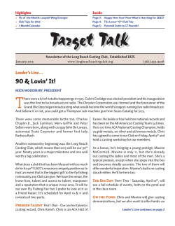
TREPAR-1349; No. of Pages 9
Review
The past, present, and future of
Leishmania genomics and
transcriptomics
Cinzia Cantacessi1, Filipe Dantas-Torres2,3, Matthew J. Nolan4, and
Domenico Otranto3
1
Department of Veterinary Medicine, University of Cambridge, Cambridge, UK
Departamento de Imunologia, Centro de Pesquisas Aggeu Magalha˜es, Fiocruz-PE, Brazil
3
Dipartimento di Medicina Veterinaria, Universita` degli Studi di Bari, Bari, Italy
4
Royal Veterinary College, University of London, North Mymms, UK
2
It has been nearly 10 years since the completion of the
first entire genome sequence of a Leishmania parasite.
Genomic and transcriptomic analyses have advanced
our understanding of the biology of Leishmania, and
shed new light on the complex interactions occurring
within the parasite–host–vector triangle. Here, we review these advances and examine potential avenues for
translation of these discoveries into treatment and control programs. In addition, we argue for a strong need to
explore how disease in dogs relates to that in humans,
and how an improved understanding in line with the
‘One Health’ concept may open new avenues for the
control of these devastating diseases.
Burden of leishmaniasis and the need for a ‘One Health’
initiative
Leishmaniases are a group of diseases caused by digenetic
protozoa of the genus Leishmania, which are transmitted
by phlebotominae sand flies (Table 1). Based on recent
estimates, up to 0.4 million and 1.2 million cases of visceral
(VL) and cutaneous leishmaniasis (CL), respectively, occur
each year in 98 countries and three territories where these
diseases are endemic [1]. Despite their widespread distribution, over 90% of global VL cases occur in only six
countries (India, Bangladesh, Sudan, South Sudan, Ethiopia, and Brazil), while most cases (70–75%) of CL occur in
ten countries (Afghanistan, Algeria, Colombia, Brazil,
Iran, Syria, Ethiopia, North Sudan, Costa Rica, and Peru)
[1]. In most cases, leishmaniases are zoonoses, affecting
the poor in rural and natural areas, where a plethora of
domestic and wild reservoir hosts and sand fly vectors
maintain the infection [2]. For instance, 13 out of the
21 human-infective Leishmania have also been reported
in domestic dogs, the latter having a major role in maintaining and transmitting the infection to other receptive
Corresponding author: Cantacessi, C. ([email protected]).
Keywords: leishmaniases; Leishmania infantum; high-throughput sequencing;
genome; transcriptome; bioinformatics; sand fly; metazoonosis; host-parasite interactions; One Health.
1471-4922/
ß 2015 Elsevier Ltd. This is an open access article under the CC BY license (http://
creativecommons.org/licenses/by/4.0/). http://dx.doi.org/10.1016/j.pt.2014.12.012
hosts via the sand fly vectors [3] (Table 1). In accordance
with the concept of ‘One Health’, defined as ‘a movement to
forge co-equal, all inclusive collaborations between physicians, [. . .], veterinarians and other scientific-health and
environmentally related disciplines [. . .] to improve and
defend the health and well-being of all species’ (http://
www.onehealthinitiative.com), successful control strategies
against human leishmaniases must include preventative
measures focussed on the human and animal hosts and
arthropod vectors, as well as on the environments where
the latter perpetuate [3]. To achieve these goals, a thorough
understanding of the host–pathogen–vector triangle, and
particularly of their intimate interactions at the molecular
level, is imperative. Recent advances in -omics technologies,
including genomics and transcriptomics, together with the
considerable decrease in the cost of these techniques, provide exciting opportunities to reveal details of the intimate
relations between Leishmania parasites, human and animal hosts, and sand fly vectors. In this review, we provide an
overview of a range of milestone studies that have used
genomics and transcriptomics techniques to improve current understanding of the biology of Leishmania, as well as
of the molecular interactions between this parasite and its
vertebrate and arthropod hosts. In addition, given the intimate relations between human and canine leishmaniases in
endemic areas, and in line with the ‘One Health’ movement,
we argue that current and future efforts should be directed
towards integrating -omics technologies (i.e., genomics,
transcriptomics, proteomics, metabolomics, and interactomics) to achieve a better understanding of the similarities
and differences between human and canine infections, with
the ultimate aim of developing new diagnostics, and treatment and control strategies against this devastating group
of diseases.
The fight against leishmaniasis: how can -omics help?
The control of leishmaniases generally relies on the early
diagnosis and treatment of human cases, vector control,
and, in some cases, management of reservoir hosts (i.e.,
treatment and/or elimination) [3]. However, the control of
leishmaniases, as with any vector-borne disease, is not
trivial due to challenges relating to intervention programs,
Trends in Parasitology xx (2015) 1–9
1
TREPAR-1349; No. of Pages 9
Review
Trends in Parasitology xxx xxxx, Vol. xxx, No. x
Table 1. Principal causative agents of human leishmaniases
Leishmania species
Leishmania aethiopica
Leishmania amazonensis
Principal
tropism a
C
C
Leishmania archibaldi d
Leishmania braziliensis
V
C, MC
Leishmania colombiensis
Leishmania donovani
C
V
Leishmania garnhami d
Leishmania guyanensis
C
C
Leishmania infantum
V, C
Leishmania
Leishmania
Leishmania
Leishmania
killicki d
lainsoni
lindenbergi
major
C
C
C
C
Leishmania mexicana
C
Leishmania naiffi
Leishmania panamensis
C
C, MC
Leishmania
Leishmania
Leishmania
Leishmania
C
C
C
C
peruviana
pifanoi d
shawi
tropica
Leishmania venezuelensis
C
Geographical distribution b
Notes on the infection in dogs c
Old World: Ethiopia, Kenya
New World: Argentina, Bolivia, Brazil, Colombia, Ecuador,
French Guiana, Peru, Suriname, Venezuela
Old World: Ethiopia, Kenya, Lebanon, Sudan
New World: Argentina, Belize, Bolivia, Brazil, Colombia, Costa
Rica, Ecuador, Guatemala, French Guiana, Honduras, Mexico,
Nicaragua, Panama, Paraguay, Peru, Venezuela
New World: Colombia, Panama, Venezuela
Old World: Bangladesh, Bhutan, China, Cyprus, Djibouti,
Ethiopia, India, Iraq, Israel, Kenya, Nepal, Saudi Arabia,
Somalia, Sri Lanka, Sudan, Ukraine, Uganda, Yemen
New World: Costa Rica, Venezuela
New World: Argentina, Bolivia, Brazil, Colombia, Ecuador,
French Guiana, Guyana, Peru, Suriname, Venezuela
Old World: Afghanistan, Albania, Algeria, Armenia, Azerbaijan,
Bosnia and Herzegovina, Bulgaria, Central African Republic,
China, Cyprus, Croatia, Egypt, France, Gambia, Georgia, Greece,
Iraq, Iran, Israel, Italy, Libyan Arab Jamahiriya, Jordan,
Kazakhstan, Kirgizstan, Lebanon, Macedonia, Malta, Morocco,
Mauritania, Monaco, Montenegro, Oman, Pakistan, Palestine,
Portugal, Syria, Romania, Senegal, Saudi Arabia, Slovenia,
Spain, Sudan, Tunisia, Turkmenistan, Turkey, Ukraine,
Uzbekistan, Yemen. NEW WORLD: Argentina, Bolivia, Brazil,
Colombia, Costa Rica, El Salvador, Guatemala, Honduras,
Mexico, Nicaragua, Paraguay, Venezuela
Old World: Algeria, Libyan Arab Jamahiriya, Tunisia
New World: Bolivia, Brazil, French Guiana, Peru, Suriname
New World: Brazil
Old World: Afghanistan, Algeria, Azerbaijan, Burkina Faso,
Cameron, Chad, Egypt, Ethiopia, Georgia, Ghana, Guinea,
Guinea-Bissau, India, Iraq, Israel, Libyan Arab Jamahiriya,
Jordan, Kazakhstan, Kenya, Kuwait, Mali, Morocco, Mauritania,
Mongolia, Niger, Nigeria, Oman, Pakistan, Palestine, Saudi
Arabia, Syria, Iran, Senegal, Sudan, Tunisia, Turkmenistan,
Uzbekistan, Yemen
New World: Belize, Colombia, Costa Rica, Ecuador, Guatemala,
Mexico, United States
New World: Brazil, French Guiana,
New World: Colombia, Costa Rica, Ecuador, Guatemala,
Honduras, Nicaragua, Panama
New World: Peru
New World: Venezuela
New World: Brazil
Old World: Afghanistan, Azerbaijan, Egypt, Ethiopia, Greece,
India, Iraq, Israel, Iran, Jordan, Kenya, Morocco, Namibia,
Pakistan, Palestine, Saudi Arabia, Syria, Turkmenistan, Turkey,
Uzbekistan, Yemen
New World: Venezuela
VL cases in Brazil
VL cases in Sudan
CL cases in Argentina, Bolivia,
Brazil Colombia, Peru, and
Venezuela
VL in a dog in Venezuela
Dogs are commonly infected in
some countries (e.g., Sudan),
but their role as reservoirs is
unknown
CL cases in Colombia
VL cases usually found in areas
where human cases are
reported. Autochthonous cases
reported in dogs in the USA (no
human cases reported so far)
CL in Egypt and Saudi Arabia
CL in Ecuador and USA
CL in Ecuador and Colombia
CL in Peru
CL in Ecuador
CL cases in India, Iran, Israel,
Morocco, and Syria
a
Abbreviations: C, dermotropic; MC, mucotropic; V, viscerotropic.
b
Based on [63,64].
c
Based on [54,65,66]. In addition, Leishmania arabica has been reported in dogs in Saudi Arabia [67]. Moreover, other Leishmania species (e.g., Leishmania equatorensis
and Leishmania utingensis) [68,69] have been described from wildlife and/or sand flies, but have not yet been detected in humans or dogs.
d
Species status is under discussion [63,70].
mostly in developing countries, where the burden of disease is heavier (due to a combination of factors including,
but not limited to, a lack of political will, of human
resources, and of infrastructure). In addition, our limited
knowledge of the host–pathogen–vector triangle, particularly of their intimate interactions at the molecular level,
impairs the development of more affordable and effective
control tools, such as antivector vaccines and more effective
chemotherapeutics.
2
-Omics technologies are increasingly being applied to
investigations of determinants of disease phenotype [4],
mode of action of current drugs [5], and parasite biology
[6]. These studies have improved our understanding of the
pathogenesis of disease in humans and possible mechanisms of resistance to antileishmanial drugs. Without a
doubt, -omics approaches are likely to reveal details of the
intimate relations between hosts, parasites, and vectors.
This refined knowledge will foster the development of new
TREPAR-1349; No. of Pages 9
Review
control tools (e.g., antivector vaccines) that could assist the
fight against leishmaniases. The determination of the
whole genome sequences of a range of Leishmania parasites causing both VL and CL represents the first step
towards these goals, providing the scientific community
with a solid infrastructure for postgenomic investigations
of the parasite biology, pathogenicity, and mastery mechanisms of manipulation of both insect and vertebrate hosts.
The Leishmania genomes: a ‘toolbox’ to understand
host–parasite interactions
Efforts to determine the whole genome sequence of key
Leishmania species infecting humans were consolidated in
1994 in Rio de Janeiro (Brazil), with the establishment of
the Leishmania Genome Network (LGN) initiative. Not
only did this network represent the researchers’ first move
to expand existing knowledge of the fundamental molecular biology of this parasite, with a view towards promoting
the discovery of novel treatment and control strategies, but
it also saw the support of the FIOCRUZ and UNICEF/
UNDP/World Bank/WHO Special Programme for Research
and Training in Tropical Diseases [7]. In 2005, these efforts
proved successful, with the publication of the first complete
genome sequence of Leishmania major (causing CL) [8],
soon followed by those of Leishmania infantum (causing
VL) and Leishmania braziliensis (causing mucocutaneous
leishmaniasis; MCL) [9]. In recent years, the advent of
high-throughput sequencing technologies (Box 1) has
assisted relentless progress in the genomics of human
leishmaniases, with the completion of the whole genome
sequences of Leishmania mexicana (CL; [10]), Leishmania
donovani (VL; [11]) and Leishmania amazonensis (CL;
[12]) (Box 1). The availability of these genome sequences
has provided unprecedented opportunities to perform detailed comparative analyses of Leishmania species associated with different human diseases at a scale previously
unimaginable [9,12].
The genomes of Leishmania vary from 29 Mb (L. amazonensis; [12]) to 33 Mb in size (L. major, L. infantum and
L. braziliensis; [9]) and are organised into a variable
number of chromosomes (i.e., 34 in L. amazonensis and
Trends in Parasitology xxx xxxx, Vol. xxx, No. x
L. mexicana, 35 in L. brasiliensis, and 36 in L. major, L.
donovani, and L. infantum) [12]. Despite the striking
variability in pathogenicity and tissue tropism of different
Leishmania species, their genomes are remarkably similar, displaying a high degree of conservation in gene content and architecture (synteny) [9,12]. The genomes of
Leishmania spp. are characterised by a high gene density,
the presence of long arrays of polycistronic gene clusters,
and the almost complete absence of introns [7]. However,
careful examination of protein-coding genes in Leishmania
allowed the identification of a relatively small number of
species-specific genes, the majority of which encode predicted proteins of unknown function [10]. Only a few of
these genes could be associated to specific tissue tropism.
For instance, LinJ.28.0340, a gene specific to L. infantum
and occurring as a pseudogene in L. major, L. braziliensis,
and L. mexicana [10], has been implicated in the ability of
the latter to spread and survive in visceral organs of the
vertebrate hosts [13]. Indeed, when a L. donovani gene
orthologue of LinJ.28.0340 was introduced into transgenic
L. major, the latter displayed a significantly increased
capacity to survive in visceral organs of BALB/C mice
[13]. Conversely, the spleens and livers of mice infected
with the LinJ.28.0340/L. donovani null mutant were characterised by significantly reduced parasite burdens compared with those infected with the wild type L. donovani
counterpart, thus providing solid evidence for a role of this
gene in the visceralisation of the infection [13]. Among
other genes thought to have key roles in the ability of
species within the L. donovani complex to colonize visceral
organs, those belonging to the A2 gene family are also
present as pseudogenes in L. major [14]. These genes were
first identified in L. donovani and shown to be exclusively
expressed by the amastigote stage (cf. [14]) (Figure 1);
subsequently, these genes were demonstrated to be essential for the survival of L. donovani in visceral organs, while
transgenic L. major expressing A2 genes displayed increased survival in the spleens of infected mice [14]. Despite the evidence for a role of A2 genes in the pathogenesis
of VL, molecules encoding A2 proteins have also been
identified in Leishmania species responsible for CL, such
Box 1. DNA sequencing technologies and Leishmania genomics: a decade of progress
In 2005, the Leishmania major genome was published in the journal
Science [8]. The successful completion of the first whole genome
sequence of a Leishmania species resulted from a collaboration
between the Wellcome Trust Sanger Institute (UK), the WTSIcoordinated EULEISH consortium, and the Seattle Biomedical Research Institute (USA) [8]. The sequencing strategy utilised a
combination of high- and low-coverage large insert clone sequencing
and a whole chromosome shotgun approach, preceded by purification of single or co-migrating chromosomes using pulse-field gel
electrophoresis (PFGE) [8]. In 2007, the genomes of two additional
Leishmania species, those of Leishmania infantum and Leishmania
braziliensis, were completed by a multi-institutional network of
scientists led by the WTSI [9]; to generate an approximate sixfold
coverage of the complete genome sequences of these two species,
researchers utilised whole-genome shotgun sequencing of plasmid
clones containing genome fragments of variable length (up to 4 kb).
Four years later, a similar approach was used to sequence the
genome of Leishmania mexicana [10]. In the same year, the reference
genome of Leishmania donovani was the first to be completed using
a next-generation sequencing strategy [11]; in particular, using a
pyrosequencing approach (454 Roche), Downing and colleagues
generated a total of 1.29 million single-end and 3.58 million pairedend reads [with an average length of 167 base pairs (bp)], 96% of
which were assembled into an initial set of reference contigs and
scaffolds [11]; subsequently, 17 clinical isolates of L. donovani
obtained from Nepalese and Indian patients with VL (including the
isolate from which the reference genome sequence was obtained),
were sequenced using high-throughput Illumina sequencing, generating a total of 41 Gb of sequence data [11]. Then, the 76-bp pairedend reads from each isolate were mapped to the reference genome
sequence for SNP analysis, which resulted in new insights into
Leishmania population genetics and mechanisms of emerging drug
resistance [11]. The most recent Leishmania genome sequence (i.e.,
that of Leishmania amazonensis [12]) was generated using a
combination of 454 and Illumina sequencing. The approximately
179 000 454 reads corresponded to a twofold coverage of the L.
amazonensis genome and were assembled into 27 856 contigs that,
together with the 4411 scaffolds derived from the assembly of
approximately 37 million 76-bp paired-end Illumina reads, resulted
in the final assembly of 2627 scaffolds [12].
3
TREPAR-1349; No. of Pages 9
Review
Trends in Parasitology xxx xxxx, Vol. xxx, No. x
Amasgotes of viscerotropic Leishmania express
A2 genes, implicated in visceralisaon of infecon [15]
3
2
4
Mammalian host
1
5
Simultaneously, Cox2 genes overexpressed by
infected macrophages promote parasite
survival [25]
6
Peritrophins and chins expressed by the sand fly midgut
serve as a barrier to the migraon of Leishmania to the thoracic
midgut, unl their degradaon by proteolyc enzymes [36]
7
Sand fly
Salivary components, such as maxadilan and
hyaluronidases, exacerbate parasite infecvity
and promote the infecon process [71]
8
9
TRENDS in Parasitology
Figure 1. Life cycle of Leishmania spp. and examples of molecules putatively involved in parasite infectivity and visceralisation of infection. Phlebotominae sand flies
release Leishmania infective stages (i.e., metacyclic promastigotes) to the mammalian hosts during blood feeding (1); the parasites invade macrophages and granulocytes
(2 and 3) and develop to amastigotes inside the phagolysosome (4); the amastigote stages replicate within the phagolysosome by simple division (5); then, amastigotecontaining macrophages are ingested by susceptible sand flies during the blood meal (6); the parasites are released from the infected macrophages within the sand fly
midgut (7), where they transform into procyclic promastigotes and divide. Then, the parasites migrate towards the stomodeal valve (anterior midgut) and transform into
different promastigote subtypes that ultimately form metacyclic promastigotes (8). These infective stages are then released into a new mammalian host during a
subsequent blood meal (9) [15,25,36,71]. Abbreviation: Cox2, prostaglandin-endoperoxide synthase 2.
as L. amazonensis and L. mexicana [10,12]. While the
presence of these proteins in Leishmania parasites with
skin tropism has been attributed to functional divergence
between Old World and New World species [14], their role
in the pathogenesis of CL is yet to be ascertained. Interestingly, a recent comparative analysis of the genomes
and transcriptomes of two phenotypically distinct substrains of L. donovani (i.e., one causing VL and one responsible for a large number of cases of CL in Sri Lanka)
revealed an increased copy number of A2 genes in L.
donovani causing VL, which was also associated with
significant upregulation in A2 mRNA transcription and
protein expression in strains causing VL [15]. In the same
study, Zhang and colleagues [15] identified the presence of
several nonsynonymous SNPs in genes from the L. donovani CL strain. Among these was a molecule encoding a
ras-like small GTPase-RagC protein; insertion of the corresponding orthologous gene from the L. donovani VL isolate into the CL counterpart resulted in a significant
increase in parasite burdens in the spleen of infected mice
4
[15]. These data provided evidence of the impact of SNPs
on gene function and phenotype, thus refining current
understanding of their potential impact on the pathogenicity of different strains of Leishmania.
While comparative analyses of the whole genome
sequences of Leishmania species causing CL and VL represent a solid basis for in-depth investigations of the intimate mechanisms of host–parasite interactions that result
in different courses of infection, studies of the regulation of
parasite gene expression throughout its life cycle in both
the vertebrate hosts and the sand fly vectors are likely to
contribute to a better understanding of the pathogenesis of
disease. Clearly, the availability of an array of genomes,
together with an explosion in microarray and highthroughput transcriptomic sequencing technologies, have
facilitated such studies (e.g., [16–19]). However, the same
organisation into polycistronic transcription units that
makes the genomes of CL- and VL-causing Leishmania
so strikingly similar [7] has been deemed responsible for
the lack of extensive gene expression regulation at the
TREPAR-1349; No. of Pages 9
Review
transcriptional level [20]. Indeed, most Leishmania genes
have been shown to be constitutively expressed throughout
the transition from promastigote to amastigote stage [21],
with post-transcriptional events, including mechanisms
that control the abundance of mRNAs, translation rates
and post-translational protein stability, hypothesised to
have key roles in the regulation of protein abundance
[21]. However, the marked variation in chromosome and
gene copy numbers among strains of L. infantum, L. mexicana, L. braziliensis, and L. major unveiled, for the first
time, a degree of aneuploidy in the genomes of these
parasites [10]. Accordingly, unstable ploidy among strains
of L. infantum, as well as variable chromosomal contents
among cells, revealed that the Leishmania genome is
characterised by ‘mosaic aneuploidy’ [11,22]. Therefore,
‘genome plasticity’ and ‘gene dosage’, rather than differential expression of single genes and gene products, are
increasingly being considered as two of the keys to the
different tissue tropism of Leishmania spp. [22].
Transcriptomics unveils Leishmania-mediated
regulation of host gene expression
Several transcriptomic studies have investigated Leishmania-induced regulation of gene expression in infected
tissues with the aim to link such responses to disease
outcome. As an example, for VL-causing Leishmania, Beattie and colleagues [23] used whole-genome array technologies to compare the gene expression profiles of liverresident macrophages (Kupffer cells) from mice infected
by L. donovani to those of uninfected macrophages exposed
to inflammatory stimuli. The authors showed significant
upregulation of genes within the retinoid X receptor a
pathway (i.e., Rxra), linked to lipid metabolism, in uninfected macrophages exposed to inflammation compared
with the infected counterpart [23]; pharmacological perturbation of the activity of this pathway in Kupffer cells
resulted in an increased resistance of these cells to
Leishmania infection, which led to speculation that either
this pathway has a role in the usage of lipids and cholesterol by the parasite, or that Leishmania lipids regulate
the activation of innate immune responses that follow the
infection [23]. For CL-causing Leishmania, Maretti-Mira
and colleagues [24] utilised high-throughput RNA-Seq
technologies to characterise and compare the transcriptomes of tissue fragments obtained from human subjects
with CL and MCL caused by L. braziliensis [24]. The outcomes from this study highlighted significant upregulation
of genes involved in biological pathways linked to the
recruitment and activation of immune cells (including
lymphocytes, granulocytes, natural killer cells, and antigen-presenting cells) and to regulation of inflammatory
responses in tissues from subjects with CL [24]. This suggested that the inability of the host to mount effective
immune responses against the parasite at the site of
cutaneous infection is linked to the progression of disease
[24]. In an effort to characterise differences in macrophage
gene expression that might contribute to the ability of
different Leishmania spp. to cause localised (CL) or systemic infections (VL), Gregory and colleagues [25] used a
DNA microarray approach to perform comparative analyses of the transcriptomes of murine macrophages infected
Trends in Parasitology xxx xxxx, Vol. xxx, No. x
by L. major and L. donovani. Interestingly, both parasites
induced a similar differential regulation of relatively
small numbers of macrophage genes, with most of these
genes unsurprisingly linked to the development of
immune responses [25]. The only noticeable difference in
gene expression profiling between L. major- and L. donovani-infected macrophages was a remarkable increase in
levels of transcription of mRNAs encoding prostaglandinendoperoxide synthase (Cox2) in the latter, which led to
speculation that this pathway is involved in the pathogenesis of VL [25] (Figure 1).
Clearly, the availability of high-throughput transcriptomic technologies has resulted in rapid expansion of the
already substantial plethora of knowledge of the molecular
interactions occurring between Leishmania and the human host; nevertheless, significant variation in host
responses to infection has been described in several studies
(cf. [26]), although a review of this variation is beyond the
scope of the present article. However, these technologies
have also enabled progress towards the exploration of the
molecular relationships between the parasite and the sand
fly vector and the patterns of Leishmania development into
its infective, nondividing metacyclic form [27].
Transcriptomics in Leishmania–sand fly interactions
Transmission of Leishmania from an infected to a susceptible host requires development of the parasites in the
midgut of a competent sand fly vector. Macrophages
containing Leishmania amastigotes are ingested by sand
fly vectors via a blood meal and, once released in the insect
midgut, develop through several developmental stages
into infective, metacyclic promastigotes [26] (Figure 1).
The reproductive mode of Leishmania parasites has traditionally been considered clonal, based on strong linkage
disequilibrium (cf. [28]); however, several studies have
provided solid evidence of the occurrence of genetic exchange between species and/or strains of Leishmania (i.e.,
L. major and L. infantum) during growth and development
in the sand fly vector, with successful transmission of
the hybrid progeny to a susceptible vertebrate host
[28–32]. The range of vertebrate and invertebrate host
species that Leishmania can infect, as well as the multiple
forms of disease that it causes, have been partly attributed
to the ability of this parasite to undergo genetic exchange
in the sand fly vector (cf. [29]). Clearly, the molecular
interactions that occur at the parasite–sand fly interface
are key processes that determine the successful development and transmission of Leishmania; therefore, a detailed understanding of these mechanisms has become a
priority. Previous studies had used Sanger sequencing of
cDNA libraries from the midgut of sand fly vectors of
both CL- and VL-causing Leishmania (i.e., Phlebotomus
papatasi, vector of L. major and Lutzomyia longipalpis,
vector of L. infantum; [33,34]) to identify molecules putatively involved in the development of the parasites in
their insect vectors. While sand fly infections by L. major
and L. infantum were consistently associated with downregulation of molecules encoding microvilli-like proteins
and chymotrypsin and upregulation of trypsin-encoding
transcripts, the transcription profiles of peritrophinlike molecules were inconsistent between P. papatasi
5
TREPAR-1349; No. of Pages 9
Review
and Lu. longipalpis [33,34]. Peritrophins are the protein
component of the peritrophic matrix (PM), an extracellular
chitin-containing structure that encapsulates the blood
meal following its ingestion by the sand fly [35]. The
formation of the PM (immediately following the blood
meal) has long been considered advantageous for Leishmania, because the parasites are thought to be protected
from the action of the sand fly proteolytic enzymes during
the vulnerable time of development to promastigotes
[35]. Several key investigations have contributed to
further understanding of the relations between Leishmania promastigotes and the sand fly PM (e.g., [36]). In particular, while previous studies hypothesised a role of
Leishmania chitinases in the disintegration of the sand
fly PM (cf. [36]), current evidence supports the theory
that the breakdown of the PM is independent from the
activity of Leishmania enzymes and that parasite promastigotes escape the PM by migrating through a posterior
opening that forms irrespective of the infection status of
the sand fly [36]. In the same study, Sadlova and Volf
[36] showed that the anterior plug of the PM serves as a
‘barrier’ for parasite migration to the thoracic midgut,
until its degradation from sand fly proteolytic enzymes
is complete [36]. The elucidation of patterns of sand fly
gene expression during the disintegration of the PM in
the presence (or not) of Leishmania parasites, and
during migration of the latter from the abdominal to the
thoracic midgut, may help to either confirm or confute this
point.
Together with studies of the midgut of sand flies, other
investigations used transcriptomic technologies to shed
light on the molecular mechanisms that govern the development of Leishmania parasites into their infective metacyclic stage [37]. While little information is available on
sand fly molecular pathways acting as trigger of Leishmania metacyclogenesis, recent studies highlighted the role of
key genes and gene products in the differentiation of
promastigote stages into metacyclic forms in the sand fly
vector. Among these molecules, a hydrophilic acylated
surface protein (HASPB) and a small hydrophilic endoplasmic reticulum (ER)-associated protein (SHERP)
showed increased expression in the metacyclic stages
[38]; in addition, creation of HASPB and SHERP null
mutants in L. major resulted in the accumulation of
non-infective parasite stages in the midgut of the sand
fly vector, thus providing evidence for the essentiality of
these molecules for parasite development [38]. Investigations of patterns of gene transcription during Leishmania
metacyclogenesis in vitro have led to the identification of
genes and gene products potentially related to parasite
infectivity (e.g. [37,39–41]). For instance, recent functional
studies of essential molecules in L. major metacyclic promastigotes highlighted major roles of mitogen-activated
protein kinases (i.e., MAPK4) and metallopeptidases of the
M24A family in the establishment of intracellular macrophage infections [40] and proliferation in infected cells
[41]. These data provided a solid basis for the exploration
of the role of these molecules as novel targets for intervention strategies.
In recent years, the search for new and effective preventative measures against Leishmania transmission has
6
Trends in Parasitology xxx xxxx, Vol. xxx, No. x
Box 2. Sand-fly salivary gland secreted proteins and
Leishmania: examples from a successful partnership
The salivary glands of sand fly vectors secrete several proteins
known to facilitate the process of host invasion by Leishmania
[27]. Among these proteins, maxadilan, a known vasodilator, was
first described in Lutzomyia longipalpis and shown to exacerbate
the infectivity of Leishmania major via the inhibition of lymphocyte
proliferation [47]. Since this first report, a range of other molecules
has been described from the salivary glands of an array of sand fly
species that contribute to the successful establishment of Leishmania infections. Among these molecules, protein homologues of
salivary hyaluronidases secreted by Phlebotomus papatasi were
demonstrated to have a role in the severity of skin lesions caused by
L. major [71]. While the mechanisms that result into this outcome
are yet to be elucidated, Volfova and colleagues [71] hypothesised
that the neutrophils recruited through the enzymatic activity of sand
fly hyaluronidases (via the presence of hyaluronan fragments) may
be exploited by Leishmania as ‘Trojan horses’, thus facilitating
macrophage infection [71]. Interestingly, a secreted endonuclease
identified from the sialotranscriptome of Lu. longipalpis has been
recently implicated in the ability of Leishmania to evade killing by
neutrophils [47]. This protein was shown to interfere with the
microbicidal activity of neutrophils recruited at the site of a sand fly
bite by disintegrating the neutrophil extracellular traps (NETs) that
ensnare Leishmania parasites, thus significantly contributing to the
immunoevasive strategies of the parasite and enhancing its
infectivity [47].
also involved the characterisation of key components of the
saliva of the sand fly vectors (e.g., [27,42–46]). The interest
of the scientific community in salivary gland transcriptomes (‘sialotranscriptomes’) is mainly derived from
knowledge that selected saliva proteins have crucial roles
in facilitating the successful establishment of Leishmania
parasites in vertebrate hosts, including the regulation of
the immune response at the site of bite [27,46,47]. Therefore, sialotranscriptomes of several competent sand fly
vector species are now available (e.g., [43–45,48–51]),
which, in some cases, have led to the selection of key sand
fly molecules that are involved in the blood-feeding process
and that may assist the immunoevasive strategies of
Leishmania [47,52] (Box 2). For instance, a potent vasodilator (maxadilan) abundantly detected in the saliva of Lu.
longipalpis [53] has not been identified in transcriptomic
data sets from the salivary glands of Lutzomyia ayacuchensis [44]. Similarly, a maxadilan homologue identified
in Lutzomyia intermedia showed only 34% identity to
maxadilan from Lu. longipalpis [50]. It is worth noting
that both Lu. intermedia and Lu. ayacuchensis are vectors
of dermotropic Leishmania species, whereas Lu. longipalpis, whose saliva contains large amounts of maxadilan, is
the main vector of the viscerotropic L. infantum in the New
World [54] (Figure 1). While these observations suggested
a role of maxadilan in visceralisation of L. infantum infection [55], the absence of maxadilan homologues from the
saliva of sand fly vectors of VL in the Old World raises
questions about the role/s of other salivary components in
disease progression. Indeed, other enzymes, such as hyaluronidases and apyrases, have been identified using transcriptomic and proteomic technologies from several sand
fly vectors of VL in both the Old and New Worlds
[45]. These enzymes have been shown to positively contribute to the spread of Leishmania parasites by promoting
TREPAR-1349; No. of Pages 9
Review
the enlargement of the feeding lesion and the diffusion of
other salivary active compounds (hyaluronidases) and preventing haemostasis (apyrases) [45].
Besides containing components essential to the infection process, the saliva of sand flies contains molecules that
can elicit specific immune responses that are indicative of
host exposure to sand fly bites (e.g., [56,57]). In particular,
three proteins from the saliva of P. perniciosus (i.e., two
yellow proteins and an apyrase), expressed in recombinant
form, were shown to be useful in determining the intensity
of exposure to sand fly bites in experimentally bitten mice
and dogs [57]. While cross-reactivity between anti-P. perniciosus antibodies and those from closely related sand fly
species was not assessed [57], the authors hypothesised
that this may occur. Both yellow proteins and apyrases
have been detected in the saliva of a range of sand fly
species. However, subtle differences in sequence may result in varying immunogenic properties; future investigations using transcriptomic and proteomic technologies may
assist elucidating this point via, for instance, the generation of whole transcript and/or protein data sets from sand
fly vector species, with the ultimate aim of identifying
suitable targets for the development of commercial diagnostic tools to assess the risk of human and canine transmission in both endemic and nonendemic areas, and the
evaluation of the effectiveness of antivector campaigns
[56]; this improved knowledge could also aid current efforts
aimed at developing recombinant vaccines containing immunogenic components from both the parasite and the
sand fly vectors. In addition, thus far, no data are available
on the effects of Leishmania infections on the global transcriptional profiles of sand fly vectors. Future studies
could, for instance, utilise RNA-Seq technologies to investigate differences in gene expression profiling of Leishmania-infected and uninfected sand flies. Exploring and
identifying molecular pathways involved in the parasite–vector–host interactions may lead to the identification
of new molecular pathways implicated in the infection
process, which would be instrumental for refining current
control strategies against sand flies. Undoubtedly, some
challenges exist in performing large-scale transcriptomic
studies of species for which a reference genome is unavailable; among these challenges, the de novo assembly of fulllength transcripts in absence of reference sequences is one
of the most significant [58]. Nevertheless, other resources,
such as the genomes and transcriptomes of selected mosquito species [59] that are phylogenetically related to sand
fly vectors of Leishmania [60], could be exploited for the
accurate reconstruction of (at least) a proportion of fulllength sand fly transcripts, thus reducing overall project
costs and limiting potential biases introduced by de novo
assembly.
Concluding remarks and research needs
Over the past decade, advances in genomics and transcriptomics technologies have contributed to considerably enhance our knowledge of the set of molecular interactions
that occur within the host–parasite–vector triangle. However, some gaps still exist in our understanding of the
similarities and/or differences between human leishmaniases and the disease in animal reservoir hosts. Dogs, for
Trends in Parasitology xxx xxxx, Vol. xxx, No. x
instance, represent the most important host reservoir for
L. infantum (causing VL) [3,61]. Therefore, differences and
similarities between human and canine infections should
be comprehensively analysed. However, most studies of
Leishmania immunobiology and genetics, as well as of
host–parasite interactions, utilise murine models of infection as ‘mirrors’ of human disease [61]. Given that transmission of key Leishmania species (e.g., L. infantum) to
humans strictly relies on the circulation of the parasite
among canine populations, elucidating whether dog leishmaniasis serves as a model for human infections should
become a priority. This could provide avenues for studies
aimed, for instance, at evaluating the ‘translatability’ of
novel treatment and vaccine strategies from humans to
dogs and vice versa. The availability of in vivo canine
models of leishmaniasis [62], together with advances in
genomics and/or transcriptomics, proteomics, and metabolomics technologies, may assist this quest. For instance,
RNA-Seq and high-throughput proteomics platforms provide a golden opportunity to monitor changes in host gene
transcription and protein expression throughout the
course of canine and human infections, thus enabling
one to draw parallels between them. Similarly, large-scale
analyses of metabolites produced during the course of
infection, both by the parasite and the vertebrate host,
may represent a gold mine for the identification of novel
diagnostic biomarkers, as well as of potential new Leishmania ‘Achilles’ heels’ that could assist current programs
aimed at breaking the transmission cycle of human and
canine leishmaniases.
Acknowledgements
Part of this article was conceived within the framework of of the
EurNegVec COST Action TD1303. Funding from the Isaac Newton Trust/
Wellcome Trust ISSF/University of Cambridge Joint Research Grants
Scheme to C.C. is gratefully acknowledged.
References
1 Alvar, J. et al. (2012) Leishmaniasis worldwide and global estimates of
its incidence. PLoS ONE 7, e35671
2 Ready, P.D. (2013) Biology of phlebotomine sand flies as vectors of
disease agents. Annu. Rev. Entomol. 58, 227–250
3 Otranto, D. and Dantas-Torres, F. (2013) The prevention of canine
leishmaniasis and its impact on public health. Trends Parasitol. 29,
339–345
4 McCall, L.I. and McKerrow, J.H. (2014) Determinants of disease
phenotype in trypanosomatid parasites. Trends Parasitol. 30, 342–349
5 Kaur, G. and Rajput, B. (2014) Comparative analysis of the omics
technologies used to study antimonial, amphotericin B, and
pentamidine resistance in Leishmania. J. Parasitol. Res. 2014,
726328
6 Pawar, H. et al. (2014) Neglected tropical diseases and omics science:
proteogenomics analysis of the promastigote stage of Leishmania
major parasite. OMICS 18, 499–512
7 Uliana, S.R.B. et al. (2008) Leishmania genomics: where do we stand?
In Bioinformatics in Tropical Disease Research: A Practical and CaseStudy Approach (Gruber, A. et al., eds), National Library of Medicine,
National Center for Biotechnology Information Chapter B02
8 Ivens, A.C. et al. (2005) The genome of the kinetoplastid parasite,
Leishmania major. Science 309, 436–442
9 Peacock, C.S. et al. (2007) Comparative genomic analysis of three
Leishmania species that cause diverse human disease. Nat. Genet.
39, 839–847
10 Rogers, M.B. et al. (2011) Chromosome and gene copy number variation
allow major structural change between species and strains of
Leishmania. Genome Res. 21, 2129–2142
7
TREPAR-1349; No. of Pages 9
Review
11 Downing, T. et al. (2011) Whole genome sequencing of multiple
Leishmania donovani clinical isolates provides insights into
population structure and mechanisms of drug resistance. Genome
Res. 21, 2143–2156
12 Real, F. et al. (2013) The genome sequence of Leishmania (Leishmania)
amazonensis: functional annotation and extended analysis of gene
models. DNA Res. 20, 567–581
13 Zhang, W.W. and Matlashewski, G. (2010) Screening Leishmania
donovani-specific genes required for visceral infection. Mol.
Microbiol. 77, 505–517
14 Zhang, W.W. et al. (2003) Comparison of the A2 gene locus in
Leishmania donovani and Leishmania major and its control over
cutaneous infection. J. Biol. Chem. 35508–35515
15 Zhang, W.W. et al. (2014) Genetic analysis of Leishmania donovani
tropism using a naturally attenuated cutaneous strain. PLoS Pathog.
10, e1004244
16 Cohen-Freue, G. et al. (2007) Global gene expression in Leishmania.
Int. J. Parasitol. 37, 1077–1086
17 Saxena, A. et al. (2007) Evaluation of differential gene expression in
Leishmania major Friedlin procyclics and metacyclics using DNA
microarray analysis. Mol. Biochem. Parasitol. 129, 103–114
18 Alcolea, P.J. et al. (2010) Transcriptomics throughout the life cycle of
Leishmania infantum: high down-regulation rate in the amastigote
stage. Int. J. Parasitol. 40, 1497–1516
19 Rastrojo, A. et al. (2013) The transcriptome of Leishmania major in the
axenic promastigote stage: transcript annotation and relative
expression levels by RNA-Seq. BMC Genomics 14, 223
20 Soysa, R. et al. (2014) A dual luciferase system for analysis of posttranscriptional regulation of gene expression in Leishmania. Mol.
Biochem. Parasitol. 195, 1–5
21 Requena, J.M. (2011) Lights and shadows on gene organization and
regulation of gene expression in Leishmania. Front. Biosci. 16, 2069–
2085
22 Sterkers, Y. et al. (2012) Novel insights into genome plasticity in
Eukaryotes: mosaic aneuploidy in Leishmania. Mol. Microbiol. 86, 15–23
23 Beattie, L. et al. (2013) A transcriptomic network identified in
uninfected macrophages responding to inflammation control
intracellular pathogen survival. Cell Host Microbe 14, 357–368
24 Maretti-Mira, A.C. et al. (2012) Transcriptome patterns from primary
cutaneous Leishmania braziliensis infections associated with eventual
development of mucosal disease in humans. PLoS Negl. Trop. Dis. 6, e1816
25 Gregory, D.J. et al. (2008) Comparison of the effects of Leishmania
major or Leishmania donovani infection on macrophage gene
expression. Infect. Immun. 76, 1186–1192
26 Kima, P.E. (2014) Leishmania molecules that mediate intracellular
pathogenesis. Microbes Infect. 16, 721–726
27 Oliveira, F. et al. (2009) Sand flies, Leishmania, and transcriptomeborne solutions. Parasitol. Int. 58, 1–5
28 Akopyants, N.S. et al. (2009) Demonstration of genetic exchange during
cyclical development of Leishmania in the sand fly vector. Science 324,
265–268
29 Inbar, E. et al. (2013) The mating competence of geographically diverse
Leishmania major strains in their natural and unnatural sand fly
vectors. PLoS Genet. 9, e1003672
30 Calvo-Alvarez, E. et al. (2014) First evidence of intraclonal genetic
exchange in trypanosomatids using two Leishmania infantum
fluorescent transgenic clones. PLoS Negl. Trop. Dis. 8, e3075
31 Ravel, C. et al. (2006) First report of genetic hybrids between two very
divergent Leishmania species: Leishmania infantum and Leishmania
major. Int. J. Parasitol. 36, 1383–1388
32 Volf, P. et al. (2007) Increased transmission potential of Leishmania
major/Leishmania infantum hybrids. Int. J. Parasitol. 37, 589–593
33 Ramalho-Ortigao, M. et al. (2007) Exploring the midgut transcriptome
of Phlebotomus papatasi: comparative analysis of expression profiles of
sugar-fed, blood-fed and Leishmania major-infected sandflies. BMC
Genomics 8, 300
34 Jochim, R.C. et al. (2008) The midgut transcriptome of Lutzomyia
longipalpis: comparative analysis of cDNA libraries from sugar-fed,
blood-fed, post-digested and Leishmania infantum-chagasi–infected
sand flies. BMC Genomics 9, 15
35 Pimenta, P.F. et al. (1997) A novel role for the peritrophic matrix in
protecting Leishmania from the hydrolytic activities of the sand fly
midgut. Parasitology 115, 359–369
8
Trends in Parasitology xxx xxxx, Vol. xxx, No. x
36 Sadlova, J. and Volf, P. (2009) Peritrophic matrix of Phlebotomus
duboscqi and its kinetics during Leishmania major development.
Cell Tissue Res. 337, 313–325
37 Dostalova, A. and Volf, P. (2012) Leishmania development in sand flies:
parasites–vector interactions overview. Parasit. Vectors 5, 276
38 Sadlova, J. et al. (2010) The stage-regulated HASPB and SHERP
proteins are essential for differentiation of the protozoan parasite
Leishmania major in its sand fly vector, Phlebotomus papatasi. Cell
Microbiol. 12, 1765–1779
39 Cunha, J. et al. (2013) Characterization of the biology and infectivity of
Leishmania infantum viscerotropic and dermotropic strains isolated
from HIV+ and HIV– patients in the murine model of visceral
leishmaniasis. Parasit. Vectors 6, 122
40 Dacher, M. et al. (2014) Probing druggability and biological function of
essential proteins in Leishmania combining facilitated null mutant
and plasmid shuffle analyses. Mol. Microbiol. 93, 146–166
41 Norris-Mullins, B. et al. (2014) LmaPA2G4, a homolog of human Ebp1,
is an essential gene and inhibits cell proliferation in L. major. PLoS
Negl. Trop. Dis. 8, e2646
42 Dostalova, A. et al. (2011) The midgut transcriptome of Phlebotomus
(Larroussius) perniciosus, a vector of Leishmania infantum:
comparison of sugar fed and blood fed sand flies. BMC Genomics
12, 223
43 Rohousova, I. et al. (2012) Salivary gland transcriptomes and
proteomes of Phlebotomus tobbi and Phlebotomus sergenti, vectors of
leishmaniasis. PLoS Negl. Trop. Dis. 6, e1660
44 Kato, H. et al. (2013) Analysis of salivary gland transcripts of the sand
fly Lutzomyia ayacuchensis, a vector of Andean-type cutaneous
leishmaniasis. Infect. Genet. Evol. 13, 56–66
45 Vlkova, M. et al. (2014) Comparative analysis of salivary gland
transcriptomes of Phlebotomus orientalis sand flies from endemic
and non-endemic foci of visceral leishmaniosis. PLoS Negl. Trop.
Dis. 8, e2709
46 Hostomska, J. et al. (2009) Analysis of salivary transcripts and
antigens of the sand fly Phlebotomus arabicus. BMC Genomics 10, 282
47 Chagas, A.C. et al. (2014) Lundep, a sand fly salivary endonuclease
increases Leishmania parasite survival in neutrophils and inhibits
XIIa contact activation in human plasma. PLoS Pathog. 10, e1003923
48 Abdeladhim, M. et al. (2012) Updating the salivary gland
transcriptome of Phlebotomus papatasi (Tunisian strain): the search
for sand fly-secreted immunogenic proteins for humans. PLoS ONE 7,
e47347
49 Abrudan, J. et al. (2013) The characterisation of the Phlebotomus
papatasi transcriptome. Insect Mol. Biol. 22, 211–232
50 de Moura, T.R. et al. (2013) Functional transcriptomics of wild-caught
Lutzomyia intermedia salivary glands: identification of a protective
salivary protein against Leishmania braziliensis infection. PLoS Negl.
Trop. Dis. 7, e2242
51 Oliveira, F. et al. (2013) Sand-fly saliva–Leishmania–man: the trigger
trio. Front. Immunol. 4, 375
52 Prates, D.B. et al. (2011) Lutzomyia longipalpis saliva drives apoptosis
and enhances parasite burdens in neutrophils. J. Leukoc. Biol. 90,
575–582
53 Valenzuela, J.G. et al. (2004) Identification of the most abundant
secreted proteins from the salivary glands of the sand fly Lutzomyia
longipalpis, vector of Leishmania chagasi. J. Exp. Biol. 207, 3717–3729
54 Dantas-Torres, F. et al. (2012) Canine leishmaniosis in the Old and
New Worlds: unveiled similarities and differences. Trends Parasitol.
28, 531–538
55 Warburg, A. et al. (1994) Saliva of Lutzomyia longipalpis sibling species
differs in its composition and capacity to enhance leishmaniasis.
Philos. Trans. R. Soc. Lond. B: Biol. Sci. 345, 223–230
56 Vlkova, M. et al. (2011) Canine antibody response to Phlebotomus
perniciosus bites negatively correlates with the risk of Leishmania
infantum transmission. PLoS Negl. Trop. Dis. 5, e1344
57 Drahota, J. et al. (2014) Recombinant antigens from Phlebotomus
perniciosus saliva as markers of canine exposure to visceral
leishmaniases vector. PLoS Negl. Trop. Dis. 8, e2597
58 Cantacessi, C. et al. (2012) Key strongylid nematodes of animals:
impact of next-generation transcriptomics on systems biology and
biotechnology. Biotechnol. Adv. 30, 469–488
59 Severson, D.W. and Behura, S.K. (2012) Mosquito genomics: progress
and challenges. Annu. Rev. Entomol. 57, 143–166
TREPAR-1349; No. of Pages 9
Review
60 Aransay, A.M. et al. (2000) Phylogenetic relationships of phlebotomine
sandflies inferred from small subunit nuclear ribosomal DNA. Insect
Mol. Biol. 9, 157–168
61 Loria-Cervera, E.N. and Andrade-Narvaez, F.J. (2014) Animal models
for the study of leishmaniasis immunology. Rev. Inst. Med. Trop. Sao
Paulo 56, 1–11
62 Costa, D.J. et al. (2013) Experimental infection of dogs with Leishmania
and saliva as a model to study canine visceral leishmaniasis. PLoS ONE
8, e60535
63 World Health Organization (2010) Control of the Leishmaniasis,
WHO
64 Pratlong, F. et al. (2013) Geographical distribution and epidemiological
features of Old World Leishmania infantum and Leishmania donovani
foci, based on the isoenzyme analysis of 2277 strains. Parasitology 140,
423–434
65 Dantas–Torres, F. (2009) Canine leishmaniosis in South America.
Parasit. Vectors 2 (Suppl. 1), S1
Trends in Parasitology xxx xxxx, Vol. xxx, No. x
66 Baneth, G. et al. (2014) Mucocutaneous Leishmania tropica infection in a
dog from a human cutaneous leishmaniasis focus. Parasit. Vectors 7, 118
67 Peters, W. et al. (1986) Leishmania infecting man and wild animals in
Saudi Arabia. 2. Leishmania arabica n. sp. Trans. R. Soc. Trop. Med.
Hyg. 80, 497–502
68 Grimaldi Ju´nior, G. et al. (1992) Description of Leishmania equatorensis
sp. n (Kinetoplastida: Trypanosomatidae), a new parasite infecting
arboreal mammals in Ecuador. Mem. Inst. Oswaldo Cruz 87, 221–228
69 Braga, R.R. et al. (2003) Leishmania (Viannia) utingensis n. sp., a
parasite from the sandfly Lutzomyia (Viannamyia) tuberculata in
Amazonian Brazil. Parasite 10, 111–118
70 El Baidouri, F. et al. (2013) Genetic structure and evolution of the
Leishmania genus in Africa and Eurasia: what does MLSA tell us.
PLoS Negl. Trop. Dis. 7, e2255
71 Volfova, V. et al. (2008) Hyaluronidase of bloodsucking insects and its
enhancing effect on Leishmania infection in mice. PLoS Negl. Trop.
Dis. 2, e294
9


© Copyright 2026