
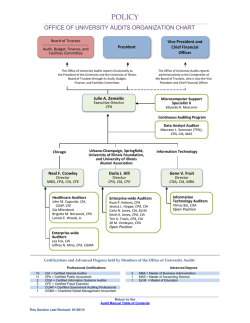
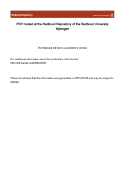
Quantitative In Vivo Assay of Human Granulocyte Colony
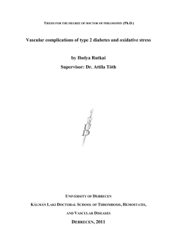
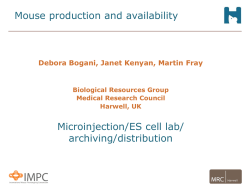
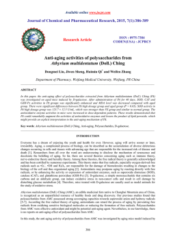
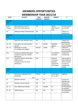
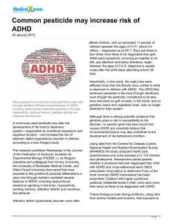
From www.bloodjournal.org by guest on February 6, 2015. For personal use only.
Quantitative In Vivo Assay of Human Granulocyte Colony-Stimulating Factor
Using Cyclophosphamide-Induced Neutropenic Mice
By Kunihiro Hattori, Koji Shimizu, Mayumi Takahashi, Masahiko Tamura, Masayoshi Oheda,
Nakaaki Ohsawa, and Masayoshi Ono
Administration of human granulocyte colony-stimulating
factor (hG-CSF) to mice with cyclophosphamide (CPAIinduced neutropenia for 4 consecutive days from the day
after the CPA dosing (100 mg/kg) resulted in a dosedependent increase in the peripheral blood neutrophil
count 6 hours after the final hG-CSF injection. Within the
hG-CSF dose range of 0.1 to 10 pg per mouse per day,
there was a strong linear relationship ( r > .9) between the
logarithm of the dose and the peripheral blood neutrophil
count in the treated mice. Using the same hG-CSF preparation, 38 experiments indicated that the regression lines are
highly reproducible. Such an association never occurred
with intact mice, and 100 mg/kg of CPA induced the
highest response to hG-CSF. This linear relationship between the two variables allows us to determine the
biologic potency of a test hG-CSF preparation relative to a
reference standard using a parallel line assay, with a
coefficient of precision of around .2. When assayed by this
bioassay procedure, which we have termed CPA-mouse
assay, natural hG-CSF and recombinant hG-CSF (produced
by Chinese hamster ovary cells) were nearly equipotent in
specific biologic activity. These results confirm the CPAmouse assay as an especially useful assay method for
quantifying the in vivo activity of hG-CSF.
0 1990 by The American Society of Hematology.
C
This method is demonstrated by an experiment on CPAinduced neutropenic mice.
OLONY-STIMULATING factors (CSFs) are classified into four subclasses according to the type of
colonies formed: (1) multi-CSF (interleukin-3),’ (2) granulocyte-macrophage-CSF (CJM-CSF),~(3) granulocyte-CSF
(G-CSF),3 and (4) macrophage-CSF (M-CSF).4 The activities of these CSFs are quantitatively assayed using a colonyforming technique that is in widespread use as a routine
laboratory procedure. The activity of human (h) G-CSF can
also be quantified using human and mouse marrow cells.
Recent research has led to the development and availability
of methods for the quantitative assay of CSFs using factordependent cell
Among these cell lines, NFS-607.*has
made it possible to estimate levels of hG-CSF with great
ease. Unfortunately, no reliable assay method has been
established for quantitatively determining these factors in
vivo. Before these factors can be used successfully for clinical
purposes, their effects on the hematopoietic mechanism in
vivo must be assessed. It is impossible to determine all of the
effects on the hematopoietic mechanism in vivo from the in
vitro effects on hematopoietic cells, since in vitro models
cannot adequately simulate the in vivo hematopoietic mechanism. Moreover, the potencies of these CSFs are apparently
under the influence of the pharmacokinetics and metabolism
in vivo, as they are all glycoproteins. It is necessary for
clinical purposes to consider the effects on the biologic
mechanism other than hematopoiesis, including homeostasis.
This report describes an in vivo high precision, quantitative
assay for hG-CSF with good reproducibility and simplicity.
From the Fuji-Gotemba Laboratory, Chugai Pharmaceutical Co
Ltd, Gotemba, and the First Department of Internal Medicine.
Faculty for Medicine. Osaka Medical College, Osaka, Japan.
Submitted December 15.1988; accepted November 15,1989.
Address reprint requests to Masayoshi Ono, PhD. Central Research Laboratories, Chugai Pharmaceutical Co Ltd, 41-8, Takada
3-chome, Toshima-ku. Tokyo. 171, Japan.
The publication costs of this article were defrayed in part by page
charge payment. This article must therefore be hereby marked
“advertisement” in accordance with 18 U.S.C.section 1734 solely to
indicate this fact.
@ 1990 by The American Society of Hematology.
0006-4971/90/7506-0005$3.00/0
1228
MATERIALS AND METHODS
Mice. We used 7- to 9-week-old male C57BL/6N mice (Cler
Japan Inc, Tokyo, Japan) raised under specific pathogen-free (SPF)
conditions.
Natural hG-CSF and recombinant hG-CSF. As described
previously: natural hG-CSF (nhG-CSF) was purified from medium
conditioned by a squamous cell line (CHU-2) that constitutively
produced CSFs. Purified recombinant hG-CSF (rhG-CSF) was
prepared from medium conditioned by Chinese hamster ovary
(CHO) cells transfected with vectors containing full-length cDNA
for hG-CSF.’ The purified hG-CSFs were diluted in phosphatebuffered saline (pH 7.4) containing 1% human serum albumin
(Green Cross Co, Osaka, Japan) and 0.01% Tween-20 (Nakarai
Chemicals, Kyoto, Japan). The amounts of hG-CSF in the preparations were determined by high-pressure liquid chromotography
(HPLC).
In vivo treatments. On day 0, mice were given intraperitoneally
a single injection of cyclophosphamide. (CPA; Shionogi Pharmaceuticals, Osaka, Japan). Beginning 24 hours later and for 4 consecutive
days from the day after the dosing with CPA, the mice were given
subcutaneously 0.2 mL of hG-CSF solution or control vehicle. The
mice were bled retro-orbitally for blood cell counts.
Blood cell counts. Total leukocytes were counted using a Microcellcounter (Type CC-l80A, Toa Medical Electronics, Japan).
Leukocyte differentials were determined by enumerating 200 cells
on Giemsa-stained smears.
Statistical analysis. Four or five mice were assayed individually
for each group. (The actual numbers of mice used in each experiment are given in the table or figure legends.) The probability of
significant differences between groups was determined using Student’s t test or Aspin-Welch’s method.
RESULTS
Eflect of rhC-CSF on CPA-induced neutropenic mice.
Mice given a single injection of CPA a t a dose of 100 mg/kg
on day 0 were given 0.2,1, or 5 pg of rhG-CSF per mouse per
day, or control vehicle for 4 days from day 1 to day 4. Blood
samples for neutrophil count were obtained 6 hours after
each injection and 30 hours after the injection on day 4.
Pretreatment values were determined using normal mice. As
shown in Fig 1, in control mice a nadir neutrophil count 50%
Blood, Vol 75, No 6 (March 15). 1990: pp 1228-1233
From www.bloodjournal.org by guest on February 6, 2015. For personal use only.
1229
QUANTITATIVE IN VIVO ASSAY OF HG-CSF
CPA
102L
'
0
rhG-CSF or Control vehicle
I
'
1
L
I
2
3
5
4
Time after CPA injection (days)
Fig 1.
Recovery from CPA-induced neutropenia. Mice received
a single intraperitoneal injection of CPA (100 mg/kg) on day 0.
Beginning 24 hours later, mice were injected subcutaneously daily
for 4 days with 0.2 pg (C.4).
1 fig (A---A), 5 pg (A----A) of
rhG-CSF. or vehicle (O---€)) in 0.2 mL. Blood samples were
obtained 6 hours after each injection and 30 hours after the
injection on day 4. Each point is the mean of four mice with the
SEM indicated by bars. *, P < .05: **. P < .01, ***, P
.W1,
as
compared with the control mice.
-=
below the pretreatment value occurred on days 1 to 4. In
contrast, in mice given daily injections of rhG-CSF beginning 1 day after the CPA, there was a slight nadir and an
accelerated, dose-dependent recovery of neutrophil levels.
Construction of dose-response curves. rhG-CSF was
given to CPA-induced (100 mg/kg) neutropenic mice for 4
days at graded dose levels of 0.005 pg per mouse per day, up
to 40 pg per mouse per day (increasing in a common ratio of
2). Then peripheral blood neutrophil counts obtained 6 hours
after the last hG-CSF injection were plotted against the
logarithm of the dose (Fig 2). As is obvious from the graphic
representation of the data, a sigmoid curve was formed with
an almost linear portion over the dose range of 0.078 to 10 pg
per mouse per day. A significant increase in the neutrophil
count, compared with the control group, appeared at a dose
level of only 0.005 pg per mouse per day (P< .05). A plateau
may have been achieved by the injection of 10 pg of rhG-CSF
per mouse per day, with a 70- to 80-fold rise in neutrophil
levels compared with the control group. Given that the
relationship between the two variables (rhG-CSF dosage and
neutrophil count) is well-represented by a straight line over
the indicated range, a regression line was calculated as Y =
131 + 85.2 x log X, where Y is the peripheral blood
neutrophil count ( x lo2 cells/pL) and X is the dose of
rhG-CSF (pg per mouse per day). The correlation coefficient
(r) was .963.
The lymphocyte count also tended to increase under the
influence of rhG-CSF, but this change was almost negligible
compared with the neutrophil count (data not shown). Thus,
the neutrophil count may be reasonably regarded as representing the total leukocyte count inclusive of lymphocytes. This
eliminates the need for leukocyte differential counts. The
regression of rhG-CSF dosage on total leukocytes was
expressed as: Y = 162 + 86.5 x log X, r = .954, where Y is
the total leukocyte count ( x lo2cells/pL).
Reproducibility of the regression line. To examine the
reproducibility of the regression line of dosage and total
leukocyte count, the assays using the same rhG-CSF preparation were repeated 38 times. In one assay, we obtained the
regression line by using 15 to 20 mice for 3 or 4 doses of
rhG-CSF. Figure 3 shows the Y intersection (a) and the
slope (b) of each regression line. The respective coefficients of
variation of the Y intersections (the leukocyte counts induced
by 1 pg of rhG-CSF per mouse per day) and the slopes were
6.1% and 14.1%.
This striking and highly reproducible linear relationship
allows us to determine by parallel line assay the biologic
potency of a test hG-CSF preparation relative to a reference
standard. We call this the CPA-mouse assay.
Relationship between the dose of CPA and the response to
rhC-CSF. Table 1 shows the increase of total leukocyte
counts in peripheral blood by the 4-day dosing with rhG-CSF
in intact mice and in mice treated with 100 mg/kg of CPA.
In intact mice, there was an rhG-CSF dose-dependent
increase in leukocyte count, but the increasing ratio was
much lower than it was in CPA-treated mice. This suggests
that the treatment with CPA induces the higher response to
rhG-CSF.
Next we examined the relationship between the dose of
CPA and the response to rhG-CSF. Mice were given 50,70,
100, 140, or 200 mg/kg of CPA. Then varying doses of
rhG-CSF or control vehicle were given daily for 4 days from
1 day after the CPA treatment, and 6 hours after the last
dose of rhG-CSF, the peripheral blood total leukocytes were
counted. The relationship between the CPA dosage and
increased peripheral blood leukocytes with increasing rhGCSF dosage is shown in Fig 4. In the range of CPA dosage
tested, the mice receiving 70 or 100 mg/kg of CPA responded most to rhG-CSF. This shows that 100 mg of CPA
per kilogram is adequate for the CPA-mouse assay.
Comparison of nhC-CSF and rhC-CSF. The in vivo
activity of these two forms of hG-CSF was determined and
compared on the premise that in a CPA-mouse assay, a
linear relationship exists between the logarithm of the dose of
hG-CSF and the leukocyte count in peripheral blood over the
dose range of approximately 0.1 to 10 pg per mouse per day
Fig 5. Computation of the data thus obtained yielded the
following linear regression formulas: (nhG-CSF) Y =
From www.bloodjournal.org by guest on February 6, 2015. For personal use only.
1230
HAlTORI ET AL
200
150
Y =131+85.2
100
IogX
r ~0.963
50
s-fl
0
W
F
/
I
I
1
1
1
I
mean offive mice with the SEM
indicated by bars.
Dose of rhG-CSF (&/mouse/day)
156 + 86.2 x log X, r = .965; and (rhG-CSF) Y =
161 + 99.0 x log X, r =.928. From these regression
equations and parallel line assay results, the recombinant:
natural (R:N) ratio was calculated as R:N = 1 . 1 1 (95%
confidence interval; range 0.822 to 1.49) with a coefficient of
precision of .198.
DISCUSSION
We reported on the in vivo effects of hG-CSF in normal
mice in our previous study." Those animal experiments
showed that hG-CSF has the following characteristic biologic effects: (1) increases the neutrophil number in peripheral blood in a few hours after administration; (2) markedly
increases the number of hematopoietic stem cells (CFU-S)
and all progenitor cell types (CFU-GM, CFU-Meg, BFU-E,
CFU-E) in spleen; and (3) increases the mature neutrophil
number in the bone marrow and spleen. Of these three
actions, the first two are exclusively in vivo ones that cannot
be adequately explained by the in vitro granulocyte colonystimulating activity of hG-CSF. Some recent s t ~ d i e s ' ~of- ' ~
.
20c
15c
h
n
d. .
-0
100
h
m
.
.
d
50
Table 1. Comparison of the Responsiveness t o rhG-CSF in Intact
Mice and CPA-Treated Mice
Dose of rhG-CSF
0
1
Fig 2. Dose-related rise in
peripheral blood neutrophil levels in CPA-treated mice. CPAinduced neutropenic mice were
given various doses of rhG-CSF
for 4 consecutive days from the
day after exposure t o CPA.
Blood samples were obtained 6
I
I
Total LeukocyteLevel ( x 10'
(ualmouseld)
Intact Mice
0
0.2
1.o
5.0
101.4 f 7.8
108.4 f 5.6
116.6 f 3.7
133.2 f 12.8
~
5
10
15
20
25
30
35
Repetition of experiments
Fig 3. Reproducibility of the regression line of rhG-CSF dosage
and total leukocyte count. The preparation of the regression line
was repeated 38 times using the same rhG-CSF solution. Each
regression line was calculated es Y = a
b x log X, where Y is
the leukocyte count ( ~ 1 0cellslpL)
'
and X is the dose of rhG-CSF
(pg per mouse per day). Each point shows the Y intersection (a, 0 )
and the slope (b, 01of each regression line.
+
/A)
CPA-Treated Mice
~~
54.2 f 4.7
105.4 f 6.8'
189.4 2 10.2*
247.8 f 9.0'
~
~~
The varying doses of rhGCSF or control vehicle were given daily for 4
days from 1 day after CPA treatment (CPA-treated mice) or nontreatment
(intact mice). The peripheral blood total leukocytes were counted 6 hours
after the last dose of rhG-CSF. Data show the mean i SEM of five mice.
'P < ,001 as compared with each control value.
From www.bloodjournal.org by guest on February 6, 2015. For personal use only.
1231
QUANTITATIVE IN VIVO ASSAY OF HG-CSF
200
h
i
E
-
1
TI
150
N
0
c
X
v
v)
+
c
: 100
0
0)
+
0"
0
Y
3
P)
5
+
50
r-"
0
I
I
I
I
1
50
70
100
140
200
Dose of CPA (mg/kg)
Fig 4. Relationship between the CPA dose and the rise of
peripheral blood leukocyte with various doses of rhG-CSF. For 4
days the mice received daily 0.125 pg (o---O),
0.5 pg (A---A), 2
pg (0-0-0) ,or 8 pg (A----A) of rhG-CSF or vehicle per mouse per
day from the day after exposure t o 50,70,100.140, or 200 mg/kg
of CPA. Blood samples were obtained 6 hours after the last dose of
rhG-CSF. The leukocyte counts in the control mice were 71.0,
54.25,37.0,25.0, and 12.25 ( x Id cells/pL) in groups given 50.70,
100,140. and 200 mg of CPA/kg. respectively, and these control
values were subtracted from the respective rhG-CSF-treated
values. Each point shows the mean of four mice with the SEM
indicated by bars.
blood neutrophil count occurred after a single dose of CPA, a
potent anticancer drug, in mice. When hG-CSF was given to
these mice for 4 consecutive days from the day after the CPA
exposure, a marked recovery from neutropenia occurred with
a quick return to the pre-CPA levels of CFU-GM and
CFU-S in the spleen." It was particularly significant that a
linear relationship existed between the neutrophil count (or
total leukocyte count) in the peripheral blood, as determined
6 hours after the last dose of hG-CSF, and the logarithm of
the dose (0.1 to 10 r g per mouse per day), as shown in Fig 2.
This correlation between the two variables was striking
( r > .9) and highly reproducible. Thus, it allows us to
establish the in vivo quantitative assay for hG-CSF (the
CPA-mouse assay).
In our preliminary studies, the measurements yielded by
this assay method were closely correlated with those yielded
by the in vitro colony-forming assay using mouse marrow
cells or by the proliferation assay using NFS-60 cells (data
not shown).
One factor influencing the precision and sensitivity of this
assay is the CPA dosage. As shown in Table 1, mice treated
with CPA induced higher response to hG-CSF. The mechanism of the rising responsiveness to hG-CSF was unclear. It
may be that in intact mice, the hematopoiesis maintains a
balance. The CPA treatment breaks down that balance and
the regulation of reponse to exogenous hG-CSF, which in
turn increases the capacity for greater hematopoiesis in the
neutrophil lineage. Another possibility is that the treatment
induces several endogenous factors, and the synergistic effect
of exogenous hG-CSF with these factors results in a marked
increase in the neutrophil level in peripheral blood. There
was an optimum dose of CPA inducing the highest response
h
250 -
0
E
E
1
$!
200-
zX
N
v
0
the second action of G-CSF have reportedly shown that
G-CSF acts in vitro on multipotent stem cells or on progenitors other than granulocytic lineage cells. But these actions
do not fully explain the phenomena listed above. There are
few reports concerning the first action of hG-CSF. It remains
uncertain whether the phenomena occur as a consequence of
a single action of hG-CSF or its multiple- independent
actions. Therefore, for the in vivo activity of hG-CSF to be
quantified efficiently and accurately, the preferred assay
system is one that permits assessment of the abovementioned actions "en bloc" and, at the same time, is as
relevant to human clinical situations as possible. One of the
clinical conditions in which hG-CSF is expected to be most
useful is neutropenia caused by chemotherapy for various
malignant t ~ m o r s . ' ~ . ' ~Therefore,
J'
we developed CPAinduced neutropenic mice as a model for this human pathologic condition.
As shown in Fig 1, an abrupt decrease in the peripheral
O
L6.1
I
I
0.5
1
5
10
Dose of hG-CSF (pg/mouse/day)
Fig 5. The comparative effects of natural (0----0)
and recomhG-CSF on peripheral blood leukocyte levels in
binant 1-(
CPA-treated mice. The mice were injected daily for 4 days with
various doses of mhG-CSF or rhG-CSF 1 day after the CPA
injection. Blood samples were obtained 6 hours after the last dose
of each form of hG-CSF. Each point shows the datum of an
individual mouse.
From www.bloodjournal.org by guest on February 6, 2015. For personal use only.
1232
HATTORI ET AL
to hG-CSF: mice receiving 70 or 100 mg/kg of CPA were
most sensitive to the rhG-CSF (Fig 4). Higher doses of CPA
may severely suppress hematopoiesis and reduce the progenitor cells of neutrophil-lineage affected by hG-CSF, resulting
in lower responsiveness to hG-CSF. Further, the lower the
dosage of CPA, the lower were the coefficients of correlation
for the regression line in Fig 4 or in our other experiments.
Thus, for the CPA-mouse assay, we usually use 100 mg of
CPA per kilogram to prepare the neutropenic mice.
This report does not refer to the influence of strain, age of
mice, or time of blood collection after final hG-CSF injection, but these factors also affect the CPA-mouse assay.
By examining the neutrophil levels in bone marrow and
spleen of CPA-treated mice (in preparation), we confirmed
that the CPA-mouse assay mainly quantifies two major
biologic effects of hG-CSF the effect of stimulating neutrophil generation (the third action of hG-CSF observed in
normal mice) and the effect of mobilizing neutrophils from
bone marrow into the peripheral blood (the first action
observed in normal mice).
Bacterial
glucocorticosteroids,’* and
catecholamines2’ are known to raise blood neutrophil levels,
while the factors that stimulate or facilitate neutrophilcolony formation in vitro, such as m ~ l t i - C S F ” -or
~ ~GM-
CSF,25-27are thought to stimulate neutrophil generation in
vivo. However, we have already found that these factors, as
assayed by the CPA-mouse assay method, were virtually
ineffective compared with hG-CSF (K. Hattori et al, manuscript in preparation).
We also reported in our previous study2* that in mice
infected with Pseudomonasaeruginosa,Serratia marcescens,
Escherichia coli, Staphylococcus aureus, or Candida albicans 4 days after being given CPA, hG-CSF administration
for 4 consecutive days from the day after the exposure to
CPA was markedly anti-infective. This showed that elevated
neutrophil levels on day 4 of hG-CSF administration can be a
reliable indicator of the effectiveness of hG-CSF.
In summary, the CPA-mouse assay, with its advantages of
accuracy, reproducibility, and simplicity, provides a useful
means of quantifying the in vivo activities of hG-CSF. In
addition, this assay system will certainly prove useful in
evaluating the effects of other cytokines or the combinations
of these factors with hG-CSF on neutrophil generation, and
in elucidating the mechanisms underlying the proliferation
and maturation of cells of the neutrophil series in hematopoietic tissues, as well as those whereby mature neutrophils are
mobilized from the bone marrow pool into the peripheral
blood.
REFERENCES
1. Yang YC, Ciarletta AB, Temple PA, Chung MP, Kovacic S,
Witek-Giannotti JS, Leary AC, Kriz R, Donahue RE, Wong GG,
Clark SC: Human IL-3 (multi-CSF): Identification by expression
cloning of a novel hematopoietic growth factor related to murine
IL-3. Cell 47:3, 1986
2. Wong GG, Witek JS, Temple PA, Wilkens KM, Leary AC,
Lexenberg DP, Jones SS, Brown EC, Key RM, Orr EC, Clark SC:
Human GM-CSF Molecular cloning of the complementary DNA
and purification of the natural and recombinant proteins. Science
228:810, 1985
3. Nagata S, Tsuchiya M, Asano S, Yamamoto 0, Hirata Y,
Kubota N, Oheda M, Nomura H, Yamazaki T The chromosomal
gene structure and two mRNAs for human granulocyte colonystimulating factor. EMBO J 5:575, 1986
4. Kawasaki ES, Ladner MB, Wang AM, Arsdell JV, Warren
MK, Coyne MY, Schweickart DL, Lee M, Wilson KJ, Boosman A,
Stanley ER, Ralph P, Mark DF: Molecular cloning of a complementary DNA encoding human macrophage-specific colony-stimulating
factor (CSF-1). Science 230:291, 1985
5. Dexter TM, Garland J, Scott D, Scolnick E, Metcalf D:
Growth of factor-dependent hemopoietic precursor cell lines. J Exp
Med 152:1036,1980
6. Metcalf D: Multi-CSF4ependent colony formation by cells of
a murine hemopoietic cell line: Specificity and action of multi-CSF.
Blood 65:357,1985
7. Weinstein Y, Ihle JN, Lavu S, Reddy P Truncation of c-myb
gene by a retroviral integration in an interleukin 3-dependent
myeloid leukemia cell line. Proc Natl Acad Sci USA 83:5010, 1986
8. Shirafuji N, Asano S, Matsuda S, Watari K, Takaku F,
Nagata S: A new bioassay for human granulocyte colonystimulating factor (hG-CSF) using myeloblastic NFS-60 cells as
targets and estimation of its levels in sera from normal healthy
persons and patients with infectious and hematological disorders.
Exp Hematol 17:116, 1989
9. Nomura H, Imazeki I, Oheda M, Kubota N, Tamura M, Ono
M, Ueyama Y, Asano S: Purification and characterization of human
granulocyte colony stimulating factor (G-CSF). EMBO J 5:871,
1986
10. Tamura M, Hattori K, Nomura H, Oheda M, Kubota M,
Imazeki I, Ono M, Ueyama Y, Nagata S, Shirafuji N, Asano S:
Induction of neutrophilic granulocytosis in mice by administration of
purified native human granulocyte colony stimulating factor (GCSF). Biochem Biophys Res Commun 142:454,1987
11. Ikebuchi K, Clark SC, Ihle JN, Souza LM, Ogawa M:
Granulocyte colony-stimulating factor enhances interleukin 3dependent proliferation of multipotential hemopoietic progenitors.
Proc Natl Acad Sci USA 85:3445,1988
12. Migliaccio G, Migliaccio AR, Adamson JW: In vitro differentiation of human granulocyte/macrophage and erythroid progenitors:
Comparative analysis of the influence of recombinant human erythropoietin, G-CSF, GM-CSF, and IL-3 in serum-supplemented and
serum-deprived cultures. Blood 72:248,1988
13. Ohara A, Suda T, Saito M, Miura Y, Okabe T, Takaku F:
Effect of recombinant human granulocyte colony-stimulating factor
on hemopoietic cells in serum-free culture. Exp Hematol 15:695,
1987
14. Suda T, Suda J, Kajigaya S, Nagata S, Asano S, Saito M,
Miura Y: Effect of recombinant murine granulocyte colonystimulating factor on granulocyte-macrophage and blast colony
formation. Exp Hematol 15:958, 1987
15. Paquete RL, Zhou JY, Yang YC, Clark SC, Koeffler H P
Recombinant gibbon IL-3 acts synergistically with recombinant
human G-CSF and GM-CSF in vitro. Blood 71:1596,1988
16. Gabrilove JL, Jakubowski A, Scher H, Sternberg C, Wong G,
Grous J, Yagoda A, Fain K, Moore MAS, Clarkson B, Oettgen HF,
Alton K, Welte K, Souza L: Effect of granulocytecolony-stimulating
factor on neutropenia and associated morbidity due to chemotherapy
for transitional-cell carcinoma of the urothelium. N Engl J Med
318:1414,1988
17. Cohen AM, Zsebo KM, Inoue H, Hines D, Boone TC, Chazin
VR, Tsai L, Ritch T, Souza LM: In vivo stimulation of granulopoie-
From www.bloodjournal.org by guest on February 6, 2015. For personal use only.
QUANTITATIVE IN VIVO ASSAY OF HG-CSF
sis by recombinant human granulocyte colony-stimulating factor.
Prw Natl Acad Sci USA 84:2484,1987
18. Dale DC, Fauci AS, Guerry D, Wolff SM: Comparison of
agents producing a neutrophilic leukocytosis in man: Hydrocortisone, endotoxin and etiocholanolone. J Clin Invest 56:808,1975
19. Quesenberry P, Morley A, Stohlman F, Rickard K, Howard
D, Smith M: Effect of endotoxin on granulopoiesis and colony
stimulating factor. N Engl J Med 286:227, 1972
20. Shadduck RK, Nunna NG, Krebs J: Granulocyte colonystimulating factor 11. Relationship to in vivo granulopoiesis. J Lab
Clin Med 7853, 1971
21. Joyce RA, Boggs DF, Haiba U, Srodes CH: Marginal
neutrophil pool size in normal subjects and neutropenic patients as
measured by epinephrine infusion. J Lab Clin Med 88:614, 1976
22. Metcalf D, Begley CG, Johnson GR, Nicola NA, Lopez AF,
Williamson DJ: Effects of purified bacterially synthesized murine
multi-CSF (IL-3) on hematopoiesis in normal adult mice. Blood
68:46, 1986
23. Metcalf D, Begley CG, Nicola NA, Johnson GR: Quantitative responsiveness of murine hemopoietic populations in vitro and in
vivo to recombinant multi-CSF (IL-3). Exp Hematol 19288,1987
1233
24. Kindler V, Thorens B, De Kossodeo S, Allet B, Eliason JF,
Thatcher D, Farben N, Vassalli P Stimulation of hematopoiesis in
vivo by recombinant bacterial murine interleukin-3. Proc Natl Acad
Sci USA 83:1001,1986
25. Metcalf D, Begley CG, Williamson DJ, Nice EC, De Lamarter J, Mermod J, Thatcher D, Schmidt A Hemopoietic responses in
mice injected with purified recombinant murine GM-CSF. Exp
Hematol lS:l, 1987
26. Donahue RE, Wang EA, Stone DK, Kamen R, Wong GG,
Sehgal PK, Nathan DG, Clark SC: Stimulation of haematopoiesis in
primates by continuous infusion of recombinant human GM-CSF.
Nature 321:872, 1986
27. Mayer P, Lam C, Obenaus H, Liehl E, Besemer J: Recombinant human GM-CSF induces leukocytosis and activates peripheral
blood polymorphonuclear neutrophils in nonhuman primates. Blood
70:206, 1987
28. Matsumoto M, Matsubara S, Tamura M, Hattori K, Nomura
H, Ono M, Yokota T: Protective effect of human granulocyte colony
stimulating factor on microbial infection in neutropenic mice. Infect
Immun 55:2715,1987
From www.bloodjournal.org by guest on February 6, 2015. For personal use only.
1990 75: 1228-1233
Quantitative in vivo assay of human granulocyte colony-stimulating
factor using cyclophosphamide-induced neutropenic mice
K Hattori, K Shimizu, M Takahashi, M Tamura, M Oheda, N Ohsawa and M Ono
Updated information and services can be found at:
http://www.bloodjournal.org/content/75/6/1228.full.html
Articles on similar topics can be found in the following Blood collections
Information about reproducing this article in parts or in its entirety may be found online at:
http://www.bloodjournal.org/site/misc/rights.xhtml#repub_requests
Information about ordering reprints may be found online at:
http://www.bloodjournal.org/site/misc/rights.xhtml#reprints
Information about subscriptions and ASH membership may be found online at:
http://www.bloodjournal.org/site/subscriptions/index.xhtml
Blood (print ISSN 0006-4971, online ISSN 1528-0020), is published weekly by the American
Society of Hematology, 2021 L St, NW, Suite 900, Washington DC 20036.
Copyright 2011 by The American Society of Hematology; all rights reserved.

© Copyright 2026