
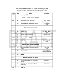
1-s2.0-S1569905614604073-main
412
Pathological validation of adjuvant anti-fibroblast growth factor receptor 3 (FGFR3) treatment
for bladder cancer
Eur Urol Suppl 2014;13;e412 Print!
Print!
Neuzillet Y. 1 , Mertens L. 1 , Shariat S. 2 , Bostrom P. 3 , Mirtti T.4 , Sagalowsky A. 2 , Ashfaq R.2 , Broeks A. 1 , Horenblas S. 1 , Hurst C.5 ,
Tomlinson D.5 , Knowles M. 5 , Bapat B. 3 , Jewett M. 3 , Zlotta A. 3 , Sanders J. 1 , Lotan Y. 2 , Van Der Kwast T.3 , Van Rhijn B. 1
1 Netherlands
Cancer Institute, Antoni Van Leeuwenhoek Hospital, Dept. of Urology & Pathology, Amsterdam, The Netherlands, 2 University
of Texas, Southwestern Medical Center, Dept. of Urology & Pathology, Dallas, United States of America, 3 University Health Network,
Princess Margaret Hospital & Mount Sinai Hospital, University of Toronto, Dept. of Urology & Pathology & Molecular Medicine, Toronto,
Canada, 4 University of Turku, Dept. of Urology & Pathology, Turku, Finland, 5 St James’s University Hospital, Leeds Institute of Molecular
Medicine, Leeds, United Kingdom
INTRODUCTION & OBJECTIVES: Activating Fibroblast Growth Factor Receptor 3 (FGFR3) mutations frequently occur in non-muscle
invasive bladder cancer (BC), whereas these mutations were poorly studied in patients with more advanced disease. In vitro studies
suggest that anti-FGFR3 treatment slows down tumour-growth. We evaluated FGFR3 mutations in superficial and invasive parts of the
same tumour at Trans-urethral resection (TUR) and in bladder and corresponding cancer positive nodes at radical cystectomy (RC) to
investigate possible heterogeneity of FGRF3 mutations.
MATERIAL & METHODS: We identified paired specimens from 61 primary TUR procedures (2 hospitals; 34 T1 and 27 T2 BC) and 494
cN0M0 RC specimens (4 hosptals). pN+ was found in 155/494 (31%) cases. Paired RC and cancer positive node samples were available
for reliable FGFR3 mutation analysis in 117 (75%) patients. DNA was isolated from formalin-fixed paraffin-embedded tissue, then FGFR3
mutation analysis was done using a primer extension-based method (SNaPShot).
RESULTS: We found 13/34 and 8/27 FGFR3 mutations in T1 and T2-TUR specimens, respectively. In T1-TUR, the same 13 mutations
were observed in superficial and invasive parts of the tumour. In T2-TUR, 4/8 mutations were not detected in the invasive part. We found
62/494 FGFR3 mutations in RC specimens, of which 53 were pN0. The presence of a FGFR3 mutation was associated with pN0 (P=0.002)
at RC. We found a FGFR3 mutation in 6 out of 117 analysed positive nodes. The same mutation was detected in the RC specimen. In the
remaining 111 cases, RC and node samples were both wild type (specificity: 100%).
CONCLUSIONS: The FGFR3 mutation was extremely rare in patients with cancer-positive nodes. FGFR3 mutation status is a promising
marker to guide decision making on adjuvant therapy after RC. Based on the results from the TUR-specimens, the deep part of the tumour
also needs to be assessed if neoadjuvant anti-FGFR3 treatment is considered.
© Copyright 2026