
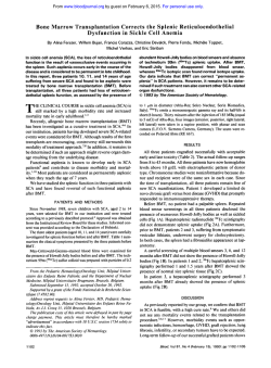
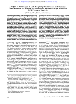
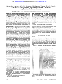
A New Method of “In-Cell Reverse Transcriptase
From www.bloodjournal.org by guest on February 6, 2015. For personal use only.
A New Method of “In-Cell Reverse Transcriptase-Polymerase Chain
Reaction” for the Detection of BCRIABL Transcript in Chronic
Myeloid Leukemia Patients
By Nicoletta Testoni, Giovanni Martinelli, Patrizia Farabegoli, Alfonso Zaccaria, Marilina Amabile,
Donatella Raspadori, Susanna Pelliconi, Eliana Zuffa, Cristina Carboni, and Sante Tura
Methods of detecting minimal residual disease (MRD) in
chronic myeloid leukemia (CML) include chromosome analysis, Southern blotting, polymerase chain reaction (PCR) and
fluorescent in situ hybridization (FISH) techniques. We report a novel method t o detect intracellular messenger RNA
(mRNA) by combining the techniques of reverse transcription (RT) and PCR performed directly inside the cells, without
extraction of the nucleic acid. We applied this method, which
we call ”in-cell RT-PCR”, t o detect hybrid BCRIABL transcript
within single cells. After cellular permeabilization and fixation of single cells in suspension, the neoplastic mRNA was
reverse transcribed into cDNA, and the cDNA was amplified
by PCR with fluorescent primers, specific for bcrlabl. Flow
cytometry was used t o detect cells positive for the amplified
DNA within the cell cytoplasm. After transferring the amplified cells onto slides by cytospin, the positive cells for BCR/
ABL cDNA were observed by fluorescent microscopy. The
technique was capable of detecting l o w abundancy signals
and distinguishing different levels of gene expression. The
amplification products were found in the cells and superna-
tants. The distribution was critically affected by the protease
digestion condition. The specificity of amplification was confirmed by a nested RT-PCR of BCRIABL performed on extracted mRNA from the same sample, and by reamplification
of supernatants. We have used the technique t o study 10
Ph’ CML patients and three normal subjects as controls.
Four patients were 100% Ph+at diagnosis time and RT-PCR’
at cytogenetic and molecular analysis, respectively. In-cell
RT-PCR showed that the residual non-neoplastic cells could
be observed in all cases. In t w o patients undergoing interferon-a (IFN-a) therapy and in four bone-marrow transplanted patients, the in-cell RT-PCR was used t o compare
the level of Ph’ positivity detected by cytogenetic analysis
with the number of cells expressing BCRIABL transcript. In
this manner, we could estimate the MRD. Our preliminary
application of the technique suggests that it is capable of
accurately identifying cells transcribing bcrlabl, and that it
may have significant clinical applications in the detection of
MRD.
0 1996 by The American Society of Hematology.
I
with a sensitivity of 1/10,000. Even though the latter technique is very sensitive, by itself, it is not capable of providing
information on the real size of the residual clone, although
some new modifications may overcome this short-coming.’”
Fluorescent in situ hybridization (FISH) could solve this
problem, but the procedure is difficult, expensive, and requires sophisticated equipment.” The results described here
are based on two methods: one for the detection of human
immunodeficiency virus (H1V)-1 DNA and mRNA both in
cell lines and in the blood of HIV-1 infected patients by insitu amplification of RNA followed by hybridization with
fluorescent probe^,'^,'^ and another for the amplification and
linkage of the rearranged immunoglobulin heavy and light
chain v-genes within single cells using PCR with specific
fluorescent primers.I4
We have devised and tested a new method described
herein, in which the hybrid mRNA is reverse transcribed
(RT) within the cells, and the resulting cDNA is amplified
using the PCR technique with fluorescent primers. We describe this easy, sensitive, and inexpensive technique, which
we call the “in-cell RT-PCR” assay.
N CHRONIC MYELOID leukemia (CML) the reciprocal
translocation between chromosomes 9 and 22, which
leads to the formation of the Philadelphia chromosome (Ph),’
allows the Abelson proto-oncogene (ABL) to move to
breakpoint cluster region (M-BCR) of chromosome 22.2-4
Consequently, a hybrid BCWABL transcription unit is created. The resultant 8.5-kb mRNA is translated into a hybrid
protein of 210 kD (p210) with increased tyrosine kinase
a~tivity.~
The presence of a cytogenetic and a molecular marker in
the leukemic clone allows a correct diagnosis of the disease
and a careful follow-up of the residual leukemic clone after
ablative treatment, such as allogenic or autologous bone marrow transplantation (BMT),~or after successful interferona (IFN-a)treatment.’ Detection of minimal residual disease
(MRD) is possible either by cytogenetics or Southern blotting methods,’ which are able to detect up to 1% to 2% of
residual disease, or by polymerase chain reaction (PCR)9
From the Institute of Haematology “L. e A. Seragnoli”, University of Bologna, Bologna, Italy.
Submitted November 21, I995; accepted December 19, 1995.
Supported by the “Associazione Italiana per la Ricerca sul Cancro (AIRC)”, by the CNR “ACRO” Grant No. 94.OI222.PF39 and
by the CNR Contract No. 952206 target projects.
Address reprint requests to Giovanni Martinelli, MD, Institute of
Haematology “L. e A. Ser&gnoli”, University of Bologna, Policlinico S. Orsola, Via Massarenti 9, 40138, Bologna, Italy.
The publication costs of this article were defrayed in part by page
charge payment. This article must therefore be hereby marked
“advertisement” in accordance with I8 U.S.C. section 1734 solely to
indicate this fact.
0 I996 by The American Society of Hematology.
0006-497I/96/8709-OO02$3.00/0
3822
MATERIALS AND METHODS
Patients. To assess the specificity and the sensitivity of the “incell RT-PCR” assay in cases with different percentages of Ph’ cells
as determined by cytogenetic analysis and of mixed chimerism, we
selected three normal subjects and 10 CML patients. After obtaining
informed consent, 10 CML patients who were cytogenetic Ph’ and
RT-PCR+ for BCRIABL transcript, were admitted to the study (see
Table 1). Four patients were studied at the time of diagnosis and
showed the t(9;22)(34;qll) in all the observed metaphases (patients
I through 4 in Table 1). Two patients (patients 5 and 6) were analyzed after 6 months of IF”-a therapy’ with different degrees of
Ph+ metaphases (96% and 60%. respectively). Two other patients
(patients 7 and 8) were studied for cytogenetic relapse after BMT
Blood, Vol 87, No 9 (May 1). 1996: pp 3822-3827
From www.bloodjournal.org by guest on February 6, 2015. For personal use only.
IN-CELL RT-PCR
3823
Table 1. Cases, Phase of Disease, Karyotypic Molecular Analysis of CML Patients
~~
Patient
No.
Phase of Disease
1
2
3
4
5
6
7
8
9
10
11
12
13
Diagnosis
Diagnosis
Diagnosis
Diagnosis
IFN therapy
IFN therapy
Allo. BMT relapse
Allo. BMT relapse
Allo. BMT relapse
Allo. BMT relapse
Normal
Normal
Normal
No. Metaphases
Ph'Ph- (%Ph')
1510 (100)
2010 (100)
5810 (100)
2510 (100)
48/2 (96)
30120 (60)
31123 (57.4)
5/43 (10.4)
0160 (0)
0120 (0)
Negative
Negative
Negative
RT-PCR
+
+
+
+
+
+
+
+
+
-neg
-neg
-neg
-neg
Type of
Transcript
No. of Cells Detected by in Cell
RT-PCR Cell+/Cell-
b2-a2
b2-a2
b3-a2
b3-a2
b2-a2
b3-a2
b2-a2
b3-a2
b3-a2
b3-a2
neg
neg
neg
29314
19518
596124
7513
486114
116126
35117
471205
11106
01100
01133
01100
01148
Cell+ (%)
98.70
96.10
96.10
96.20
97.20
81.70
67.30
18.60
0.90
0
0
0
0
Comparison of cytogenetic, RT-PCR, and "in-cell" RT-PCR techniques on the samples from 10 different patients and three normal subjects.
Patients 1 to 4 were CML patients at diagnosis, patients 5 and 6 were studied after IFN-a treatment, and patients 7 to 10 previously underwent
BMT. The number of cases, phase of the disease, number of metaphases expressed as ratio between Ph' and Ph- and as percentage of Ph+
cells are reported. The result of each classical RT-PCR, the type of transcript detected at diagnosis in all cases, and the number of cells detected
by in-cell RT-PCR expressed as ratio of positive (+) cellslnegative (-) cells and as percentage (%) of positive cells are also reported.
performed from sibling bone marrow donors. At the time of RTPCR and in-cell RT-PCR, they were on IFN-(Ytherapy and had 31
of 54 and 5 of 45 Ph+ metaphases (57.4% and 10.4%). respectively.
Patients 9 and 10 were also analyzed after allogeneic BMT during
a complete clinical and cytogenetic remission: while patient 10 was
also negative by nested primer PCR analysis and in molecular remission, patient 4 was still nested RT-PCR+ 6 months after BMT (molecular relapse).
Cell preparation. Mononuclear cells from human bone marrow
and peripheral blood samples were separated and isolated by centrifugation on Ficoll-Hypaque. Collected cells were washed twice in
phosphate buffered saline (PBS) pH 7.2 and pelleted at 1,500 rpm
for 10 minutes.
We assessed the best conditions to perform the in-cell RT-PCR
test, avoiding clamping or intracellular amplified release. The cells
were resuspended and fixed in 4% paraformaldehyde solution (50
pL for 400,000cells) for 15 minutes at room temperature. Different
concentrations of paraformaldehyde (2%, 4%, 10%);different fixing
times (2, 5, 10, and 30 minutes) and temperature were analyzed for
assessing the reproducibility of the test (data not shown). Overfixation and proteinase K over-digestion have been reported to give
rise, respectively, to low amounts of amplified products and higher
intracellular amplified product release resulting in a higher final
background fluorescence. The method was then performed on the
samples at the same fixing conditions mentioned above. Fixed cells
were again washed twice in PBS and pelleted at 1,500 rpm for 10
minutes and resuspended in 50% ethanol and a final concentration
of 1 to 2 x lo6/mL. They were stored in I-mL diquots at -20°C
for up to 3 months. An aliquot was pelleted at 1,500 rpm for 5
minutes and washed in PBS, resuspended in 50 pL of proteinase K
(1 pg/mL) (Boehringer Mannheim GmbH, Mannheim, Germany) in
0.1 moVL Tris HCI, 50 mmoVL EDTA buffer (pH 8), and incubated
at 37°C for 10 minutes. Protease digestions of 5, 15, 30, 45, or 60
minutes were tested for optimal signal preservation. After digestion,
cells were pelleted and washed as above, and underwent reverse
transcription.
In-cell reverse transcription. A total of 38 pL of a reaction
mixture containing 100 U of reverse transcriptase, 90 mmoVL KCI,
100 mmollL Tris HCl (pH 8.3), 1.5 mmovL. MgCl, 200 mmoVL
each of deoxyadenosine 5'-triphosphate (dATP), deoxycytidine 5'-
triphosphate (dCTP), deoxyguanosine 5'-triphosphate (dGTP) and
deoxythymidine 5' triphosphate (dlTP), 40 U of RNAsin RNase
inhibitor and 50 pmol of the AZ FAM primer, were added to each
sample. Samples were incubated at 37°C overnight (13 to 17 hours)
and then placed in ice.
In-cell DNA ampli$ca!ion. Cells were resuspended in 160 pL
of a PCR reaction mixture containing 90 mmoVL KCl, 100 mmoY
L Tris HCl (pH 8.3). 1.5 mmoVL MgCI, 200 mmoVL each of dATP,
dCTP, dGTP, and dTTP, 100 pmol of the EA 122 FAM and 50 pmol
of AZ FAM primers and 1 p L (5 U) of Taq polymerase. The PCR
process is covered by United States patents owned by HoffmannLaRoche Inc Perkin Elmer Cetus. Samples were then cycled in an
automated thermocycler (PCR Thermocycler Perkin Elmer 480, a
commercially available machine; Branchburg, NJ), which was programmed for 25 cycles of thermal denaturation (94°C for 30 seconds), annealing (58°C for 30 seconds), and extension (72°C for 2
minutes).
Slide preparation and amplijkation detection. After the in-cell
RT-PCR step, cells were squashed onto glass slides. In each case,
one slide was stained with Giemsa to evaluate cellular morphology
after RT-PCR. Another slide was counterstained with propidium
iodide (PI) in antifade, a coverslip sealed with nail varnish. The
slides were examined and photographed by fluorescence microscopy
with a dual pass filter specific for fluoroisothicyanate (FITC) and
PI. With this filter, the amplified DNA exhibited a yellow green
signal, and the nucleus was stained red. We used 640 ASA film
(Scotch-3M) and double exposure to photograph the representative
cells (magnification x 1,800).
In some cases data acquisition from cytofluorimetric analysis was
performed using Lysis I1 software (Becton Dickinson, San Jose,
CA) by FACScan (Becton Dickinson). A total of 10,OOO cells were
analyzed. To monitor cell integrity and to assess the level of cell
damage, cell loss, and release of intracellular amplified product due
to the squashing process, cell integrity and morphology were
checked after the cytospin. A high proportion of cells (-20%) were
found to be disrupted after the in-cell RT-PCR steps by centrifugation; we counted only those cells in which integrity and clear morphology were maintained. Furthermore, to monitor cell integrity and
to assess the level of considerable cell damage due to the steps of
fixation, digestion, reverse transcription, PCR, and cell transfer to
From www.bloodjournal.org by guest on February 6, 2015. For personal use only.
TESTONI ET AL
3824
A
7
Fig 1. Intracellularlocalizationof amplified BCRlABL cDNA in positive cells by in-cell PCR. Intracellular localization of BCR/ABL by incell PCR in positive cells, counterstained with PI and visualized by
fluorescence microscopy with dual pass filter for FlTC and PI. The
cells show a yellow fluorescent cytoplasm around the red nucleus
(original magnification x 1,800).
the coverslide, we performed the same procedures on samples from
K562 cells, a bcr/abl expressing cell line (data not shown). In conclusion, to assess the possible preferential damage of neoplastic or
normal cells by the in-cell RT-PCR procedures, we performed the
same experiments on different and serial samples of normal plus
K562 cells and selective damage of neoplastic cells was excluded
(data not shown).
Fluorescent primers. The sequences of the fluorescent primers
used in the in-cell RT-PCR were based on the sequences of the bcd
ab1 gene15 and were synthesized using an Applied Biosystem 394
DNA synthesizer. The fluorescent phosphorammidite (FAM Amidite, Applied Biosystem, Milan, Italy) was used to perform the 5'
end-labeling of synthetic oligonucleotides. The sequences used were:
5'-GTTTCAGAAGCTTCTCCCTG-3'for EA1 22FAM and 5'CCA7TTTTGGTITGGGCTTCACACCATTCC-3' for AZFAM.
Cytogenetic analysis. The chromosome analysis of bone marrow
cells was performed on short-term culture without stimulation. Gbanding with Wright stain was employed. Chromosome identification was performed in accordance with International System Cytogenetic Nomenclature.'6
RT-PCR of BCRLABL transcript. RT-PCR analysis on RNA
samples (or supernatants) was performed according to methods previously described." Only the primers employed for reverse transcription and amplification were modified on sequences: we used the
antisense abl-based sequence primer (AZ) 5'-CCATTTTTGGTTTGG GCTTCACACCATTCC-3' and the sense bcr sequence based
primer (EA122) 5'-GTTI%AGAAGC7TCTCCCTG-3'for EA130
and for EA12, respectively."
PCR and cytogenetic analysis. To assess the sensitivity of
the technique, we compared the findings from the in-cell
RT-PCR with those from the cytogenetic and classical RTPCR (Table 1). In all the samples analyzed, the cells showed
variable signal intensity, possibly indicating differential message expression; the intensity of the fluorescence signal in
positive cells permitted their distinction from negative cells
in all instances. We found that clamping of the cells during
the fixation step may lead to underestimation of positive
cells, and we assessed the lowest concentration and time
necessary for fixing the cells, avoiding clamping artefacts
(4% paraformaldehyde solution, 50 pL for 400,000 cells for
15 minutes at room temperature).
RT-PCR is a qualitative assay, while in-cell RT-PCR is a
qualitative-quantitative assay, although we found that the
latter was able to detect the normal cell (bcr/abl negative)
not assessed by RT-PCR in all four patients (patients 1, 2,
3, and 4 in Table l), while cytogenetic analysis was unable
to detect these cells without expression of the BCRIABL
transcript. This suggests that the sensitivity of in-cell RTPCR was higher than cytogenetic analysis, as far as quanti&
cation of nonleukemic residual clones is concerned.
Two patients (patients 5 and 6 in Table 1) were analyzed
after 8 months of IFN-a therapy. In patient 5, the bone
marrow karyotype showed a minor cytogenetic conversion:
48 of 50 (96%) metaphases were Ph'. In-cell RT-PCR, however, showed a higher percentage of positive cells: 486 of
500 (97.2%). In patient 6, who apparently had 60% Ph+
bone marrow cells, we found 116 (81.2%) positive cells by
in-cell RT-PCR. Furthermore, in these two cases in-cell RTPCR confirmed the presence of negative cells for BCRIABL
transcript expression. Did it always agree with RT-PCR of
RNA purified samples.
Four patients had received BMT. Two patients, (patients
7 and 8 in Table l), were studied after karyotypic relapse.
At the time of classical RT-PCR and in-cell RT-PCR analysis, they were receiving LFN-a therapy and were mixed chimeric, with 57.4% and 10.4% Ph+ metaphases, respectively.
The in-cell RT-PCR analysis showed a higher degree of bcr/
ab1 expressing cells in both patients (67% and 18%, respectively). Patients 9 and 10 (Table 1) were analyzed during a
complete clinical and cytogenetic remission after BMT:
while patient 10 was found to be negative by classical RT-
RES ULTS
Fluorescence microscopy after PI counterstaining showed
that the leukemic cells containing the amplified hybrid DNA
from the 10 CML patients exhibited a yellow-green fluorescent cytoplasm around the red nucleus (Fig 1). Only the red
nucleus was visible in the negative cells (Fig 2).
Examination of the Giemsa stained cells showed that approximately 20% had been damaged during the technique.
In the remaining cells, the cellular morphology was partially
preserved, as the cytoplasm is smaller than in normal cells.
Comparative sensitivities of in-cell RT-PCR, classical RT-
-3
-1
Fig 2. Intracellularlocalization of amplified BCR/ABL cDNA in positive cells by in-cell PCR. As in Fig 1, negative cells from a control
subject: only the red nucleus is visible.
From www.bloodjournal.org by guest on February 6, 2015. For personal use only.
3825
IN-CELL RT-PCR
natant, it is possible that they may diffuse nonspecifically
into bystander cells. We examined this possibility by performing in-cell RT-PCR in CML patients (patients 5 to 8 in
Table 1) after a partial degree of cytogenetic Ph+ conversion.
Before this step, IFN-a therapy or an allogeneic BMT had
been performed, with subsequent cytogenetic and molecular
relapse. In all cases the levels of in-cell RT-PCR were higher
than cytogenetic detection of Ph+ cells (ie, 67.3% v 57.4%
in patient 7), although influence of amplification diffusion
could not be excluded. The only case with real molecular
MRD (patient 9 in Table 1) was detected and quantified
by the in-cell RT-PCR assay, suggesting that the methods
involving PCR may be more sensitive than cytogenetic analysis.
DISCUSSION
Fig 3. Flow cytometric fiuorescent profile of amplified BCRlABL
cDNA after in-cell PCR. Flow cytometric fluorescent profile of mRNA
sequences obtained from a normal subject CTRI and from a Phf CML
patient (POS).
PCR analysis (molecular remission about 9 years after
BMT), patient 9 was still RT-PCR' (about 6 months after
BMT). Using in-cell RT-PCR analysis, we confirmed both
the RT-PCR results and quantified the positive result (1 positive cell and 106 negative cells, ie, 0.9%).
The remaining three subjects were healthy bone marrow
donors, in which in-cell RT-PCR was negative in 133, 100,
and 148 observed cells, respectively.
In some cases, flow cytometry was used to analyze mRNA
sequences in the cells' cytoplasm. Figure 3 shows representative histograms for the RNA of the cell population from a
bone marrow donor and a Ph+ CML patient, respectively.
Negative controls for ' 'in-cell RT-PCR. " Because false
positivity is a common pitfall in many PCR procedures, we
performed a number of control experiments to ensure that the
strong bcr/abl expression was not an artefact. The absence of
signal when the reverse transcriptase was omitted in RTPCR of the supernatants, showed that the in-cell RT-PCR
was truly detecting the synthesized cDNA, rather than genomic DNA. Furthermore, the great distances on DNA between
genomic sequences of bcr and ab1 genes involved in translocation were unlike DNA amplification. The absence of a
detectable signal after cDNA synthesis alone indicated the
necessity of message amplification: this observation was
confirmed when Taq polymerase was omitted from the PCR
reaction. Moreover, the absence of a signal in the cells of
the three normal subjects employed as negative controls confirmed the fidelity of the in-cell RT-PCR assay (Table 1).
Signal difJsion and speciJicity of in-cell RT-PCR. Because in-cell RT-PCR products were detectable in the super-
Intracellular nucleic acid reactions have strong potential
for clinical diagnostics, prognostics, monitoring of therapy,
and research. FISH is a highly specific method that gives
molecular information on both the genotype and gene expression of individual cells, without the need to extract DNA
or RNA. However, FISH is relatively insensitive, and the
results are often difficult to interpret. Furthermore, the success of FISH protocols is often dependent on the sequence
being investigated. An obvious way of overcoming FISH
is to amplify the cDNA by PCR or by other nucleic acid
amplification techniques. DNA amplification has traditionally been performed in vitro, and recently, several different
amplification strategies and protocols using in-situ PCR have
been d e s ~ r i b e d . ' ~ . Intracellular
~'~'~
reverse transcription is
possible and is relatively quick and simple.
We also have developed a method for in-cell sequence
transcription and amplification of the neoplastic BCRIABL
transcript in CML patients. Cell preparations on suspension
are fixed and then either gently digested with proteinase
K before use, or cryopreserved for long periods. Alcoholic
fixation has also been reported by other investigators to give
excellent results.20~21
Instead of an alcoholic fixation, we employed a paraformaldahyde fixation method, trying to avoid the reported
cross-linking effect of the fixative on membranes and on
Taq polymerase. We keep permeabilization to the absolute
minimum needed to retain the right balance between getting
reagents into cells, keeping them there, and preventing the
egress of amplified nucleic acids (over-permeabilization necessitates subsequent cell fixation to prevent product leakage). This optimizes morphologic preservation, allows
nested reactions to be performed, and may also inactivate or
exclude nucleic acid-degrading enzymes maintaining cellular membrane compartmentalization.
In the first application of the method,14 the FAM generic
oligonucleotides were substituted by our one-step PCR with
sequence specific primers made fluorescienated during synthesis. This reduces the time needed for in-cell analysis. The
two-round in-situ PCR with outer flanking primers for the
first round in-situ nucleic acid reactions, followed by inner
flanking primers for the second round in-situ nucleic acid
reactions (so called nested in-situ nucleic acid reaction^)^^.^^
From www.bloodjournal.org by guest on February 6, 2015. For personal use only.
TESTONI ET AL
3826
is probably more sensitive and specific, but requires more
time. When the amplification is complete, the cell suspensions are placed on slides and are ready for visualization. In
the future, image analysis2' and the highly optimized microscopic environment (HOME) system" may provide exciting
possibilities for quantifying the histological evaluation, both
for signal intensity and number of positive cells identified.
The method described here was used to detect mRNA
expression from bcr/abl single-copy gene in CML Ph+ cells.
It seems to provide a good tool for a qualitative and quantitative analysis of the MRD after either BMT or IFN-cr treatment of Ph-positive CML patients.
Selective, intracellular amplification of hybrid BCRIABL
transcript, which is not present in normal cells, allows the
detection of single leukemic cells among a normal cell population. This method appears more sensitive than cytogenetics
and Southem blotting, which can detect up to 1% to 2% of
MRD, depending on the number of metaphases scored and
the characteristics of the DNA employed, respectively.' The
sensitivity of the technique may be amplified by the use of
a cytofluorimeter able to count several thousand cells in a
short time. Because the target can be exponentially amplified
with PCR, in-cell PCR enables the researcher to detect and
localize low-abundance DNA and RNA sequences. A very
high sensitivity is expressed by the classic RT-PCR technique, which does not quantify the amount of residual transcript inside the single cell.I3 Quantitative PCR has recently
been described and allows calculation of the relative size of
the abnormal clone," with a rather high degree of confidence.
On the contrary, in-cell RT-PCR, as we have called our
method, is easy, can be done quickly, and does not require
nucleic acid extraction and purification. We used it for detecting the MRD in allogeneic bone marrow transplanted
patients with cytogenetically undetectable MRD. Furthermore, at diagnosis, we were able to detect the presence of
residual nonneoplastic cells and to quantify the number cells
of not expressing the BCRIABL transcript. In our opinion, it
is the only method that can permit this, while avoiding the
necessity of analyzing a large number of metaphases by
karyotypic analysis. In conclusion, in-cell RT-PCR extends
the researcher's ability to detect and localize specific RNAtargets cells and t i s s ~ e * ~ ' ~and
~ ' *in~ the
* ~ future
~ ~ ~ may be
used for in-cell detection of gene level expressions. The
technique appears reproducible, reliable, inexpensive, and
sensitive enough to become a valid alternative to traditional
MRD-detecting methods.
ACKNOWLEDGMENT
The authors are grateful to Robin Cooke for checking through the
English version of the manuscript and to Dr M.J. Embleton, (Patterson Institute, Manchester, UK) and Dr D. DiGiusto (R.P.I. Inc,
Boulder, CO) for helpful discussion on the results.
REFERENCES
1. Rowley JD: A new consistent chromosomal abnormality in
chronic myelogenous leukemia identified by quinacrine fluorescence
and Giemsa staining. Nature 24:290, 1973
2. Heisterkamp N, Stam K, Groffen J, de Klein A, Grosveld
G: Structural organization of the bcr gene and its role in the Phl
translocation. Nature 315:758, 1985
3. Groffen J, Stephenson JR, Heisterkamp N, de Klein A, Bartram
CR, Grosveld G: Philadelphia chromosome breakpoints are located
within a limited region, bcr, on chromosome 22. Cell 36:93, 1984
4. Kurzrock R, Gutterman JU, Talpaz M: The molecular genetics
of Philadelphia chromosome positive leukemias. N Engl J Med
319:990, 1988
5. Ben Neriah Y, Daley GQ, Mes-Masson AM, Witte ON, Baltimore D: The chronic myelogenous leukemia specific p210 protein
is the product of the bcr/abl gene. Science 233:212, 1986
6. Guerrasio A, Martinelli G, Saglio G, Rosso C, Zaccaria A,
Rosti G , Testoni N, Ambrosetti A, Izzi T, Sessarego M, Frassoni F,
Gasparini P, Chiamenti A, Di Bartolomeo P, Pignatti PF: Minimal
residual disease status in transplanted chronic myelogenous leukemia patients: Low incidence of polymerase chain reaction positive
cases among 48 long disease-free subjects who received unmanipulated allogeneic bone marrow transplants. Leukemia 6:507,
1992
7. Tura S, Baccarani M, Zuffa E, Russo D, Fanin R: Interferon
Alfa-2a as compared with conventional chemotherapy for the
treatment of chronic myeloid leukemia. N Engl J Med 330:820,
1994
8. Southem ED: Detection of specific sequences among DNA
fragment separated by gel electrophoresis. J Mol Biol 98:503,
1975
9. Saiki RK, Scharf F, Faloona F, Mullis KB, Horn GT, Erlich
HA, Arnheim N: Enzymatic amplification of betaglobin genomic
sequences and restriction site analysis for diagnosis of sickle cell
anemia. Science 230:1350, 1985
10. Cross NCP, Feng L, Chase A, Bungey J, Hughes T, Goldman
JM: Competitive polymerase chain reaction to estimate the number
of BCR-ABL transcripts in chronic myeloid leukemia patients after
bone marrow transplantation. Blood 82:1929, 1993
11. Tkachuk DC, Westbrook CA, Andreeff M, Donlon TA,
Cleary ML, Suryanarayan K, Home M, Redner A, Gray J, Pinkel
D: Detection of bcr-ab1 fusion in chronic myelogenous leukemia by
in situ hybridization. Science 250:559, 1990
12. Bagasra 0,Hauptman SP, Lischner H W ,Sachs M, Pomerantz
RJ: Detection of human immunodeficiency virus type 1 provirus in
mononuclear cells by in situ polymerase chain reaction. N Engl J
Med 326:1385, 1992
13. Patterson BK, Till M, Otto P, Goolsby C, Furtado MR,
McBride LJ, Wolinsky: Detection of HIV-1 DNA and messenger
RNA in individual cells by PCR-driven in situ hybridization and
flow cytometry. Science 260:976, 1993
14. Embleton MJ, Gorochov G , Jones FT, Winter G: In-cell PCR
from mRNA: Amplifying and linking the rearranged immunoglobulin heavy and light chain V-genes within single cells. Nucleic Acids
Res 20:3831, 1992
15. Ausubel FM, Brent R, Kingstone RE, Moore DD, Seidman
DD, Smith JA, Struhl K: Current Protocols in Molecular Biology.
Greene and Wiley-Interscience, New York, NY, 1990
16. Intemational System for the Human System Nomenclature:
Birth Defect: Original Article Series 21, No. 1, March of Dimes
Birth Defects Foundation, New York, 1985
17. Martinelli G , Sessarego M, Gasparini M, Ambrosetti A, Frassoni F, Chiamenti AM, Pignatti P F An assessment of chimeric
transcript detection in CML patients after bone marrow transplantation. Bone Marrow Transplant 7:21, 1991
18. Nuovo GJ, Gallery F, McConnell P, Becker J, Bloch W:
An improved technique for the in situ detection of DNA after
From www.bloodjournal.org by guest on February 6, 2015. For personal use only.
IN-CELL RT-PCR
polymerase chain reaction amplification. Am J Pathol 139: 1239,
1991
19. Haase AT, Retzel EF, Staskus KA: Amplification and detection of lentoviral DNA inside cells. Proc Natl Acad Sci USA
87:4971, 1990
20. Ray R, Smith M, Sim R, Bruce I, Wakefield A: In situ hybridization detection of short viral amplicon sequences within cultured
cells and body fluids after the in situ polymerase chain reaction. J
Virol Methods 52:247, 1995
3827
21. Ray RA, Cooper PJ, Wakefield AJ: The era of intracellular
nucleic acid technology. Biotechnology 1 3 4 5 , 1995
22. Tecott LH, Barchas JD, Eberwine JH: In situ transcription:
Specific synthesis of complementary DNA in fixed tissue sections.
Science 240:1661, 1988
23. Ried T, Baldini A, Rand TC, Ward DC: Simultaneous visualization of seven different DNA probes by in situ hybridization using
combinatorial fluorescence and digital imaging microscopy. Proc
Natl Acad Sci USA 89:1388, 1992
From www.bloodjournal.org by guest on February 6, 2015. For personal use only.
1996 87: 3822-3827
A new method of "in-cell reverse transcriptase-polymerase chain
reaction" for the detection of BCR/ABL transcript in chronic myeloid
leukemia patients
N Testoni, G Martinelli, P Farabegoli, A Zaccaria, M Amabile, D Raspadori, S Pelliconi, E Zuffa, C
Carboni and S Tura
Updated information and services can be found at:
http://www.bloodjournal.org/content/87/9/3822.full.html
Articles on similar topics can be found in the following Blood collections
Information about reproducing this article in parts or in its entirety may be found online at:
http://www.bloodjournal.org/site/misc/rights.xhtml#repub_requests
Information about ordering reprints may be found online at:
http://www.bloodjournal.org/site/misc/rights.xhtml#reprints
Information about subscriptions and ASH membership may be found online at:
http://www.bloodjournal.org/site/subscriptions/index.xhtml
Blood (print ISSN 0006-4971, online ISSN 1528-0020), is published weekly by the American
Society of Hematology, 2021 L St, NW, Suite 900, Washington DC 20036.
Copyright 2011 by The American Society of Hematology; all rights reserved.


© Copyright 2026