
Molecular Analysis of T-cell Receptor Vp Chains of Human T
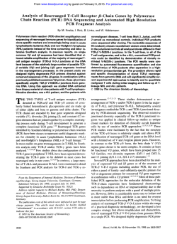
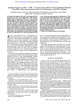
From www.bloodjournal.org by guest on February 6, 2015. For personal use only.
Molecular Analysis of T - c e l l Receptor Vp Chains of Human T - c e l l Chronic
Lymphocytic Leukemia Does Not Show Intraclonal Variability:
Implications for Immunotherapy
By Fabienne Picard, Thierry Martin, Fabienne Legras, Bruno Lioure, and Jean-Louis Pasquali
Human T-cell chronic lymphocytic leukemia (T-cell CLL) is
a heterogeneous disease characterized by a monoclonal
malignant proliferationof T cells in which the T-cell receptors (TCRs) can be, when expressed, considered to be
membrane tumor-specific antigens. Owing to the increasing number of available monoclonal antihuman TCR reagents, it could be of interest to evaluate the feasability of
anti-TCR treatment during T-cell CLL. To test the therapeutic potentiality of anti-TCR monoclonal antibodies, w e first
analyzed the intraclonal variability in two terminally ill patients suffering from TCRaj3-positive cell CLL bearing dif-
ferent immunophenotypes. The cDNA corresponding to
the variable regions of the TCRj3 chains originating from
the malignant T cells were amplified, cloned into M13
phages, and sequenced. The sequence analysis of multiple
independent clones showed no intraclonal variability, with
no evidence for ongoing hypermutation in the Vj3 region
genes. The relevance of these findings with regard to an
anti-VB therapy and the comparison with similar analysis
during 6-cell monoclonal lymphoproliferations are discussed.
0 1993 by The American Society of Hematology.
T
tic avenues depend on the intraclonal homogeneity of the
TCR products bound by the malignant T cells.
Confirming the known low frequency of somatic mutations affecting the VP chains bound by normal T cells,’ we
found no intraclonal diversity in two patients suffering from
TCRap-positive T-cell chronic lymphocytic leukemia (Tcell CLL).
HE MAMMALIAN immune system is able to produce
T-cell receptors (TCRs) that bind a vast array of antigenic peptides. Three mechanisms are crucial to this process
of generating diversity: ( 1) the use of families of rearranging
gene segments (V, D, and J regions),’ ( 2 )the combination of
two polypeptide chains (a, /3, or 6) to form a functional
protein,2and (3) the junctional diversity created by deletion
of nucleotides, template-independent addition of nucleotides, and inverted repeats of n~cleotides.~
The resulting
TCRs are highly characteristic of T-cell clones and can be
considered to be clonal markers during malignant proliferations of these cells.4 In these cases, the membrane TCR,
being a tumor-specific antigen, could theoretically constitute a target for a specific immune therapy. Two different
approaches are possible. On one hand, the clonotypic or
idiotypic approach dictates the use of antibodies directed
mainly to the highly variable complementarity determining
region 3 (CDR3) region of a and/or /3 chains. This first
approach, although highly specific for the malignant clone,
should necessitate the production of individual antibodies.$
On the other hand, an anti-VP treatment will specifically
target I of the 24 VP families, not only the malignant clone,
but without compromising the host immunity.6 This second approach relies on the availability of a limited number
of anti-VP antibodies, and, therefore, seems technically
more suitable. However, both of these theoretical therapeu-
From the Laboratoire d’lmmunopathologie, Centre de Recherche
d’lmmunohhnatologie, H6pital Central, and the Service d’Oncohkmatologie, H6pital de Hautepierre, H6pitaux Universitaires de
Strasbourg, Strasbourg, France.
Submitted January 27, 1993; accepted June 15, 1993.
Supported by the Association pour la Recherche contre le Cancer
and the Ligue Nationale contre le Cancer.
Address reprint requests to Jean-Louis Pasquali, MD, PhD, Laboratoire d’lmmunopathologie et Unite d’lmmunologie Clinique,
H6pital Central, H6pitailx Universitaires de Strasbourg, 6 7091
Strasbourg, France.
The publication costs of this article were defrayed in part by page
charge payment. This article must therefore be hereby marked
“advertisement” in accordance with 18 U.S.C.section 1734 solely to
indicate this fact.
0 I993 by The American Society of Hematology.
0006-49 71/93/820 7-0026$3.00/0
2152
MATERIALS AND METHODS
Patients
Patient no. 1. Patient no. I was a 59-year-old woman diagnosed
3 years ago as having a T-cell CLL on the basis of a white blood cell
count showing 13,500 lymphoid cells/pL. These cells were 90%
CD3+, 75% CD8+, and 86% TCRap-positive. The lymphocytosis
was stable under oral chemotherapy over the last 3 years. She died
of an infectious complication 3 months after the blood was withdrawn.
Patient no. 2. Patient no. 2 was a 60-year-old man diagnosed as
Table 1 . Oligonucleotide Primers Corresponding
to Cj3 and Vg Genes
TTTTGGGTGTGGGAGATCTC
CCAAGCTTTTCTGATGGCTCAA ACAC
GGGAATTCTTCCCTAGGTCTGGAGACCTCTCT
GGGAATTCTTTCAGGCCACAACTATGTTTTG
GGGAATTCGATATGGACCATGAA A ATATGTTC
GGGAATTCACGATCCAGTGTCAAGTCGA
GGGAATTCCTGATCAAAACGAGAGGACAGCA
GGGAATTCTCAGGTGTGATCCA ATTTC
GGGAATTCCAACATATGGGGCACAGGGCAATG
GGGAATTCGAGGTCACAGAGATGGGACA
GGGA ATTCGAACAAAATCTGGGCCATGATACT
GGGAATTCGGTCCTATAAAAGCACATAGTTAT
GGGAATTCTCTCAAACCATGGGCCATGACATGACAAA
GGGAATTCCTGAGATGTCACCAGACTGAG
GGGAATTCGCATGACACTGCAGTGTGCCC
GGGAATTCACCCAAGATACCTCATCACAG
GGGAATTCTCTCAGACTAAGGGTCATGATAGA
GGGAATTCGACCCAATTTCTGGACATGATAAT
GGGA ATTCGAACAGAATTTGAACCACGATGCC
GGGAATTCAGCCCAATGAAAGGACACAGTCAT
GGGAATTCACCCCCGAAAAAGGACATACTTTT
GGGAATTCGAGGGAACATCAAACCCCAACCTA
EcoRl and Hindlll cloning sites are underscored. These primers were
shown to correctly show the main 20 Vp human families (data not
shown).
Blood, Vol 82, No 7 (October 1). 1993: pp 2152-2156
From www.bloodjournal.org by guest on February 6, 2015. For personal use only.
2153
V GENE EXPRESSION IN T-CELL CLL
JP
D
VP
L
CP
+
Family specific
Southern
PT 22
probe
primers
zT9C,rl
OD 128
Ci3E
Fig 1. PCR-based strategy of the amplification of the variable region of the B chains. CBE, external primer; CBI, internal primer.
L MgCI,) containing 10%NP40. RNA were prepared as described.R
ethanol precipitated. and stored in sterile water (20 pL).
having a T-cell CLL on the basis ofa white blood cell count showing
12.600 lymphocytes/pL; 93% of the cells were CD3'. 5% CD4+,9%
CD8'. and 87% [email protected] clinical course was more aggressive and the patient died despite chemotherapy with central
nervous system involvement 6 months after the blood sample was
withdrawn.
Prepururion of TCR fi C h i n cDNA
To analyze the Vg gene segments. 2 pL of total mRNA was reverse transcribed into first-strand cDNA using avian myeloblastosis
virus (AMV) reverse transcriptase (Molecular Genetic Resources.
Tampa. FL) and a C@primer OD I28 (Table I and Fig I ) in a 50 pL
reaction volume.
Sorling e/ Tirmor Cdls
Peripheral blood mononuclear cells were separated from heparinized blood through Ficoll Hypaque (Pharmacia. Uppsala, Sweden)
sedimentation, washed three times in RPMI, and subsequently frozen in 80% RPMI 1640, IO% fetal calf serum (FCS). and IO% dimethylsulfoxide (DMSO) at -80°C until the time of the study.
Cells were thawed quickly 2 hours before sorting. After washing
twice in RPMI. cells were resuspended in I mL of phosphate-buffered saline (PBS)-I% bovine scrum albumin (BSA) and allowed to
react with different monoclonal antibodies for single- and doublelabeling experiments: fluorescein isothiocyanate (FITC)-labeled or
phycoerythrin (PE)-labeled antihuman CD4 (Dako. Glostrup.
Denmark), PE-labeled antLCD8 (Dako). FITC-labeled antLCD3
(Dako), and FITC-labeled anti-pan TCRng (T Cell Diagnostics.
Cambridge. MA). Cells were labeled for 30 minutes at 4°C cells.
washed twice in I mL of PBS-I % RSA. and sorted on an ATC-3.000
cell sorter (ODAM. Wissembourg. France).
Polvmeruse C h i n Reaction (PCR)
Second-strand synthesis and amplification were performed with
PCR. For the first set of PCR. we used O D 128 as a 3' end primer
and 20 5'V@-familyspecific ( V a l through VP20) primers (Table 1
and Fig I ) specific for the V@genes' first framework region at a final
concentration of I pmol/L each. I p L of cDNA corresponding to
approximately IO' cells. and Taq DNA polymerase ( 1 U: PerkinElmer Cetus. Nonvalk, CT) or VENT polymerase ( I U: New England Biolabs, Beverly. MA) in a 50 pL reaction volume. The thermal cycler (Perkins Elmer Thermal) was set for 25 cycles with the
following temperatures: melting. 94°C for I O seconds: annealing.
50°C for I minute and I5 seconds: extension. 72°C for 45 seconds.
with a final extension at 72°C for 5 minutes. One microliter of the
product of the first PCR was mixed with a 3' more internal CP
primer, OH 29 (Table I and Fig I ) and the same 20 5'primers. The
thermal cycler was set for 25 cycles (melting. 94°C for IO seconds:
annealing, 65°C for 45 seconds: extension, 72°C for 45 seconds).
The amplification products were analyzed on ethidium-bromidestained 1% agarose gels for 45 minutes at 100 V.
Total mRNA Isolation
The sorted cells were centrifuged and resuspended in cold lysis
buffer ( I O mmol/L Tris-HCI. pH 7.99: IO mmol/L NaCI, 2 mmol/
MW1
rearranged
Fig 2. Amplification
monoclonal of
V(D)J
the
regions of the @ chains originating from patients no. 1 (A) and 2
(E). Analysis on an ethidium
bromide-stained 1% agarose
gel.
2
3
4
5
6
7
8
9
1
0
11
1213 1415 16 17 1 8 1 9 2 0
"
i
I
From www.bloodjournal.org by guest on February 6, 2015. For personal use only.
PICARD ET AL
2154
52
seq VB 8.1 germline
patient 1
seq 1
patient 1
seq 2
seq 3
patient 1
seq 4
patient 1
seq 5
patient 1
patient 1
seq 6
patient 1
seq 7
patient 1
Seq 8
111
Seq VB 8.1
P1
s1
P1
s2
P1
s3
P1
s4
P1
s5
P1
56
P1
57
P1
58
seq VB 8.1
P1
s1
P1
s2
P1
s3
P1
s4
P1
s5
P1
56
P1
57
P1
S8
Seq VB 8.1
P1
s1
P1
s2
P1
s3
P1
s4
P1
s5
P1
56
P1
s7
P1
S8
s1
P1
P1
P1
P1
P1
P1
P1
P1
s2
s3
s4
s5
56
s7
S8
VB
110
AAGTGACTCTGAGATGTAAACCAATTTCAGGCCACACAACTCCCT~TCTGGTACAGACAG
...........................................................
____-_______________--------------------------------------____________________--------------------------------------____________________--------------------------------------...........................................................
____-___________________________________------------------...........................................................
...........................................................
VB
176
ACCATGATGCGGGGACTGGAGTTGCTCATTTACTTTAACATTTACTTTAACAAC~CGTTCCGATAGATGA~CAGGG
..................................................................
..................................................................
-----------------------------------------------g------------_-----
..................................................................
---------------------------------c--------------------------------
..................................................................
-T---------------------------------------------------------------..................................................................
177
VB
252
ATGCCCGAGGATCGATTCTCAGCTAAGATGCCT~TGCATCATTCTCCACTCTGAAGATCCAGCCC
..................................................................
..................................................................
..................................................................
___-_______---_____-____________________------------------------------------------------------------------g------------------------
----------------------------_--------------c----------------------
-------------------------------------------c----------------------
-------------------------------------------------------G----------
253
VB
-DBTCAGAACCCAGGGACTCAGCTGTGTACTTCTGTGCCAGCAGT
........................................
A
A
........................................
A
........................................
A
........................................
A
A
A
........................................
A
........................................
........................................
........................................
JB
310
CTCCTACAATGAGCAGT
____-________-____
_________________
_________________
_________________
_ _ _ _ _ _ _ _ _ _ _ L _ _ _ _ _
____________-____
-__-----___-----
311
JB
TCTTCGGGCCAGGGACACGGCTCACCGTGCTAG
Fig 3. Nucleotide sequences
of 8 randomly selected V(D)J
regions of the monoclonal Vb
originating from patient no. 1.
The sequences are aligned
under the germline V88.1
(EMBL data base).
-------_-------_-_---------------
_________________________________
-------------------______________
.................................
.................................
--------_-_--------_-------------
.................................
Southern Blot Analysis
N~CI,
~~l~ were denatured in 0.5 mol/^ N ~ O Hplus 1.5 m o ] / ~
neutralized in o.5 mol/L Tris-HC1PIUS .5mol/L NaCI, and transfemed ovemiKhtin 20x ssc (sscis 3 mol/L NaC1,0,3 mol/L Na,
citrate 2 H,O) to nitrocellulose filter (Nitro Plus; MSI, Westboro,
MA). The membrane was put in 2X SSC for 5 minutes and then
treated with UV light. The filter was then prehybndized in 5X SSC,
I X Denhardt's 50% formamide, and 100 pg/mL salmon sperm
DNA at 56°C for 6 hours. The filter was hybridized overnight using
the same solution plus a "P-labeled CP probe, PT 22, more internal
than OH 29 (Table 1 and Fig 1). Filters were washed twice for 20
minutes with 1X SSC and 0.1% sodium dcdecyl sulfate (SDS) at
room temperature. Autoradiography at -80°C followed.
,
DNA ligase, or into pBluescript vector (Stratagene, La Jolla, CA).
Single- and doubIe-stranded DNA sequencing were performed by
the dideoxytermination method following the manufacturer's recommendations, using the T7 sequencing kit (Pharmacia).
-
Cloning and DNA Sequencing
PCR products were digested with EcoRI and Hind111 restriction
enzymes and ligated into M13 mp 19 sequencing vector using T4
RESULTS
Tumor cell PhenotYPk
C,tofluorometfic analys.s showed that patient no. 1*sTcell CLL was the result of the expansion of CD3+, CD4-,
CD8+, TCRap-positive T cells.
Patient no. 2's T-cell CLL was related to the expansion of
CD3+, CD4-, CD8-, TCRorp-positive T cells (data not
shown).
Double-positive (CD3, CD8) cells were sorted from the
peripheral blood lymphocytes (PBL)of patient no. 1. CD3+
cells were sorted from patient no. 2.
From www.bloodjournal.org by guest on February 6, 2015. For personal use only.
21 55
V GENE EXPRESSION IN T-CELL CLL
S e q VB 6 . 9 g e r m l i n e
Patient 2
seq 1
Patient 2
seq 2
Patient 2
Seq 3
patient 2
Seq 4
patient 2
seq 5
49
VB
109
CAGAATGTAACTTTCAGGTGTGATCCAATTTTCTGAACACAATCC~CTTTATTGGTACCGAC
.............................................................
.............................................................
.............................................................
.............................................................
.............................................................
110
S e q VB 6 . 9
P2
s1
P2
S2
s
3
P2
s4
P2
P2
S5
......................................................................
-----------------------------------------G---------------G-----------......................................................................
_________----___________________________---------T
------------G-------------------------------------
180
S e q VB 6 . 9
P2
s1
P2
s2
P2
s3
Fig 4. Nucleotide
squences
S e q VB 6 . 9
of 5 randomly selected V(D)J p2
s1
regions of the monoclonal Vj3 P2
52
s3
originating from patient no. 2. P2
The sequences are aligned
under the germline Vj36.9.
(EMBL data base). The substitution C to T at position 150 introduced an Hindlll site.
p2
p2
P2
S1
52
s3
179
VB
AGACCCTGGGGCAGGGCCCAGAGTTTCTGACTTACTTACTTCCAG~TGAA~TC~CTAG~TCAAGGCT
T
249
VB
G C T C A G T G A T C G G T T C T C T G C A G A G A G G C C T A A G G G A T C T G
......................................................................
......................................................................
---------c------------------------------------------------------------
250
VB
CAGGGGGACTCGGCCATGTATCTCTGTGCCAGCAGCTT
....................................
__--________________________________
....................................
-DBJB
TCCAGGGGTCG
---------------------
311
AGAGACCCAGTACTT
-----------------------------
312
JB
CTGGCCAGGCACGCGGCTCCTGGTGCTCG
.............................
----________--____-__________
PCR Amplijication of the TCRp Variable Regions
The cDNA originating from approximately 103 Sorted T
cells and corresponding to the p chain of rearranged TCR
was synthesized and amplified using the 20 5' Vp familyspecific primers (Vpl through Vp20) in combination with
an internal 3t cpprimer (Fig l). The amplified products
were analyzed in an ethidium bromide agarose gel. The resuits are shown in Fig 2 . The monoclonal expansions of the
Vag family in patient no. 1 and 7 the Vp6 family in patient
no. 2 were further confirmed by the Southern analysis using
a Cp probe (data not shown). It should be mentioned that
the Same amplification procedures starting from 105 sorted
T cells resulted in both patients in multiple vp family
bands.
Ana1ysis of the Intrac1ona1variabi1ity Of the Rearranged
Vp Chains
To analyze the homogeneity of the Vp chains produced
by the malignant T cells, we cloned the monoclonal VP-amplified products into EcoRI-HindIII-digested MI 3 mp19
phages and transformed XL Blue bacterias. We randomly
selected 8 clones for patient no. 1 and 5 clones for patient
no. 2 and sequenced the single-strand DNAs.
Patient no. 1. The 8 rearranged V(D)J regions are reported in Fig 3. These sequences were compared with
known germline sequences (EMBL data base, Heidelberg,
Germany). Two ofthese sequences reached 100%homology
with the Vp8. 1 germline gene.' The CDR3 regions ofthese 8
sequences were identical, proving the monoclonal origin of
the rearranged Vp. Only one nucleotide could be assigned to
a DO segment without N addition nor P nucleotide. Among
these sequences, 6 contained one to two nucleotide changes.
Patient no. 2. The 5. rearranged V(D)J regions are reported in Fig 4 and compared with known germline sequences. One Of our %quences reached loo% homo1ogy
with a Vp6.9 germline gene.gFour sequences contained one
or two nucleotide changes, one ofthese introducing twice an
Hind111 site that shortened the HindIII-digested vp insert.
The CDR3 region was longer than in patient no. 1 and
Probably contained a DP 1.1 gene segment (6 nucleotides)
with " and 3' N additionS.
To make sure that the changes were Taq po1ymerase-induced errors, but not intraclonal mutations (clonally related
mutants), we started two new preparations of PCR using
either the Taq polymerase or the Vent polymerase, which
has less infidelity," in patient no. 2. In these conditions, the
Hind111 digestion ofthe second PCR products did not show
any visible cut on an ethidium bromide-stained gel, suggesting that the changes that introduced the Hind111 site in S4
and S5 in Fig 4 were not reproducible. This was further
confirmed by cloning the Vent polymerase PCR product
into Bluescript and by double-stranded sequencing. The
level of the changes was considerably reduced to only one
nucleotide change, not existing in Fig 4, among the 6 randomly selected VDJ sequences (data not shown).
DISCUSSION
T-cell CLL is a heterogeneous malignant process affecting
T cells bearing different immunophenotypes, mainly CD3+,
CDV, CD4-.'' Some of the T-cell clones rearrange functional TCRs that, when membrane bound, can be consid-
From www.bloodjournal.org by guest on February 6, 2015. For personal use only.
PICARD ET AL
21 56
ered to be tumor-specific antigens. It is thus important to
define the antigenic homogeneity ofthe TCR-positive malignant cells in a late stage of the disease. Despite the fact that
we chose two end-stage T-cell CLL differing by their immunophenotypes, we did not find conclusive intraclonal
variability.
Our analysis showed only a limited number of base
changes that were scattered over the Vg gene segments. We
think that the base substitutions are certainly related to the
infidelity of the Taq polymerase for different reasons. ( I )
Under our experimental conditions (number of cycles of
PCR), the estimated error rate is not different from the experimental error rate (close to 2 for 1,000 nucleotides). (2)
The majority of the substitutions appear only once, consistent with the fact that the errors occur late in amplification.
(3) The vast majority ofthe changes concern A to G or C to
T substitutions ( 13 of 14), which is highly suggestive of Taq
polymerase introducing err0rs.~3'~
The PCR with the VENT
polymerase did not reproduce the observed changes.
Thus, the malignant process that affects the T cells during
this type of leukemia does not induce ongoing mutations,
regardless of the T-cell immunophenotype. This result is
reminiscent of the known stability of the Vp chains during
normal T-cell development7 and encourages anti-Vg-based
immunointerventions during these diseases. On the other
hand, TCR stability of T cells during ontogeny and the absence of variability during the present malignancy discourages any attempt to trace, with Vp analysis, the stage of the
T-lymphoid development in which the malignant conversion took place. This stands in contrast with the results obtained during B-cell malignancies such as B-cell CLL, multiple myeloma, and follicular l y m p h ~ m a , ' ~in
- ' ~which the
differences of the expressed variable region genes of the
monoclonal Ig and the corresponding germline genes, as
well as the intraclonal variability, can serve as markers of
the stage in which the malignancy occurs.
REFERENCES
1. Sin G, Clark SP, Yoshikai Y, Malissen M, Ganag Y, Strauss E,
Mak TW, Hood L: The human T cell antigen receptor is encoded
by variable, diversity and joining gene segments that rearrange to
generate a complete V gene. Cell 37:393, 1984
2. Saito H, Kranz DM, Takagaki Y, Hayday MC, Ersen HN,
Tonegawa S: Complete primary structure of a heterodimeric T-cell
receptor deduced from cDNA sequences. Nature 309:757, 1984
3. Lieber MR: The mechanism of V(D)J recombination: A balance of diversity, specificity and stability. Cell 70:873, 1992
4. Foa R, Pelicci PG, Migane N, Lauria F, Pizzolo G, Flug F,
Knowles DM, Dalla-Favera R: Analysis of T-cell receptor beta
chain (TO) gene rearrangements demonstrates the monoclonal nature of T-cell chronic lymphoproliferative disorders. Blood 67:247,
1986
5. Janson CH, Tehrani MJ, Mellstedt H, Wigzell H: Anti-idiotypic monoclonal antibody to a T-cell chronic lymphatic leukemia.
Cancer Immunol Immunother 28:225, 1989
6. Kanagawa 0: In vivo T-cell tumor therapy with monoclonal
antibody directed to the V@chain of T-cell antigen receptor. J Exp
Med 170:1513, 1989
7. Ikuta K, Ogura T, Shimizu A, Honjo T: Low frequency of
somatic mutation in @-chainvariable region genes of human T-cell
receptors. Proc Natl Acad Sci USA 82:7701, 1985
8. Maniatis T, Fritsch EF, Sambrook J: Molecular Cloning. Cold
Spring Harbor, NY, Cold Spring Harbor Laboratory, 1989
9. Kimura N, Toyonaga B, Yoshikai Y, Du RP, Mak T W Sequences and repertoire of the human T-cell receptor 01 and 0 chain
variable region genes in lymphocytes. Eur J Immunol 17:375, 1987
10. Mattila P, Korpela J, Tenkanen T, Pitkilnen K: Fidelity of
DNA synthesis by the Thermococcus litoralis DNA polymeraseAn extremely heat stable enzyme with proofreading activity. Nucleic Acids Res 19:4967, 1991
11. Bennet JM, Catovsky D, Daniel MT, Flandrin G, Galton
DAG, Gralnick HR, Sultan C: The French-AmencawBritish
(FAB) Cooperative Group: Proposals for the classification of
chronic (mature) B and T lymphoid leukaemias. J Clin Pathol
42367, 1989
12. Ennis PD, Zemmour J, Salter RD, Parham P: Rapid cloning
of HLA-A, B cDNA by using the polymerase chain reaction: Frequency and nature of errors produced in amplification. Proc Natl
Acad Sci USA 87:2833, 1990
13. Meeker TC, Grimaldi JC, O'Rourke R, Loeb Y, Juliusson G,
Einhorn S: Lack of detectable somatic hypermutation in the V region of the IgH chain gene of a human chronic B lymphocytic
leukemia. J Immunol 141:3994, 1988
14. Kipps TJ, Tomhave E, Chen PP, Carson DA: Autoantibodyassociated kappa light chain variable region gene expressed in
chronic lymphocytic leukemia with little or no somatic mutation.
Implications for etiology and immunotherapy. J Exp Med 167:840,
1988
15. Bakkus MHC, Heinnan C, Van Riet I, Van Camp B, Thielemans K: Evidence that multiple myeloma Ig heavy chain VDJ
genes contain somatic mutations but show no intraclonal variation.
Blood 80:2326, 1992
16. Levy R, Levy S, Cleary M, Carrol W, Kon S, Bird J, Sklar J:
Somatic mutation in human B-cell tumors. Immunol Rev 96:43,
1987
From www.bloodjournal.org by guest on February 6, 2015. For personal use only.
1993 82: 2152-2156
Molecular analysis of T-cell receptor V beta chains of human T-cell
chronic lymphocytic leukemia does not show intraclonal variability:
implications for immunotherapy
F Picard, T Martin, F Legras, B Lioure and JL Pasquali
Updated information and services can be found at:
http://www.bloodjournal.org/content/82/7/2152.full.html
Articles on similar topics can be found in the following Blood collections
Information about reproducing this article in parts or in its entirety may be found online at:
http://www.bloodjournal.org/site/misc/rights.xhtml#repub_requests
Information about ordering reprints may be found online at:
http://www.bloodjournal.org/site/misc/rights.xhtml#reprints
Information about subscriptions and ASH membership may be found online at:
http://www.bloodjournal.org/site/subscriptions/index.xhtml
Blood (print ISSN 0006-4971, online ISSN 1528-0020), is published weekly by the American
Society of Hematology, 2021 L St, NW, Suite 900, Washington DC 20036.
Copyright 2011 by The American Society of Hematology; all rights reserved.


© Copyright 2026