
Basic Bladder Neurophysiology
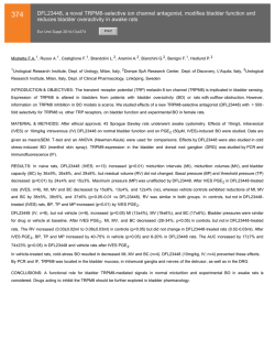
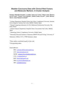
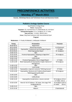
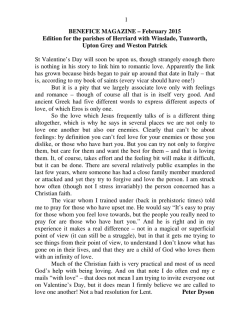
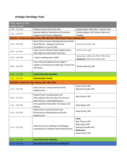
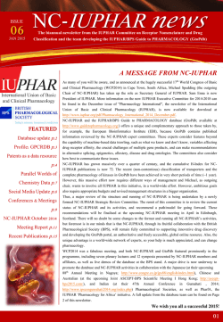
Basic Bladder
N e u ro p h y s i o l o g y
J. Quentin Clemens, MD, MSCI
KEYWORDS
Normal lower urinary tract function requires the
storage of urine at low intravesical pressure,
without leakage. Intermittently, this storage function is interrupted by the voluntary and complete
expulsion of urine. These processes (storage and
voiding) are unique in that they involve the coordination of the peripheral autonomic, peripheral
somatic, and central nervous systems (CNS).
This article provides an overview of the basic principles that are recognized to regulate these functions, although many of these processes remain
poorly understood. Furthermore, much of this
knowledge has been obtained from in vivo animal
models, and the relevance of these findings to
human physiology is not always clear.
low-pressure urine storage across a wide range
of bladder volumes. This process is measured by
calculating the bladder compliance (change in
bladder volume/change in intravesical pressure),
which is expressed in mL/cm H2O. Normal compliance is maintained throughout filling by reorientation of the detrusor smooth muscle fibers and
connective tissue so that they are parallel to the
lumen, thinning of the lamina propria, and flattening of the urothelium.4 Bladder accommodation
is a complex and poorly understood process that
can be altered because of neurologic damage or
changes in the ECM content. For instance, an
increase in type III collagen has been found in
bladders with decreased compliance.5
BLADDER ANATOMY AND BIOMECHANICS
GENERAL NEUROANATOMY
The bladder base refers to the trigone and bladder
neck, whereas the body consists of the supratrigonal portion.1 The bladder wall consists (from the
outside, away from the bladder lumen) of the
serosa, smooth muscle and extracellular matrix
(ECM) (in approximately 50-50 distribution), the
lamina propria, and the urothelium. Like other
smooth muscles, the detrusor muscle is oriented
in a seemingly random fashion, is not attached to
tendon or bone, is able to maintain steady tension
over a wide range of muscle lengths, and has
a slower contraction velocity than the skeletal
muscle.2,3 The detrusor muscle uses the ECM as
a scaffold to generate tension, which produces
a bladder contraction.
Bladder accommodation refers to the changes
that occur during bladder filling to permit
The CNS consists of the brain and spinal cord, and
the peripheral nervous system (PNS) consists
of the sensory (afferent) and motor (efferent)
neurons that communicate with the CNS. The
PNS is divided into the somatic and the autonomic
nervous systems. The somatic nervous system is
responsible for regulating structures that are under
conscious control (such as the striated external
urethral sphincter and the levator muscles in the
pelvic floor). The autonomic nervous system
controls visceral and endocrine functions,
including bladder contraction and relaxation. The
autonomic nervous system is subdivided into the
parasympathetic and sympathetic nervous
systems. Sympathetic and parasympathetic are
anatomic terms that indicate the location from
where the nerve fibers emanate. Parasympathetic
Disclosures: Merck, stock ownership; Pfizer, consultant, investigator; Lilly, consultant; Afferent Pharmaceuticals, consultant; Medtronic, proctor.
Division of Neurourology and Pelvic Reconstructive Surgery, Department of Urology, University of Michigan
Medical Center, 1500 East Medical Center Drive, Taubman Center 3875, Ann Arbor, MI 48109-5330, USA
E-mail address: [email protected]
Urol Clin N Am 37 (2010) 487–494
doi:10.1016/j.ucl.2010.06.006
0094-0143/10/$ e see front matter Ó 2010 Elsevier Inc. All rights reserved.
urologic.theclinics.com
Neuroanatomy Physiology Lower urinary tract
Pharmacology
488
Clemens
fibers emerge from the cranial and sacral
segments of the spinal cord, whereas sympathetic
fibers emerge from the thoracic and lumbar
segments of the spinal cord.
CROSS-SECTIONAL ANATOMY OF THE
SPINAL CORD
Peripheral sensory information is carried via
afferent nerves fibers, which enter the dorsal
(posterior) aspect of the spinal cord and then travel
upward to the central processing centers in the
CNS (Fig. 1). Afferent cell bodies are located in
the dorsal root ganglia. The white matter of the
spinal cord contains bundles of myelin-coated
neurons, whereas the gray matter contains the
cell bodies of interneurons and efferent motor
neurons. Within the gray matter, nerve cell bodies
are generally organized into functional clusters
called nuclei (such as Onuf nucleus). Axons within
the white matter are functionally grouped into
tracts. Efferent motor axons exit from the ventral
root of the spinal cord.
RELEVANT NEUROANATOMY: PNS AND
SPINAL CORD
Preganglionic parasympathetic efferent nerves
exit from the sacral segment of the spinal cord at
S2 through S4. The axons travel a long distance
within the pelvic nerve to the ganglia (pelvic
plexus) that are located immediately adjacent to
the end organ (bladder) (Fig. 2). These fibers
modulate bladder contractions. The primary
neurotransmitter for both pre- and postganglionic
parasympathetic fibers is acetylcholine (ACh).6
DORSAL
DRG
Preganglionic sympathetic efferent nerves exit
from the thoracolumbar segment of the spinal
cord at T10 through L2. The ganglia for these
nerves have variable locations: some are next to
the vertebrae (paraganglia), some are between
the vertebrae and the target organ (preganglia),
and some are located with the end organ (peripheral ganglia).7 Sympathetic efferent nerves to the
lower urinary tract are located within the hypogastric nerve. The sympathetic efferent nerves modulate contractions of the urethral smooth muscle
and bladder outlet and inhibit parasympathetic
activity that promotes bladder contraction. The
primary neurotransmitter for postganglionic
sympathetic fibers is norepinephrine, but the
primary neurotransmitter for preganglionic sympathetic fibers is ACh.
Preganglionic somatic efferent nerves exit from
the sacral segment of the spinal cord at S2 through
S4. Nerve bodies for these nerves are located in
the Onuf nucleus, along the lateral border of the
ventral gray matter in the sacral region of the spinal
cord. The nerve fibers travel within the pudendal
nerve to the external urethral sphincter, where
they modulate striated (voluntary) sphincter
contraction.8
In humans and animals, afferent nerves have
been identified in the detrusor muscle and the suburothelium.9,10 The suburothelial afferent nerve
fibers form a plexus that lies immediately beneath
the urothelial lining, with some nerve terminals extending into the urothelium itself. This plexus is
more prominent in the trigone and bladder neck
and relatively sparse in the bladder dome. Afferent
nerve fibers from the lower urinary tract travel
within the pelvic, hypogastric, and pudendal
DRG
Sensory
(Afferent) In
GRAY
MATTER
WHITE
MATTER
VENTRAL
Motor
(Efferent) Out
Fig. 1. Cross section of the sacral
segment of the spinal cord.
Sensory nerve fibers enter the
dorsal spinal cord. Cell bodies of
these sensory nerves are located
in the dorsal root ganglia (DRG).
The white matter contains
bundles of neurons, whereas the
gray matter contains the cell
bodies of interneurons and
efferent motor neurons. Motor
nerve fibers exit the ventral spinal
cord in the ventral root.
Basic Bladder Neurophysiology
Fig. 2. Parasympathetic efferent nerves exit from the sacral region of the spinal cord at S2 through S4 and travel
within the pelvic nerve. Parasympathetic activity promotes bladder emptying by causing contraction of the detrusor and relaxation of the bladder outlet. Sympathetic efferent nerves exit from the thoracolumbar segment
of the spinal cord at T10 through L2 and travel within the hypogastric nerve. These nerves modulate contractions
of the urethral smooth muscle and bladder outlet and inhibit parasympathetic activity that promotes bladder
contraction. Somatic efferent nerves exit from the sacral segment of the spinal cord at S2 through S4 and travel
within the pudendal nerve to the external urethral sphincter, where they modulate striated (voluntary) sphincter
contraction. Afferent nerve fibers from the lower urinary tract travel within the pelvic, hypogastric, and pudendal
nerves. Therefore, these peripheral nerves carry bidirectional (afferent and efferent) information between the
end organs and the spinal cord.
nerves.11,12 Therefore, these peripheral nerves
carry bidirectional (afferent and efferent) information between the end organs and the spinal cord.
The sensory fibers enter the spinal cord via the
dorsal root, and the nerve cells bodies are located
within the dorsal root ganglia. Afferent nerves
release
numerous
neurotransmitters
(eg,
substance P, neurokinins, calcitonin geneerelated
polypeptide, vasoactive intestinal polypeptide).13
Most sensory innervation of the bladder and
urethra originates in the thoracolumbar region of
the spinal cord and travels within the pelvic
nerve.12 Within the pelvic nerve, 2 types of bladder
afferent nerves have been identified, myelinated
Ad fibers and unmyelinated C fibers. The Ad fibers
respond to normal bladder distention and are
thought to be the primary functional afferent
nerves during normal micturition.12 Conversely,
the C fibers respond to chemical irritation (nociception) or to cold, and most of these fibers are
inactive during normal micturition.14,15 However,
during certain pathologic states (eg, inflammation,
suprasacral spinal cord injury), these “silent” C
fibers appear to activate, become mechanosensitive, and modulate pathologic voiding reflexes.16
RELEVANT NEUROANATOMY: BRAINSTEM
AND ABOVE
Conclusive experimental evidence using brainlesioning techniques, electric stimulation, and
axonal tracing studies indicate that an area of the
pons (the pontine micturition center [PMC] or Barrington nucleus) mediates the normal micturition
reflex by coordinating the activity of the detrusor
and urethral sphincter muscles.17e20 Therefore,
spinal cord lesions below this level often result in
discoordination between the detrusor and urethral
sphincter (detrusor-sphincter dyssynergia). The
PMC receives input from multiple higher brain
centers, including the basal ganglia, periaqueductal gray, thalamus, and hypothalamus.21 Brain
imaging studies in healthy volunteers suggest
a model of supraspinal bladder control, in which
afferent signals from the lower urinary tract are
received in the periaqueductal gray and relayed
489
490
Clemens
via the thalamus to the insula (which makes the
sensations accessible to conscious awareness).22
According to this model, the cortex (via the anterior cingulate gyrus) monitors and controls micturition reflexes and also makes voluntary voiding
decisions (via the prefrontal cortex).
NEURAL CONTROL OF THE LOWER URINARY
TRACT
During the storage phase of micturition, bladder
filling activates myelinated Ad afferent nerve fibers
in the bladder wall. This afferent input results in
stimulation of sympathetic efferent activity (via
the hypogastric nerve), leading to contraction of
smooth muscles in the bladder base and proximal
urethra (via activation of a-adrenergic receptors)
and relaxation of the detrusor (via activation of
b-adrenergic receptors in the bladder body).
Somatic efferent activity (via the pudendal nerve)
also increases, resulting in increased tone of the
striated external urethral sphincter. These
responses occur by spinal reflex pathways organized in the lumbosacral region of the spinal cord
and represent guarding reflexes, which promote
continence.23e25 The parasympathetic system is
largely inactive during urine storage, which may
partly be because of the sympathetic inhibition of
parasympathetic transmission at the ganglia level.
The voiding phase of micturition is initiated
voluntarily by signals from the cerebral cortex.
The initial event is relaxation of the striated
external urethral sphincter, caused by inhibition
of somatic efferent activity. There is inhibition of
sympathetic efferent activity, with concomitant
activation of parasympathetic outflow to the
bladder and urethra.26 Bladder contraction is
mediated via muscarinic receptors in the bladder
body, and urethral smooth muscle relaxation is
mediated through the release of nitric oxide
(NO).27 Maintenance of the voiding reflex is
a complicated phenomenon that is mediated via
communication between the spinal cord and the
pons (spinobulbospinal reflex), with the involvement of midbrain structures such as the periaqueductal gray.21
PHARMACOLOGY OF THE LOWER URINARY
TRACT
Cholinergic Mechanisms
In certain neurons of the CNS and PNS, the neurotransmitter ACh is synthesized from the essential
nutrient choline by the enzyme choline acetyltransferase. In response to various stimuli, ACh is
released into the synaptic cleft, where it either
binds to cholinergic receptors or is broken down
by acetylcholinesterase. ACh is released from
postganglionic parasympathetic neurons, preganglionic autonomic neurons (sympathetic and parasympathetic), and somatic neurons. Two main
types of cholinergic receptors exist: nicotinic and
muscarinic. Nicotinic receptors (which are responsive to ACh and nicotine) are ligand-gated ion
channels and are found on the skeletal muscle
motor end plates, on the autonomic ganglia, and
in the CNS. The nicotinic receptors seem to have
a limited role in the control of micturition. Muscarinic receptors (which are responsive to ACh and
muscarine) are G proteinecoupled receptors that
activate ion channels via second-messenger
cascades. These receptors are found on all autonomic effector cells (eg, bladder, sweat glands,
bowel) and in the CNS. Five subtypes of muscarinic receptors (M1 through M5) have been identified, and the M2 and M3 subtypes predominate in
the bladder.28 Although M2 receptors are the
most plentiful in the detrusor (70% M2 vs 30%
M3 receptors), in vitro studies indicate that the
M3 receptors are responsible for detrusor muscle
contraction.29e32 The functions of the detrusor
M2 receptors are less clear. Muscarinic receptors
are also found on presynaptic nerve terminals in
the bladder and elsewhere, where they may play
a regulatory role via feedback inhibition.33,34
Excitation-contraction Coupling
Excitation-contraction coupling refers to the
process whereby binding of a ligand to a receptor
causes force generation (muscle contraction)
(Fig. 3).35 In the detrusor smooth muscle, the
ligand is ACh and the receptor is the M3 receptor.
At rest, there is a very low concentration of free
calcium ions (Ca21) in the smooth muscle cell.
Binding of ACh to the M3 receptor triggers a G proteinemediated process, which causes Ca21
release from the sarcoplasmic reticulum as well
as Ca21 influx from transmembrane ion channels.
The free Ca21 binds to calmodulin, and the
Ca21-calmodulin complex then activates the
enzyme myosin light chain kinase, which phosphorylates the light chain of the contractile protein,
myosin. This phosphorylation causes the myosin
light chain to change shape and interact with actin,
causing force generation.35 Alongside this
process, alternate methods are at work to facilitate
subsequent muscle relaxation. The Ca21-calmodulin complex activates transmembrane Ca21
pumps to remove free Ca21 from the cell, the
ligand-receptor complex is degraded, and excess
extracellular ACh is degraded by acetylcholinesterase. This enzyme is abundant in the synaptic
cleft, and its role in rapidly clearing free ACh
Basic Bladder Neurophysiology
Nerve Terminal
ACh
ACh
ACh
ACh
ACh
ACh
ACh
Ca2+
ACh
G Protein M3
ACh
PLC
IP3
Ca2+
CONTRACTION
SR
Actin
Ca2+
MLCK
Ca2+
My osin
Calmodulin
Fig. 3. Excitation-contraction coupling. Ach is released from postganglionic parasympathetic nerve terminals into
the synaptic cleft, where it binds to the M3 receptor. This ligand-receptor complex triggers a cascade of signaling
events that involves a G protein, phospholipase C (PLC), and inositol triphosphate (IP3). This signaling cascade
leads to the release of free calcium (Ca21) into the cytosol by the sarcoplasmic reticulum (SR) as well as a direct
influx of Ca21 via transmembrane ion channels. The free Ca21 binds with calmodulin, and the resulting
Ca21-calmodulin complex activates the enzyme myosin light chain kinase (MLCK). The MLCK causes changes in the
structure of the myosin molecule, which allows the myosin to interact with actin, leading to a muscle contraction.
from the synapse is essential for proper muscle
function.
where they cause detrusor smooth muscle
relaxation.38
Adrenergic Mechanisms
Receptor Distribution in the Lower Urinary
Tract
Adrenergic receptors are G proteinecoupled
receptors that bind catecholamines (most
commonly norepinephrine and epinephrine) that
are released from postganglionic sympathetic
neurons. Stimulation of a-adrenergic receptors
causes vasoconstriction and smooth muscle
contraction, whereas stimulation of b-adrenergic
receptors causes increased myocardial contractility and smooth muscle relaxation. There exists
multiple subtypes of adrenergic receptors.
a-Adrenergic receptors were initially divided into
a1 (postsynaptic) and a2 (presynaptic) receptors,
but a2 receptors were subsequently also found in
postsynaptic locations. There are 3 subtypes of
a1 receptors (a1A, a1B, and a1D). The a1A subtype
is the primary subtype in the prostate and urethra,
where it mediates contraction of the bladder
outlet.36,37 The role of a2 receptors in the lower
urinary tract is not clear. There are 3 subtypes of
b-adrenergic receptors (b1, b2, and b3). b1 Receptors are located in the heart, whereas b2 and b3
receptors are located in the lower urinary tract,
The actions of the various neurotransmitters on the
lower urinary tract are largely a function of receptor
location.39,40 There is a higher density of muscarinic receptors in the bladder body than in the
base, and therefore activation of these receptors
results in detrusor contraction. Adrenergic receptors are distributed such that a-adrenergic receptors (muscle contraction) predominate in the
bladder base and urethra, whereas b-adrenergic
receptors (muscle relaxation) predominate in the
bladder body. Therefore, activation of the sympathetic nervous system promotes urine storage
(relaxation of the bladder body and contraction
of the outlet).
Nonadrenergic Noncholinergic Mechanisms
of Bladder Excitation
In various mammalian species, complete blockage
of cholinergic receptors with atropine does not
pre-vent detrusor muscle contraction,27 indicating
that nonadrenergic noncholinergic (NANC)
491
492
Clemens
mechanisms exist, which at least partially mediate
bladder contractions. In these nonhuman
mammals, there is good evidence that the transmitter responsible for the NANC component of
detrusor contraction is adenosine triphosphate
(ATP) acting on purinergic receptors.41,42
However, the component of NANC activation in
the normal human detrusor seems to be small. It
is possible, however, that NANC mechanisms
may become more predominant in certain pathologic states (eg, bladder outlet obstruction, overactive bladder, neurogenic bladder, interstitial
cystitis).43,44
Nitric Oxide
Stimulation of cholinergic receptors in the urethra
causes contraction, rather than relaxation, of the
urethral smooth muscle.45 Therefore, parasympathetic pathways that promote voiding must elicit
urethral relaxation via noncholinergic mechanisms. Experimental studies indicate that NO,
released from postganglionic parasympathetic
nerves, is the major inhibitory neurotransmitter
that causes the urethral smooth muscle relaxation
during voiding.46,47 The mechanism of relaxation is
similar to that observed in corporal smooth muscle
(increased production of intracellular cyclic guanosine monophosphate and so forth). The functional
role of NO in the detrusor has not been
established.
Vanilloids
Vanilloids are a class of compounds denoted by
the presence of a vanillyl functional group. Two vanilloids (capsaicin and resiniferatoxin [RTX]) are
known to bind to receptors on C-fiber afferent
neurons. This binding causes desensitization, so
that the neurons no longer respond to stimuli. As
discussed previously, C-fiber afferent neurons
are considered to mediate pathologic bladder
reflexes. Therefore, intravesical treatment with
capsaicin and RTX has been examined as a potential therapy for conditions such as neurogenic detrusor overactivity and interstitial cystitis.48,49
Additional Neurotransmitters and Receptors
Multiple additional neurotransmitters and their
associated receptors have been found to be
synthesized, stored, and released in the human
lower urinary tract, including tachykinins (eg,
substance P, neurokinins), prostanoids (prostaglandins and thromboxanes), and endothelins.13,35
However, the functional roles of most of these
agents have not been established, and animal
experiments suggest that these roles may vary
across species.
SENSORY ROLE OF THE UROTHELIUM
The urothelium has traditionally been viewed as
a passive barrier that prevents passage of urinary
toxins into the underlying bladder interstitium.
However, there is increasing evidence that the urothelium also has sensory and signaling properties
that allow it communicate with nearby nerve and
muscle tissue, suggesting that it is actively
involved in the storage and voiding phases of
micturition.50,51 For instance, the urothelium has
been shown to release chemical mediators (ATP,
ACh, and NO) and to express numerous receptors
(eg, muscarinic, adrenergic, purinergic, and tachykinin receptors).35,52 The mechanisms responsible
for urothelial ATP release have been the subject of
considerable study because intravesical administration of ATP induces detrusor overactivity and
mice deficient in the ATP receptor P2X3 exhibit
decreased voiding frequency and increased
bladder capacity.53,54 Therefore, further understanding of these findings may provide additional
therapeutic targets for lower urinary tract
disorders.
REFERENCES
1. Elbadawi A, Schenk EA. Dual innervation of the
mammalian urinary bladder, a histochemical study
of the distributions of cholinergic and adrenergic
nerves. Am J Anat 1966;119:405e27.
2. Uvelius B. Length-tension relations of in vitro urinary
bladder smooth muscle strips. J Pharmacol Toxicol
Methods 2001;45:87e90.
3. Uvelius B, Gabella G. Relation between cell length
and force production in urinary bladder smooth
muscle. Acta Physiol Scand 1980;110:357e65.
4. Chang SL, Chung JS, Yeung MK, et al. Roles of the
lamina propria and the detrusor in tension transfer
during bladder filling. Scand J Urol Nephrol Suppl
1999;201:38e45.
5. Landau EH, Jayanthi VR, Churchill BM, et al. Loss of
elasticity in dysfunctional bladders: urodynamic and
histochemical correlation. J Urol 1994;152:702e5.
6. de Groat WC, Booth AM. Synaptic transmission in
pelvic ganglia. In: Maggi CA, editor. Nervous control
of the urogenital system: the autonomic nervous
system, vol. 3. London: Harwood Academic; 1993.
p. 291e347.
7. Kihara K, de Groat WC. Sympathetic efferent pathways projecting to the bladder neck and proximal
urethra in the rat. J Auton Nerv Syst 1997;62:
134e42.
8. Thor KB, Morgan C, Nadelhaft I, et al. Organization
of afferent and efferent pathways in the pudendal
nerve of the female cat. J Comp Neurol 1989;288:
263e79.
Basic Bladder Neurophysiology
9. Dixon JS, Gilpin CJ. Presumptive sensory axons of
the human urinary bladder: a fine structural study.
J Anat 1987;151:199e207.
10. Gabella G, Davis C. Distribution of afferent axons in
the bladder of rats. J Neurocytol 1998;27:141e55.
11. Yoshimura N, de Groat WC. Neural control of the
lower urinary tract. Int J Urol 1997;4:111e25.
12. Janig W, Morrison JFB. Functional properties of
spinal visceral afferents supplying abdominal and
pelvic organs, with special emphasis on visceral nociception. Prog Brain Res 1986;67:87e114.
13. Andersson KE. Bladder activation: afferent mechanisms. Urology 2002;59(Suppl 5A):43e50.
14. Habler HJ, Janig W, Koltzenburg M. Activation of
unmyelinated afferent fibres by mechanical stimuli
and inflammation of the urinary bladder in the cat.
J Physiol 1990;425:545e62.
15. Fall M, Lindstrom S, Mazieres L. A bladder-tobladder cooling reflex in the cat. J Physiol 1990;
427:281e300.
16. de Groat WC, Kawatani M, Hisamitsu T, et al. Mechanisms underlying the recovery of urinary bladder
function following spinal cord injury. J Auton Nerv
Syst 1990;30:S71e7.
17. Barrington FJ. The effects of lesions of the hind and
midbrain on micturition in the cat. Q J Exp Physiol
1925;15:81e102.
18. Mallory BS, Roppolo JR, de Groat WC. Pharmacologic modulation of the pontine micturition center.
Brain Res 1991;546:310e20.
19. Holstege G, Griffiths D, De Wall H, et al. Anatomical
and physiological observations on supraspinal
control of the bladder and urethral sphincter
muscles in the cat. J Comp Neurol 1986;250:
449e61.
20. Nadelhaft I, Vera PL, Card JP, et al. Central nervous
system neurons labeled following the injection of
pseudorabies virus into the rat urinary bladder. Neurosci Lett 1992;143:271e4.
21. Fowler CJ, Griffiths G, de Groat WC. The neural
control of micturition. Nat Rev Neurosci 2008;9:
453e66.
22. Griffiths D, Tadic SD. Bladder control, urgency, and
urge incontinence: evidence from functional brain
imaging. Neurourol Urodyn 2008;27:466e74.
23. de Groat WC, Booth AM, Yoshimura N. Neurophysiology of micturition and its modification in animal
models of human disease. In: Maggi CA, editor.
Nervous control of the urogenital system: the autonomic nervous system, vol. 3. London: Harwood
Acadmeic Publishers; 1993. p. 227.
24. de Groat WC, Vizzard MA, Araki I, et al. Spinal interneurons and preganglionic neurons in sacral autonomic reflex pathways. Prog Brain Res 1996;107:
97e111.
25. Park JM, Bloom DA, McGuire EJ. The guarding
reflex revisited. Br J Urol 1997;80:940e5.
26. de Groat WC. Organization of the sacral parasympathetic reflex pathways to the urinary bladder and
large intestine. J Auton Nerv Syst 1981;3:135e60.
27. Andersson KE. Pharmacology of lower urinary tract
smooth muscles and penile erectile tissues. Pharmacol Rev 1993;45:253e308.
28. Bschleipfer T, Schukowski K, Weidner W, et al.
Expression and distribution of cholinergic receptors
in the human urothelium. Life Sci 2007;80:2303e7.
29. Harriss DR, Marsh KA, Birmingham AT, et al. Expression of muscarinic M3 receptors coupled to inositol
phospholipid hydrolysis in human detrusor smooth
muscle cells. J Urol 1995;154:1241e5.
30. Yamaguchi O, Shishido K, Tamura K, et al. Evaluation
of mRNAs encoding muscarinic receptor subtypes in
human detrusor muscle. J Urol 1996;156:1208e13.
31. Hegde SS, Choppin A, Bonhaus D, et al. Functional
role of M2 and M3 muscarinic receptors in the
urinary bladder of rats in vitro and in vivo. Br J Pharmacol 1997;120:1409e18.
32. Lai FM, Cobuzzi A, Spinelli W. Characterization of
muscarinic receptors mediating the contraction of
the urinary detrusor muscle in cynomolgus monkeys
and guinea pigs. Life Sci 1998;62:1179e86.
33. D’Agostino G, Bolognesi ML, Lucchelli A, et al. Prejunctional muscarinic inhibitory control of acetylcholine release in the human isolated detrusor:
involvement of the M4 receptor subtype. Br J Pharmacol 2000;129:493e500.
34. Somogyi GT, de Groat WC. Evidence for inhibitory
nicotinic and facilitatory muscarinic receptors in
cholinergic nerve terminals of the rat urinary
bladder. J Auton Nerv Syst 1992;37:89e97.
35. Andersson KE, Arner A. Urinary bladder contraction
and relaxation: physiology and pathophysiology.
Physiol Rev 2004;84:935e86.
36. Furuya S, Kumamoto Y, Yokoyama E, et al. Alphaadrenergic activity and urethral pressure in prostatic
zone in benign prostatic hypertrophy. J Urol 1982;
128:836e9.
37. Nasu K, Moriyama N, Fukasawa R, et al. Quantification and distribution of alpha-1 adrenoreceptor
subtype mRNAs in human proximal urethra. Br J
Pharmacol 1998;123:1289e93.
38. Yamaguchi O. Beta 3-adrenoceptors in human detrusor muscle. Urology 2002;59(Suppl 1):25e9.
39. Johns A. Alpha- and beta-adrenergic and muscarinic cholinergic binding sites in the bladder and
urethra of the rabbit. Can J Physiol Pharmacol
1983;61:61e6.
40. Levin RM, Ruggieri MR, Wein AJ. Identification of
receptor subtypes in the rabbit and human urinary
bladder by selective radio-ligand binding. J Urol
1988;139:844e8.
41. Lee HY, Bardini M, Burnstock G. Distribution of P2X
receptors in the urinary bladder and the ureter of the
rat. J Urol 2000;163:2002e7.
493
494
Clemens
42. O’Reilly BA, Kosaka AH, Chang TK, et al.
A quantitative analysis of purinoceptor expression
in human fetal and adult bladders. J Urol 2001;
165:1730e4.
43. Bayliss M, Wu C, Newgreen D, et al. A quantitative
study of atropine-resistant contractile responses in
human detrusor smooth muscle from stable, unstable
and obstructed bladders. J Urol 1999;162:1833e9.
44. Palea S, Artibani W, Ostardo E, et al. Evidence for
purinergic neurotransmission in human urinary
bladder affected by interstitial cystitis. J Urol 1993;
150:2007e12.
45. Ek A. Innervation and receptor functions of the
human urethra. Scand J Urol Nephrol Suppl 1977;
45:1e50.
46. Andersson KE, Garcia Pascual A, Persson K, et al.
Electrically-induced, nerve-mediated relaxation of
rabbit urethra involves nitric oxide. J Urol 1992;
147:253e9.
47. Bennett BC, Kruse MN, Roppolo JR, et al. Neural
control of urethral outlet activity in vivo: role of nitric
oxide. J Urol 1995;153:2004e9.
48. Fowler CJ, Beck R, Gerrard S, et al. Intravesical capsaicin for treatment of detrusor hyperreflexia. J Neurol Neurosurg Psychiatry 1994;57:
169e73.
49. Payne CK, Mosbaugh PG, Forrest JB, et al. Intravesical resiniferatoxin for the treatment of interstitial
cystitis: a randomized, double-blind, placebo
controlled trial. J Urol 2005;173:1590e4.
50. Apodaca G. The uroepithelium: not just a passive
barrier. Traffic 2004;5:117e28.
51. Ferguson DR. Urothelial function. BJU Int 1999;84:
235e42.
52. Birder LA, de Groat WC. Mechanisms of disease
involvement of the urothelium in bladder dysfunction. Nat Clin Pract Urol 2007;4:46e54.
53. Pandita RK, Andersson KE. Intravesical ATP stimulates the micturition reflex in awake, freely moving
rats. J Urol 2002;168:1230e4.
54. Cockayne DA, Hamilton SG, Zhu QM, et al. Urinary
bladder hyporeflexia and reduced pain-related
behaviour in P2X3-deficient mice. Nature 2000;
407:1011e5.
© Copyright 2026