
Progression and Survival Studies in Early Chronic
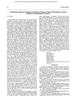
From www.bloodjournal.org by guest on February 6, 2015. For personal use only.
Progression and Survival Studies in Early Chronic Lymphocytic Leukemia
By Stefano Molica
To investigate the natural history of stage A chronic lymphocytic leukemia (CLL) we reviewed 84 such patients. Among
74 cases evaluable for disease progression, 22 (29.6%) progressed to more advanced clinical stages (9 to stage B,l3 to
stage C); the actuarial estimation of such an event at 4 years
was 30% (95% CI: 26.3% to 33.6%). Despite a linear trend
toward an increasing risk (r = .92), the hazard function
analysis showed a constant pattern of progression, suggesting a lack of correlation of such an event with time (r = .04).
Furthermore, disease progression when analyzed as a timedependent variable had a clear-cut impact on survival
(P < .OOl). With the aim of identifying a subgroup of patients
with low probability of disease progression and death, we
applied to our set of patients four different proposals for
subclassifying stage A. All methods were similar in terms of
sample size, 5-year survival rate, and disease progression
risk, suggesting that the choice between different proposals
is somewhat arbitrary. Whatever the criteria are for defining
”smoldering“ CLL, such patients (accounting in the present
study for 20.5% of overall series and 46.7% of stage A
patients) should not be treated until progression occurs.
o 1991by The American Society of Hematology.
C
more. Patients in whom enlarged lymphnodes antedated the
appearance of peripheral blood lymphocytosis were considered to
have diffuse well-differentiated lymphoma (DWLL) and were not
included in this analysis, as well as patients with prolymphocytic
leukemia’ or “prolymphocytoid” transformation of CLL.”
B-cell phenotype was demonstrated in all patients by means of
immunologic markers (IgS, E-rosettes). A panel of monoclonal
antibodies (MoAbs), CD5, CD3, and HLA-DR, was incorporated
to study the last 52 diagnosed patients.” BM biopsies were
performed only in 40 patients. Four histologic patterns were
recognized: nodular, interstitial, mixed, and difise.12
Lymphocyte doubling time (LDT), defined as the time taken to
double the lymphocyte count found at the diagnosis,was calculated
either directly (in 31 cases) or by means of a linear regression (in 43
cases). Disease progression was defined as the change to a more
advanced clinical stage than that present at diagnosis (eg, from
stage A to B or C). Both LDT and disease progression were
assessed as long as patients remained untreated.
As far as the sub-classification of stage A is concerned, four
proposals were applied to our set of patients.2s,78Table 1 depicts
different methods tested: one based on Rai’s classification: one on
Montserrat’s proposal,’ and two on the successive proposals of
French Cooperative Group on CLL.?.*
Treatmentand follow-up data. All patients were treated according to the conventionally accepted methods. Indications for treatment were progressive disease with clear B-symptoms (fever, night
sweats, or weight loss not resulting from other causes), progressive
lymphadenopathy, anemia, and thrombocytopenia with hemoglobin levels consistently less than 10 g/dL, or platelet count decreasing to less than 100 x 109/L.Fifty-three patients did not receive any
treatment after periods ranging from 6 to 204 months. An alkylating agent, usually chlorambucil associated with low doses of
corticosteroids,was the drug chosen for the remaining 31 patients
after periods ranging from 1 to 58 months (median, 16 months).
As far as survival is concerned, median duration of follow-up for
the whole series of stage A patients was 45 months (range, 6 to 204
months).
Statistical methods. End points considered in this study were
LINICAL STAGING systems have provided useful
tools for assessing the prognosis and planning therapy
in chronic lymphocytic leukemia (CLL). In patients with
advanced disease (Binet’s stage B or C, or Rai’s stage 11,111,
or IV)’z’ median survival is less than 5 t o 6 years. Generally,
these patients need treatment at the time of diagnosis or
early during the follow-up period, In patients in early
clinical stage (Binet’s stage A or Rai’s stage 0 or I) survival
can b e very long (10 years or more) and treatment can be
often delayed. However, a certain number of patients
progress to a more advanced clinical stage and eventually
die of their disease. Because a t diagnosis up t o 50% of CLL
patients are in early clinical stage, the question concerning
the nature and management of such cases is of great
importance.
T h e few studies dealing with disease progression in CLL
have yielded inconclusive results.324In 1988, Montserrat e t
al’ first proposed criteria t o identify patients with
“smoldering” CLL (namely, stage A, non-diffuse bone
marrow [BM] histology, hemoglobin level 2 13 g/dL, lymphocyte value < 30 x 109/L,lymphocyte doubling time > 12
months). More recently, in an International Workshop on
CLL meeting6 the natural history of early CLL was further
discussed. Analysis of patients followed-up a t a single
institution or entered into randomized clinical trials indicates that a definition of “smoldering” CLL is possible. This
finding is of clinical relevance because such patients should
not be treated unless progression is observed.
In this report we investigated the natural history of 84
patients with early CLL followed-up a t a single institution.
In addition, we tried to validate different proposals for
defining “smoldering” CLL by using our patients as test-set
MATERIALS AND METHODS
Patient population. Among 195 patients diagnosed as having
CLL at our institution, 84 (43%) were in Binet’s stage A.
Fifty-three were males and 31 females. Average age was 65.6 years
(SD, 8.5). Forty-one patients were classified as being in Rai’s stage
0, 13 in I, and 30 in 11, respectively. Clinical observations were
always made by the same person (S.M.).
Diagnostic criteria. CLL diagnosis was established according to
the conventionally accepted methods.’ Eighteen patients whose
absolute lymphocyte count was less than 15 x 10y/Lat the time of
diagnosis (5 to 10 X 109/Lin 10 patients, 11 to 14 x 109/L in 8
patients) were included in this study. All patients who were
observed eventually showed blood lymphocytes of 15 x 10y/Lor
Blood, VOI 78, NO4 (August 15). 1991: pp 895-899
From the Diviswne di Ematologia, Ospedale Regionale ‘2.
Pugliese, ”
Catanzaro, Italy.
Submitted July 31,1990; accepted March 14, 1991.
Address reprint requests to Stefano Molica, MD, via Casalinuovo,
6, 88100 Catunzaro, Italy.
The publication costs of this article were defrayed in part by page
charge payment. This article must therefore be hereby marked
“advertisement” in accordance with 18 U.S.C. section I734 solely to
indicate this fact.
0 I991 by The American Sociey of Hematology.
0006-4971/91/7804-0129$3.00/0
895
From www.bloodjournal.org by guest on February 6, 2015. For personal use only.
STEFAN0 MOLICA
896
Table 1. Different Proposals for Defining "Smoldering" CLL
Rai et al'
1.Hb r l l g/dL
2. PLT 2100 x 10g/L
3. No involved areas
Montserrat et a15
French Group'
1. Stage A
2. Non-diffuse BM histology
3. Hb r 13 g/dL
4. PB lymphocytes <30 x 10g/L
5.LDT > 12 mo
1.Hb r12g/dL
2. PB lymphocytes <30 x 107L
French Group'
1.Hb r12gldL
2. PB lymphocytes <30 x 109/L
3. BM lymphocytes <80%
4. Less than two areas involved
Abbreviations: Hb, hemoglobin; PLT, platelets; PB, peripheral blood.
21
20
Y
I
13
11
Y
S t a g e A I01
rz0.44
062.
42
g
A
010.
U
;0.08.
Y
5
VJ
0.04 .
0.02
S t a g e A [I-111
z
.-v) 0.06.
a:
0.3-
P C 0.001
0.2 .
Patients a t risk
A
72
70
60
44
36
1
2
3
4
5
25
20
6
7
years
16
0.1-
8
i
Fig 1. Hazard survival function of stage A patients. Regression
analysis for a linear function results in a correlation coefficient of I =
.44. (A) Hazard function. (W) Regression line.
progression and survival. Median time of treatment-free disease
was 28 months (range, 6 to 204 months). Ten patients who were
treated within 6 months from diagnosis were excluded from the
analysis of disease progression.
Survival curves and curves of disease progression were plotted
according to the method of Kaplan and Meier'' and compared by
using the log-rank test.14 To determine the influence of disease
progression in the patients' survival, this event was analyzed as a
time-dependent one according to the method of Mantel and Byar."
The hazard function analysis was performed as suggested by Simes
and Ze1en"to evaluate how the risk of each event varied over time.
RESULTS
Survival. At the time of this report, 20 patients had
died. Median survival for all stage A patients was not
reached, the 8-year survival rate was 52% (95% CI: 38% to
66%). More detailed information on the pattern of failure
is given by the hazard survival analysis. As it can be seen in
1
2
3
4
5
6
7
years
Fig 2. Actuarial survival curves of stage A CLL patients according
to Rai's substages. The projected survival at 10 years was 87% (95%
CI: 74% to 100%)for substage A (0)and 28% (95% CI: 13% to 59%) for
substage A (It o 11).
Fig 1, the risk of dying increases progressively as a linear
function of time (r = .44).
As far as Rai's clinical sub-stages are concerned, the
8-year rates of survival were 84% (95% CI: 74% to 99%) for
stage 0, 30% (95% CI: 6% to 54%) for stage I, and 40%
(95% CI: 18% to 62%) for stage I1 (P < .001). Due to the
lack of significant difference between patients in stage A (I)
and A (11) in the graphic representation of survival, they
were grouped together (Fig 2).
The influence on survival was also evident for LDT.
Clear-cut differences were found between patients who
doubled their initial lymphocyte count and those who did
not (Table 2).
Disease progression. Twenty-two (29.6%) of 74 stage A
patients progressed to a more advanced clinical stage (9 to
B and 13 to stage C) during the observation period. The
Table 2. Log-Rank Analysis of Survival Probability and Disease-Progression Risk
According to the Doubling Time of Peripheral Blood Lymphocytes
No. of
Patients
Observed
Expected
OiE
XZ
PValue
43
31
6
12
10.57
7.41
0.56
1.61
4.81
< .05
43
31
5
17
12.91
0.38
1.87
11.75
< ,001
Survival
LDT (no)
LDT (yes)
Disease progression
LDT (no)
LDT (yes)
Abbreviation: O/E, relative death rate.
8
9.07
From www.bloodjournal.org by guest on February 6, 2015. For personal use only.
NATURAL HISTORY OF STAGE A CLL
897
Table 3. Hazard Function and Actuarial Risk of Disease Progression for Stage A CLL Patients
Years
Follow-up
Patients
at Risk
Actuarial
Risk (tSE)
Hazard
Function ( 2 SE)
0-1
1-2
2-3
3-4
4-5
5-6
6-7
7-8
74
62
48
37
28
24
20
16
9.8% (k0.02)
19.3% (20.06)
27.4% (20.08)
30.0% (20.08)
32.7% (aO.10)
36.1% (k0.16)
36.1% (kO.19)
39.8% (20.20)
10% (20.03)
11% (20.03)
10% (k0.03)
3.5% (20.009)
4.7% (kO.01)
5.0% (20.02)
6.0% (aO.O1)
6.0% (kO.01)
Regression analysis for a linear function results in a correlation coefficient of r = .92 for the actuarial risk and of r = .04 for the hazard function
analysis, respectively.
Table 4. Characteristics of Stage A Patients: Progressed Versus Non-Progressed
No. of patients
Sex (M/F)
Age (y), mean k SD
Hb (g/dL), mean 2 SD
Lymphocytes (lOs/L)
Platelets (109/L)
LDT>12mo
E M histology (diffuse/non-diffuse)
Rai‘s substages (O/I to II)
Causes of death (CLUnon-CLL-related)
Stage A Non-Progressed
Stage A Progressed
52 (70.2%)
38/14
64.3 k 8.7
13.8 k 1.4
19.6 2 10.2
194.3 a 60.7
94%
1/29
33119
2/4
22 (29.7%)
11/11
66.5 k 8.7
12.8k 1.3
24.0 k 9.8
177.1 2 58.9
60%
3ff
3/19
11/1
P Value
NS
NS
<.01
< .05
NS
< ,0005
< .002
< .0005
< .01
Abbreviation: NS, not significant.
actuarial progression risk at 4 years was 30% (95% CI:
26.3% to 33.6%). Despite a linear trend toward an increasing risk (r = .92), the hazard function analysis showed a
constant pattern of progression, suggesting a lack of correlation of such an event with time (r = .04) (Table 3).
Table 4 depicts the main characteristics of 74 stage A
patients from the whole series that, according to their
outcome, could be classified as “progressed” and “nonprogressed,” respectively. As shown, Rai’s stage 0 accounts
for the majority of non-progressed cases (33 of 52; 63%).
Furthermore, lower hemoglobin level, higher lymphocyte
count, which progressively increased in number, and diffuse
BM disease were all associated with disease progression.
More important, perhaps, was the fact that disease progression, when analyzed as a time-dependent variable, had
clear-cut impact on survival (P < .001) (Fig 3). In this
context it was of interest that progressed stage A patients
died more frequently of CLL-related causes than nonprogressed ones (E‘ < .01).
“Smoldering” CLL. In Table 5 are displayed four ways
for sub-classifying stage A CLL patients applied to our
series. Results of different proposals were similar in terms
of sample size, 5-year survival rate, and disease progression
risk. Patients fulfilling criteria of “smoldering” CLL accounted for 20.5% of overall series and 46.7% of all stage A
cases.
highly heterogeneous and, therefore, treatment decisions
can be difficult. CLL staging systems provide useful tools
for predicting survival and planning therapy. However, they
have some limitations, the most important of which is their
inability to distinguish between patients with early disease
who will remain stable for many years and require no
therapy and those who will develop progressive disease and
require treatment.
Some investigators have attempted to address this problem by analyzing the effect of rapidly increasing lymphocyte
COUnt4,17-19
and a diffuse BM histology on the disease.”22
U
Overall series
a7
\
0.6 -
05>
m
.L
>
Mantel-Byar
Progressed
I
DISCUSSION
CLL is the most frequent type of leukemia in Western
countries for which at present there is no curative treatment. The survival probability of patients with CLL is
1
1
2
3
4
5
6
7
8
years
Fig 3. Survival of stage A patients according to the disease
progression.
From www.bloodjournal.org by guest on February 6, 2015. For personal use only.
STEFAN0 MOLICA
898
Table 5. Sample Sizes, Rates of Survival, and Disease Progression Risk According to Four Proposed Classifications of Stage A Patients
Rates of Survival
Subgroups
Rai et all
Stage 0
Stage I to I1
Montserrat et al’
Smoldering
Active
French Group’
A‘
Nt
No. of Patients ( O h )
5-year
10-year (95% CI)
Disease Progression Risk
5-year (95% CI)
41 (48.8%)
43 (51.1%)
92%
51 %
87% (95% CI: 71% to 99%)
28% (95% CI: 13% to 59%)
13% (95% CI: 12% to 14%)
47% (95% CI: 31 Yo to 62%)
40 (54%)
34 (46%)
92%
53%
85% (95% CI: 74% to 100%)
26% (95% CI: 7% to 45%)
14% (95% CI: 12% to 14%)
53% (95% CI: 32% to 74%)
39 (46.4%)
45 (53.5%)
93%
53%
87% (95% CI: 74% to 100%)
25% (95% CI: 6% to 44%)
14% (95% CI: 12% to 16%)
53% (95% CI: 33% to 73%)
39 (46.4%)
45 (53.5%)
93%
55%
87% (95% CI: 75% to 99%)
26% (95% CI: 12% to 58%)
15% (95% CI: 14% to 16%)
52% (95% CI: 33% to 69%)
French Group*
A,
A*
Han et a13reported an interesting retrospective group of 20
patients with Rai’s stage 0 who had normal karyotype and
stable disease for 6.5 to 24 years. This form of CLL has been
defined “benign monoclonal lymphocytosis.” However, in
most studies it has been considered that it is not possible to
identify patients unlikely to progress and with prolonged
survival probability after diagnosis.
Twenty-two (29.6%) of the 74 patients included in this
study progressed to a more advanced clinical stage. This
result is in agreement with 26.4% reported by Montserrat et
al’ and with 25.2% reported by the French Cooperative
Group on CLL.’ Because progression affects overall survival, it is important to define criteria to identify a subgroup of patients with low probability of disease progression and death. Montserrat et al’ first proposed the definition
of “smoldering” CLL for stage A CLL patients whose
survival was not different from that of sex- and age-matched
controls. The need of a better understanding of the natural
history of stage A CLL patients has been considered in a
recent International Workshop on CLL meeting.6 The
analysis of large series followed-up at a single institution or
entered into randomized clinical trials allowed the identification of stage A patients in whom disease may progress
and survival is likely to be shorter. Clinical and laboratory
indicators of such events were: high lymphocyte count,
short LDT, diffuse BM histology, relatively low hemoglobin
concentration, and sub-stage A (I to 11). More recently, the
results of the French Cooperative Group on CLL suggested
that a definition of “smoldering” CLL is possible, although
the choice between different proposals remains somewhat
arbitrary? In our set of patients we confirm that, whatever
the proposal used for defining “smoldering” CLL sample
size, 5-year survival rate and disease progression risk are
similar.
Although the International Workshop on CLL recommended to integrate Binet’s and Rai’s stages,23the studies
dealing with this issue are virtually absent. As shown in our
study, stage A patients are heterogeneous with regard to
prognosis and, consequently, the separation for prognostic
purpose of stage A (0) from stage A (I to 11) is mandatory.
As far as patients in Rai’s stage I are concerned, their
pattern of survival was not different from that of patients in
stage 11. RaiZ4had already observed that there are three (0,
I to 11, and I11 to IV) rather than five groups that differ with
respect to survival.
In conclusion, in patients in early CLL disease progression is a constant risk that is associated with shortened
survival. Rai’s substage 0, non-diffuse BM histology (both at
diagnosis), and high LDT as a dynamic parameter during
follow-up all identify within stage A a sub-group with low
probability of disease progression and death. In addition,
what emerges from this study is that recent proposals of
“smoldering” CLL can be successfully applied to other
series. The study of some biologic parameters such as the
cells’ karyotype may help to identify in the future those
patients who will eventually require treatment.
REFERENCES
1. Binet JL, Auquier A, Dighiero G, Chastang C, Piguet H,
Goasguen J, Vaugier G, Potron G, Colona P, Oberling F, Thomas
M, Tchernia G, Jacquillat C, Boivin P, Lesty C, Duault MT,
Monconduit M, Belabbes S, Gremy F: A new prognostic classification of chronic lymphocytic leukemia derived from a multivariate
survival analysis. Cancer 48:198,1981
2. Rai KR, Sawitsky A, Cronkite EP* Chamna
RM,
Pasternack BS: Clinical staging of chronic lymphocytic leukemia.
Blood 46:219, 1975
3. Han T, Ozer H, Gavignan M, Gajera R, Minowada J, Bloom
M, Samadori N, Sandberg AA, Gomez GA, Henderson ES: Benign
monoclonal B cell lymphocytosis: A benign variant of CLL.
Clinical, immunological, phenotypic and cytogenetic studies in 20
patients. Blood 64:244,1984
4. Tura S, Cavo M, Baccarani M: Stage 0 chronic lymphocytic
leukemia, in Gale RP, Rai KR (eds): Chronic Lymphocytic
Leukemia: Recent Progress and Future Direction. UCLA Symposia on Molecular and Cellular Biology (vol 59). New Series. New
York, NY, Liss, 1987, p 265
5. Montserrat E, Vifiolas N, Reverter JC, Rozman C: Natural
history of chronic lymphocytic leukemia: On the progression and
prognosis of early clinical stages, N~~~ R~~ F~ Haematol 30~359,
1988
6. International Workshop on CLL: Prognostic features of early
chronic lymphocytic leukemia. Lancet 2:968,1989
7. French Cooperative Group on Chronic Lymphocytic Leukemia: Effects of chlorambucil and therapeutic decision in initial
From www.bloodjournal.org by guest on February 6, 2015. For personal use only.
NATURAL HISTORY OF STAGE A CLL
forms of chronic lymphocytic leukemia (stage A): Results of a
randomized clinical trial on 612 patients. Blood 75:1414,1990
8. French Cooperative Group on Chronic Lymphocytic Leukemia: Natural history of stage A chronic lymphocytic leukaemia
untreated patients. Br J Haematol76:45,1990
9. Galton DAG, Goldman JM, Wiltshaw E, Catovsky D, Henry
K, Goldenberg GJ: Prolymphocytic leukaemia. Br J Haematol277,
1974
10. Enno A, Catovsky D, O’Brien M, Cherchi M, Kumaran TO,
Galton DAG: “Prolymphocytoid” transformation of chronic lymphocytic leukaemia. Br J Haematol41:9,1979
11. Molica S, Alberti A Investigation of nuclear clefts as a
prognostic parameter in chronic lymphocytic leukemia. Eur J
Haematol41:62,1988
12. Rozman C, Montserrat E, Rodriguez-FernandezJM, Ayats
R, Vallespi T, Parody R, Rios A, Prados D, Morey M, Gomis F,
Alcala A, Gutierrez M, Maldonado J, Gonzales G, Giralt M,
Hemandez-Nieto L, Cabrera A, Fernandez-Rafiana JM: Bone
marrow histological pattern. The best single prognostic parameter
in chronic lymphocytic leukemia: A multivariate survival analysis of
329 cases. Blood 64542,1984
13. Kaplan EL, Meier P: Non-parametric estimation of incomplete observations. J Am Stat Assoc 53:457,1958
14. Pet0 R, Pike MC, Armitage P, Breslow NE, Cox DR,
Howard SV, Mantel N, McPherson K, Pet0 J, Smith PG: Design
and analysis of randomized clinical trials requiring prolonged
observation of each patient. 11. Analyses and examples. Br J
Cancer 35:1,1977
15. Mantel N, Byar P: Evaluation of response-time data involving transient states: An illustration using heart-transplant data. J
Am Stat Assoc 69:81,1974
899
16. Simes RJ, Zelen M: Explanatory data analysis and the use of
hazard function for interpretering survival data: An investigator’s
primer. J Clin Oncol3:1418,1985
17. Galton DAG: The pathogenesis of chronic lymphocytic
leukemia. Can Med Assoc J 94:1005,1966
18. Montserrat E, Sanchez-Bison6 J, Vifiolas N, Rozman C:
Lymphocyte doubling time in chronic lymphocytic leukaemia:
Analysis of its prognostic significance.Br J Haematol62567, 1986
19. Molica S, Alberti A: Prognostic value of lymphocyte doubling time in chronic lymphocytic leukemia. Cancer 602712,1987
20. Montserrat E, Rozman C Clinicopathologic staging of
chronic lymphocytic leukemia, in Gale RP, Rai KR (eds): Chronic
Lymphocytic Leukemia: Recent Progress and Future Direction.
UCLA Symposia on Molecular and Cellular Biology (~0159).New
Series. New York, NY,Liss, 1987, p 215
21. Pangalis GA, Boussiotis VA, Kittas C: B-chronic lymphocytic leukemia. Disease progression in 150 untreated stage A and B
patients as predicted by bone marrow pattern. Nouv Rev Fr
Haematol30:373,1988
22. Raphael M, Chastang C, Binet JL Is bone marrow biopsy a
prognostic parameter in B-CLL? Nouv Rev Fr Haematol 30:377,
1988
23. International Workshop on CLL Chronic lymphocytic leukaemia: A new proposal for a revised prognostic staging system. Br
J Haematol98:365, 1981
24. Rai KR: A critical analysis of staging in CLL, in Gale RP,
Rai KR (eds): Chronic Lymphocytic Leukemia: Recent Progress
and Future Direction. UCLA Symposia on Molecular and Cellular
Biology (vol59). New Series. New York, NY,Liss, 1987, p 253
From www.bloodjournal.org by guest on February 6, 2015. For personal use only.
1991 78: 895-899
Progression and survival studies in early chronic lymphocytic
leukemia
S Molica
Updated information and services can be found at:
http://www.bloodjournal.org/content/78/4/895.full.html
Articles on similar topics can be found in the following Blood collections
Information about reproducing this article in parts or in its entirety may be found online at:
http://www.bloodjournal.org/site/misc/rights.xhtml#repub_requests
Information about ordering reprints may be found online at:
http://www.bloodjournal.org/site/misc/rights.xhtml#reprints
Information about subscriptions and ASH membership may be found online at:
http://www.bloodjournal.org/site/subscriptions/index.xhtml
Blood (print ISSN 0006-4971, online ISSN 1528-0020), is published weekly by the American
Society of Hematology, 2021 L St, NW, Suite 900, Washington DC 20036.
Copyright 2011 by The American Society of Hematology; all rights reserved.


© Copyright 2026