
Differential expression of TGF ft\, pi and /J3 genes
Development 111, 117-130 (1991)
Printed in Great Britain © The Company of Biologists Limited 1991
117
Differential expression of TGF ft\, pi and /J3 genes during mouse
embryogenesis
PETER SCHMID*, DAVID COX, GRAEME BILBE, RAINER MAIER and GARY K. McMASTER*
Ciba Geigy Ltd, Biotechnology, Department of Molecular Genetics, Basel, Switzerland
* To whom correspondence should be addressed
Summary
We have examined by Northern analysis and in situ
hybridisation the expression of TGF pi, pi and /33
during mouse embryogenesis. TGF pi is expressed
predominantly in the mesodermal components of the
embryo e.g. the hematopoietic cells of both fetal liver
and the hemopoietic islands of the yolk sac, the
mesenchymal tissues of several internal organs and in
ossifying bone tissues. The strongest TGF pi signals
were found in early facial mesenchyme and in some
endodermal and ectodermal epithelial cell layers e.g.,
lung and cochlea epithelia. TGF p3 was strongest in
Introduction
Transforming growth factor beta (TGF ft) is the name
given to a family of polypeptides that have multifunctional regulatory activities. To date, this family
consists of five closely related members of which three
mammalian isoforms exist: TGF pi, pi and pi (see
recent reviews by Roberts and Sporn, 1990a; Lyons and
Moses, 1990). TGF /3s are synthesized as inactive
precursors, consisting of a latent associated protein
(LAP) and an active mature form which is cleaved from
the carboxy-terminal of the precursor to yield the
biologically active dimeric molecule. Comparison of the
mature human TGF pi, TGF p2 and TGF p3 peptides
reveals identities of between 75 and 80%, while the
LAP sequences are only 25-35 % conserved (Dernyck
et al. 1988). The diverse biological effects of TGF pi in
vitro and in vivo have been well described in the
literature and are detailed in the above review articles
and will be briefly discussed below. In vitro TGF Pi has
mitogenic effects on bone, cartilage and connective
tissue fibroblasts. It inhibits the proliferation of
epithelial cells and stimulates the expression of extracellular matrix proteins and the integrin class of
adhesion molecules. In vivo TGF pi stimulates the
formation of granulation tissue during wound healing
and also induces bone formation. The data available
prevertebral tissue, in some mesothelia and in lung
epithelia. All three isoforms were expressed in bone
tissues but showed distinct patterns of expression both
spatially and temporally. In the root sheath of the
whisker follicle, TGF pi, pi and /O were expressed
simultaneously. We discuss the implication of these
results in regard to known regulatory elements of the
TGF P genes and their receptors.
Key words: mouse embryogenesis, TGF pi, pi, and pi, in
situ hybridisation, Northern blots.
about the bioactivities of TGF pi and TGF pi indicate
that these molecules are qualitatively similar although
quantitative differences appear to exist in some systems
(see proceedings of the Ciba Foundation Symposium
Number 157, 1990, in press). Northern blot analysis has
shown that all three TGF fi genes are expressed during
embryonic development and that the total levels of
their specific mRNAs increase with the age of the
embryo (Heine et al. 1987; Miller et al. 1989a,6).
Previous in situ hybridisation studies have shown that
TGF pi is very abundant in fetal bone and megakaryocytes (Lehnert and Akhurst, 1988; Wilcox and Derynck, 1988), which correlates with immunohistochemical investigation showing that TGF pi is closely
associated with connective tissue, cartilage, bone and
tissues derived from neural crest mesenchyme (Heine et
al. 1987). In situ hybridisation studies have demonstrated TGF pi mRNA expression in various embryonic tissues of mesenchymal origin (Pelton et al. 1989).
In the present study, we have investigated the spatial
and temporal expression patterns of TGF pi, TGF pi
and TGF pi by in situ hybridisation in histological
sections of mouse embryos from day 10.5 p.c. to day
16.5 p.c. We have used homologous
S-labelled
riboprobes, which were identical in length and were
complementary to the DNA sequences encoding the
mature forms of all three TGF p peptides.
118
P. Schmid and others
Materials and methods
Sample preparations
Embryos, placentas and whole deciduas were isolated from
RB (4.15) 4 RMA mice (Jackson Laboratories) at the times
indicated in the text. Midday of the day of vaginal plug
appearance was considered as day 0.5 post coitum (p.a).
Samples were fixed overnight in PBS at 4°C in a freshly
prepared solution of 4% paraformaldehyde and were then
placed overnight at 4°C in 0.5 M sucrose in PBS before storage
in liquid nitrogen. Prior to sectioning, embryos were
embedded in OCT compound (Miles). 10 fim cryostat sections
were placed on 3-aminopropyltriethoxy-saline-treated slides
(Rentrop et al. 1986) and stored at -70°C. Prior to
hybridisation with RNA probes, the sections were dried for
5min on a heated plated at 50°C, postfixed with 4%
paraformaldehyde in PBS for 5min, rinsed in PBS and H2O,
depurinated for 20min with 0.2 N HC1 at room temperature,
treated for 30min with 2xSSC at 70 °C, dehydrated with
increasing ethanol solutions and finally air dried. All solutions
were treated with 0.1% diethylpyrrocarbonate and autoclaved.
Preparation of probes
'Sense' and 'antisense' RNA probes were labelled with o?5SUTP (1200Cimmor\ New England Nuclear) to a specific
activity of >109disintsmin~Vg using SP6 or T7 RNA
polymerase and according to the suppliers directions (Boehringer Mannheim). The TGF p riboprobe templates were 339nucleotide long fragments, subcloned into pGEM5 (Promega,
Biotec) and corresponded to the cDNA sequences encoding
the mature forms (plus stop codon) of murine TGF pi, TGF
pi and TGF p3. The a--fetoprotein riboprobe template was a
900-nucleotide long Pstl fragment of the murine a--fetoprotein
cDNA subcloned into pSPT18 (Boehringer Mannheim).
In situ hybridisation
Prehybridisation was performed at 54 °C for 3h in 50%
formamide, 10% dextransulfate, 0.3M NaCl, 10mM Tris,
10 mM sodium phosphate pH6.8, 20 mM dithiothreitol,
0.2xDenhardt's reagent, O-lmgrnl"1 E. coli RNA, and cold
0.2 /iM Q-S-UTP. Hybridisation was carried out overnight in
the same mix supplemented with 2xl0 5 ctsmin~'/i'~ 1 a^SUTP-labelled RNA probe in a humidified chamber at 54°C.
Slides were washed in hybridisation solution without dextransulfate, RNA and 'cold' UTP containing 50% formamide and
10 mM dithiotreithol at 55 °C two times for 1 h and equilibrated
for 15 min in a buffer solution consisting of 0.5 M NaCl, 10mM
Tris, lmM EDTA pH7.5. Sections were then treated with
50 jig RNAase A in equilibration buffer for 30 min at 37°C to
remove any non-specifically bound probe. Slides were washed
in 2xSSC for l h and then in O.lxSSC for l h at 37°C.
Sections were then sequentially dehydrated in 65 %, 85 % and
95% (v/v) ethanol solutions containing 300 mM ammonium
acetate and absolute ethanol before being air dried. Following
X-ray autoradiography, the sections were coated with a 1:2
dilution of Ilford K5 photoemulsion, air dried and exposed for
two weeks in a light-safe box containing silica gel at 4°C.
Slides were developed in D19 developer (Kodak), fixed in
AGEFIX LIQUID (AGFA) and stained either with Giemsa
or with haematoxylin/eosin.
Northern blot analysis
RNA was isolated from day 13, 14 and 16 p.c. embryos and
newborn mice by homogenisation of tissues in 4 M guanidinium thiocyanate, 0.5% sarkosyl, 0.1M mercaptoethanol
and 25 mM sodium citrate pH7 using a Brinkman Polytron
(Chomczynski and Sacchi, 1987). Poly(A)+ RNA was purified
by oligo(dT)-cellulose chromatography (Maniatis et al. 1982).
Samples of mRNA (10 //g) were electrophoresed through
0.8% agarose-formaldehyde gels and blotted onto Zetraprobe membranes (BioRad) according to the protocol of the
manufacturer. Hybridisations were performed with TGF /31,
p2 and /33 cDNAs corresponding to the mature form peptide
of the mouse. Hybridisations were performed at 42°C in 40 %
formamide, 5xSSC, 1% SDS, 5x Denhardt's reagent,
200 j/g ml" 1 tRNA (Maniatis et al. 1982). After each
hybridisation the blot was stripped by heating to 90°C in
0.2xSSC/l% SDS for 15 min and rehybridised with the next
cDNA probe.
Results
Northern blot analysis
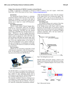
Northern blot analyses of mouse embryo polyadenylated RNAs for TGF /SI, pi and /33 are shown in Fig. 1.
The following characteristic bands were seen: TGF pi,
2.4 kb; TGF pi, multiple bands from 6 to 3 kb (a
predominant band at 4.4kb); TGF pi, 3.5 kb, thereby
confirming the specificity of the probes for each of the
three isoforms. The difference in signal intensity of
embryo day 16 RNA is due to a smaller quantity being
loaded as determined by u.v. shadowing and by probing
a constitutively expressed mRNA, the eukaryotic
protein synthesis initiation factor gene eEF-4A (Nielsen
etal. 1985).
In situ hybridisation
In situ hybridisation analysis performed with antisense
RNA probes revealed complex hybridisation patterns
indicating that all three TGF /3 genes are expressed in a
distinct and unique pattern in the developing mouse
embryo. In contrast, the sense probes that were used as
negative controls produced a weak and uniformly
distributed non-specific background signal (data not
shown). At day 10.5 p . c , TGF /SI transcripts were
clearly discernible in different tissues. We also detected
moderate levels of TGF pi expression which were
confined to the myocardium and the endocardial
cushion tissue (data not shown). No TGF /S3 hybridisation signals were observed; although, as reported by
Miller et al. (198%), Northern blots of polyadenylated
RNA from day 10.5 p.c. embryos revealed low signals.
Expression of TGF-fis in the placenta
In the placenta, only TGF /SI and TGF /33 were
expressed at all developmental stages investigated. At
day 10.5 p.c. TGF pi and TGF /33 transcripts revealed
distinct patterns of expression (Fig. 2A-C). TGF /Si
expression was strong in mesenchymal cells forming the
vascular zone near the central artery of the maternal
part of the placenta (Fig. 2A). In the fetal part of the
placenta, high levels of TGF pi expression were
detected in a small number of cells scattered throughout
the connective tissue of the chorion. These strongly
labelled cells had the appearance of blood cells and
were larger in size than the connective tissue cells of the
chorion (Fig. 2D). In contrast, high levels of TGF /S3
Expression of TGF fi genes in mouse embryogenesis
12
3
1 2
4
3
4
B
1 2
C
28S-
3
119
4
9.5
-7.5
.4.5
.2.4
1.4
0.24
•••i
Fig. 1. Northern analysis of TGF @ expression during mouse development. (A) TGF pi, (B) TGF pi, (C) TGF pi,
(D) control. 1, 2, 3 and 4 refer to days 13, 14, 16, post coitum and newborn. Positions of the ribosomal 18s and 28s
subunits are shown on the left, size markers (in kilobases) are indicated to the right.
transcripts were found exclusively in the spongiotrophoblast, a cell layer of fetal origin that forms the
junctional zone between the chorionic villi and the
maternal blood vessels (Fig. 2C). These patterns of
expression did not change during placental development although there was an apparent decrease in signal
intensity (data not shown).
The fetal liver
From day 10.5 p.c. to day 16.5 p . c , TGF pi expression
was strongest in the developing liver. To more clearly
define the cells expressing high levels of TGF pi, we
compared the pattern of TGF /SI expression with the
pattern of o--fetoprotein expression. During embryogenesis, formation of the liver becomes evident around
day 9 as a thickening and progressive stratification of a
region on the ventral side of the foregut, at which point
high levels of o--fetoprotein transcripts are present
(Schmid and Schulz, 1990), but no TGF Pi transcripts
are detected (data not shown). During the next stage of
liver development, strands of epithelial cells invade the
surrounding mesodermal mesenchyme and eventually
form a three-dimensional network. Hematopoietic
precursor cells invade the liver at this stage and become
established (Moore and Johnson, 1976). At day 10.5
p.c, both a'-fetoprotein and TGF pi were expressed at
high levels but showed a distinct cellular distribution
(Fig. 3D-E). Both a'-fetoprotein mRNA (Schmid and
Schulz, 1990) and protein (Dziadek and Adamson,
1978) are confined to the endodermal population of prehepatocytes whereas TGF pi signals were restricted to
the mesoderm. Our findings strongly suggest that the
TGF /J-expressing cell population represents mesodermal hematopoietic precursor cells, which arise in the
liver at this stage. Later in liver development the
pattern of TGF pi expression became more dispersed.
On day 12.5 p . c , megakaryocytes scattered throughout
the fetal liver were labelled strongly by the TGF pi
probe (Fig. 7A), but not by either TGF pi or TGF pi
probes. Other hematopoietic precursor cells showed
moderate levels of expression. The endodermal prehepatocytes, which represent about 40% of the liver
cells at this stage, were only weakly labelled. Between
days 14.5 and 16.5 p.c, TGF /SI expression remained
very strong in the megakaryocytes but decreased in
other cells of the fetal liver until on day 18.5 p.c TGF
Pi transcripts were detectable only in the large
megakaryocytes (data not shown). In contrast, TGF pi
and TGF pi mRNAs were not expressed either in the
hematopoietic precursor cells or in the prehepatocytes
of the fetal liver at all stages investigated; however,
moderate levels of TGF pi transcripts were visible in
the mesenteric epithelium surrounding the fetal liver on
day 12.5 p.c. (Fig. 4C).
The extraembryonic sites of hematopoiesis
At day 10.5 p.c, TGF pi expression was visible in
blood corpuscles of the visceral yolk sac. The first blood
cells are produced in extraembryonic sites as small
groups of mesodermal cells located next to the
endodermal wall of the visceral yolk sac are thought to
constitute a primitive population of erythrocytes (Dieterlen-Lievre, 1984). Afterwards, a new population of
hematopoietic stem cells arises from the intraembryonic
120
P. Schmid and others
D
.
* <"*• E
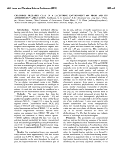
Fig. 2. Expression of TGF /Ss in the placenta and yolk sac at day 10.5 p.c. (A) Dark-field photography of a section
through the placenta hybridised with the TGF /SI riboprobe. A strong hybridisation signal is visible in the maternal
decidua. (B) Dark-field photography of an adjacent section hybridised with the TGF /52 riboprobe. Only non-specific
background signals are visible. (C) Dark-field illumination of TGF /S3 hybridisation signals in the placenta. Strong TGF /S3
expression is clearly visible in the ectoplacental glycogen rich cells of the spongiotrophoblast. (D) Bright-field photography
of TGF /SI hybridisation signals in the choronic villi. The strongly labelled cell probably represents a Hofbauer cell.
(E) Bright-field photography of a section through the blood-forming island of the yolk sac hybridised with the TGF /SI
riboprobe. A small number of hematopoietic cells are strongly labelled, eg, ectoplacental glycogen-rich cells; la, labyrinth;
md, maternal decidua; sc, subchorial clefts; he, hofbauer cell; ys, yolk sac.
mesoderm and produces a population of blood cells
which progressively substitutes for the yolk sac-derived
blood cells. The early embryonic visceral yolk sac
shares some properties with the embryonic liver during
later stages of development such as hematopoiesis and
production of cr-fetoprotein. TGF /SI is expressed in the
hematopoietic islands of the yolk sac but only in a small
number of cells; however, in these cells there is an
extremely high level of expression (Fig. 2E). It therefore appears that TGF /SI expression is restricted to a
specific cell type during early hematopoiesis. TGF /S2
and TGF /S3 transcripts were not detected.
The early neural crest mesenchyme
At day 10.5 p . c , we found TGF /SI expressing cells
scattered throughout the embryonic mesenchyme with
transcripts most abundant in the mandibular and
maxillary arches and in single cells surrounding the
somites (Fig. 3A) and the nervous tissue (Fig. 3F-G).
Due to their distribution we assume that these strongly
labelled cells are neural crest cells, which are endowed
with the ability to undertake extrensive but tightly
controlled migrations throughout the embryo. TGF 01
and TGF /S3 expression was not observed in the neural
crest mesenchyme.
The fetal thymus
The embryonic thymus contains a heterogenously
distributed population of stromal cells and T-cells at
various stages of maturation. TGF /SI transcripts were
seen in all cells of the embryonic thymus but the
hybridisation signals were interspersed with T-cell
precursors, which were apparently stronger labelled
than the endodermal cell population (Fig. 7B). TGF /S2
and TGF /S3 transcripts were not found in the
developing thymus.
The skeleton
On day 12.5 p . c , high levels of TGF /S2 and TGF /S3
transcripts were visible in the primordia of the vertebral
column and were strongest in the caudal sclerotomic
halves. TGF /S3 was expressed in all prevertebral
segments (Fig. 4C). In contrast, high levels of TGF /S2
transcripts were visible only in the thoracic sclerotomes
(Fig. 4B). TGF /SI transcripts were detected in intersegmental cells of the early spinal column and in cells lining
the cerebral ganglia (Fig. 4A).
Expression of TGF fi genes in mouse embryogenesis
121
r
Fig. 3. Expression of TGF ps and a^fetoprotein in the day 10.5 p.c. embryo. (A) Dark field photography of a section
through a 10.5 p.c. embryo hybridised with the TGF /SI riboprobe. The strongest hybridisation signals are visible in the
liver and mesenchymal cells lining the nervous tissues and the somites. (B) Dark field photography of a serial section
hybridised with the TGF p2 riboprobe. Compared to panel A only non-specific background signals are visible. (C) Darkfield photography of a serial section hybridized with the TGF p3 riboprobe. Only non-specific background signals are
visible. (D) Bright-field photography showing a--fetoprotein expression in the early fetal liver. Strands of endodermal
prehepatocytes are strongly labelled. (E) Bright-field photography showing TGF pi expression in the early fetal liver. TGF
/31 expression is confined to single cells scattered throughout the endodermal strands of prehepatocytes. (F) Bright-field
photography of a section through the telencephalon and the nasal process hybridised with the TGF pi riboprobe.
(G) Dark-field photography of the section shown in F. A strong hybridisation signal is visible in cells lining the nervous
tissue and in a small number of cells scattered throughout the nasal mesenchyme. ma, mandibular arch; tc, telencephalon;
li, liver; so, somites; nt, neural tube.
On day 14.5 p . c , the TGF /Jl probe strongly labelled
the narrow bands of osteoblasts adjacent to the
vertebrae and ribs (Fig. 5A). However, less differen-
tiated elements of the developing axial skeleton, such as
sternum and limb skeleton, were only weakly labelled
by the TGF pi probe (Fig. 5A). In contrast, TGF 03
122
P. Schmid and others
expression was observed in the perichondrium of all
cartilaginous elements of the axial skeleton but the
levels of expression varied (Fig. 5C). Within the
vertebral column, TGF /33 expression was strongest in
the intervertebral discs (data not shown). Only low
levels of TGF pi mRNA were visible in perichondreal
mesenchyme (Fig. 5B).
On day 16.5 p . c , TGF /31 expression was strong in
the vertebrae and ribs and was detected both in the
periosteal layer and in the ossification centres (Fig. 6A)
Fig. 4. Dark-field illuminations of TGF pi, pi and p3
expression in sagittal sections of a mouse embryo at day 12.5
p.c. (A) TGF )31; (B) TGF j82; (C) TGF /33; in, intestine; li,
liver; lu, lung; me, metencephalon; nt, neural tube; pv,
prevertebrae; tc, telencephalon; tr, trachea.
124
P. Schmid and others
Fig. 6. Dark-field illuminations of TGF pi and pi expression in parasagittal sections of a mouse embryo at day 16.5 p.c.
(A) TGF pi; (B) TGF p3; bp, basisphenoid; fl, forelimb; fr, frontale; ma, maxilla; ri, ribs.
where only very low levels of TGF pi transcripts were
visible (Fig. 6B). On the other hand, TGF pi was
expressed in the perichondrium of the limb skeleton
(Fig. 6B). TGF pi transcripts were only visible in
growth zones of the limb plates (data not shown).
All components of the skeleton are derived from
mesenchyme. Mesenchyme can be converted into
skeletal elements by first forming a cartilaginous
framework, which is subsequently replaced by true
bone (endochondral bone formation), or by forming
bone directly (intramembranous ossification). Most
elements of the facial skeleton, such as mandible and
maxilla, are formed by intramembranous ossification of
the cranofacial mesenchyme. Highly contrasting patterns of distribution of TGF /31 and TGF pi transcripts
were observed during formation of the facial skeleton
elements. TGF pi was expressed at very high levels in
ossifying tissues of the upper and lower jaw (Fig. 5A,
6A and 8A) whereas TGF pi expression was strongest
in the undifferentiated mesenchymal cell layers adjacent to the ossification centres (Figs 5C, 6B and 8C).
TGF pi expression was not observed in the upper and
lower jaw regions that were undergoing intramembranous ossification. However, TGF pi transcripts were
visible in the perichondreal layers of certain cartilaginous elements of the facial skeleton and also in the
nasal and mandibular mesenchyme that forms the soft
tissue components of the face (Fig. 5B and 8B).
The lung
Formation of the lung begins around day 10 p.c. with
the outgrowth of the foregut-derived endodermal
trachea into the bronchial mesoderm. As the trachea
lengthens, it bifurcates at its caudal end to form two
lung buds. These in turn continue to grow and branch,
giving rise to the bronchial trees of the lung. All three
TGF ft genes were expressed at high levels and showed
distinct spatial and temporal patterns of mRNA
distribution. At all stages investigated, TGF pi
expression was strongest in the bronchial mesoderm
(Figs 4A, 5A and 6A) whereas TGF pi transcripts were
found exclusively in the endodermal bronchiolar
epithelia where the signal became stronger in later
stages of development (Figs 4B and 5B). The TGF pi
expression pattern changed during lung development.
At day 12.5 p.c. TGF pi transcripts were found
predominantly in the tracheal mesenchyme (Fig. 4C)
but at day 14.5 p.c. TGF pi signals were visible in the
endodermal epithelia cells of the growing bronchioles
(Fig. 5C) although by day 16.5 p.c. TGF pi expression
was no longer detectable (Fig. 6B). TGF pi transcripts
were also expressed in mesodermal epithelial cells,
which later give rise to the visceral pleura (Fig. 5C).
The gut
TGF pi expression was strongest on day 14.5 p.c. in the
mesodermal cell layers of the submucosa but not in the
Expression of TGF {5 genes in mouse embryogenesis
*
125
r*
V
*
B
* •
Fig. 7. Expression of TGF /Ss in liver, thymus and cochlea. (Left side) Bright-field photographs, (right side) dark-field
photographs. (A) Section through the day 12.5 p.c. liver hybridised with the TGF /SI riboprobe. Although the majority of
fetal liver cells are clearly showing TGF /SI hybridisation signals, very high levels of TGF /SI expression are confined to
megakaryocytes scattered throughout the fetal liver. (B) Section through the day 12.5 p.c. fetal thymus hybridised with the
TGF /SI riboprobe. All cells of the fetal thymus are labelled but the level of expression is variable among individual cells.
(C) Section through the day 14.5 p.c. cochlea hybridised with the TGF /S2 riboprobe. TGF pi expression is confined to the
sensory epithelium.
intestinal epithelia (Fig. 5A) although it was visible at
later stages of gut development at decreased levels. In
contrast, TGF /32 was expressed exclusively in the
single-cell layer forming the mesodermal mesentera
(Figs 4B and 5B). On day 12.5 p.c. moderate levels of
TGF pi transcripts were visible in both the submucosal
mesenchyme and mesenteric mesoderm (Fig. 4C) but
later became confined to the mesentera (Figs 5C and
6B).
The kidney
Kidney development is the result of reciprocal inductive
interactions between the metanephric duct and the
surrounding metanephrogenic tissue. The terminal
portions of the metanephric duct induce the formation
of metanephric tubules. TGF /SI expression was
detected in the surrounding stromal mesenchyme
during formation of the metanephros at day 14.5 p.c.
(Fig. 5A). The epithelial tubules deriving from the
126
P. Schmid and others
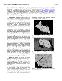
Fig. 8. Expression of TGF /SI, /S2 and /B in the upper jaw. (Left side) Bright-field photographs, (right side) dark-field
photographs. (A) Section through ethmoid cartilages and upper jaw bones of a day 16.5 p.c. embryo hybridised with the
TGF /SI riboprobe. A strong hybridisation signal is visible in the bone tissues. A weak signal is also visible in the
perichondreal layer of the cartilages. (B) Adjacent section hybridised with the TGF pi riboprobe. TGF pi is not expressed
in the bone tissue of the upper jaw although low levels of TGF pZ transcripts are visible in the perichondrium of the
ethmoid cartilages. TGF pi hybridisation signals are also discernible in nasal epithelia. (C) Adjacent section hybridised
with the TGF /S3 riboprobe. TGF /93 expression is strongest in mesenchymal cells surrounding upper jaw bones and in
periosteal layers of the ethmoid cartilages, ec, ethmoid cartilage; nc, nasal camber; pa, palatinum.
metanephrogenic mesenchyme showed only low levels
of TGF pi transcripts although no TGF /B expression
was observed.
The sensory epithelia
From day 12.5 p.c. to day 16.5 p . c , the most prominent
site of TGF pi expression was the cochlear epithelium
(Figs 5B and 7C). At day 16.5 p . c , TGF pi hybridisation signals were also visible in the olfactory epithelium
of the nasal chamber (Fig. 8B). No expression of TGF
pi or TGF pi was found in these tissues.
The skin
Early mouse embryo skin has a simple epithelium. By
16 days p.c. a second skin layer, the stratum germinativum, arises followed by an interstitial basal layer. At
this stage, only TGF pi expression was detected in the
loose mesenchyme underlying the epidermis (Fig. 6A).
Expression of TGF f5 genes in mouse embryogenesis
Within the dermis a small number of cells showed high
levels of TGF /SI expression and were scattered
between moderately or weakly labelled mesenchymal
cells. TGF pi or TGF pi signals were not detectable in
the skin.
The vibrissae
Formation of the facial hair follicles is visible as early as
day 13 p.c. when the vibrissae papillar are still
invaginated and ectoderm is proliferating in the
external nares. On day 14.5 p.c, all three TGF /S
transcripts were expressed at high levels in the
ectodermal epithelium of the external root sheath
(Fig. 5A-C). In the primordia of the dermal sheaths
only moderate TGF pi expression was detected. On
day 16.5 p . c , TGF pi, pi and pi transcripts were
visible both in the inner and outer root sheath of the
whisker hair follicles but with striking differences in
expression levels. TGF pi was expressed stronger than
TGF pi and pi. Low levels of TGF pi were also visible
in the connective tissue sheath of the hair follicle;
however, TGF pi and pi were not expressed.
Discussion
In this report, we have described the first comparative
study of TGF fi gene expression during mouse
embryogenesis where homologous gene probes of the
same length have been used. We clearly demonstrate
that TGF pi, pi and pi show distinct patterns of
expression during development.
TGF pi transcripts are expressed predominantly in
hematopoietic cells of the fetal liver, in fetal bone and
in the mesenchymal compartments of several internal
organs. These results are in agreement with the work of
Lehnert and Akhurst (1988). In addition, we have
observed on days 10.5 p.c. and 12.6 p.c. expression of
TGF Pi transcripts in mesenchymal cells of probable
neural crest origin that line the nervous tissue and
somites. Immunohistochemical studies of Heine et al.
(1987) have shown a similar distribution pattern for the
protein. In addition, a strong TGF pi signal was clearly
seen in the maternal decidua. In the fetal part of the
placenta and in the haematopoietic islands of the yolk
sac, a small number of cells snowed very strong TGF pi
expression. This cell population probably represents
precursors of the Hofbauer cells, which are believed to
be primitive macrophages. Since peripheral blood
monocytes and mature macrophages produce only the
TGF pi isoform (G. Bilbe, unpublished results), it is
likely that TGF pi is switched on at an early stage in
myeloid differentiation. Interestingly in vitro and in
vivo studies on bone marrow preparations have shown
that TGF pi inhibits the proliferation of myeloid
progenitor cells while more differentiated cells of this
lineage are not inhibited (Keller et al. 1990; Ruscetti et
al. 1990).
For the first time, our study demonstrates that TGF
pi is expressed in the mesenchymal components of the
facial region and also in bronchial epithelia. Moreover,
127
TGF pi was the only isoform expressed in the cochlear
epithelium. We were unable to confirm the general
findings of Pelton et al. (1989). The differences in
expression patterns can most likely be explained by the
choice of probes. Pelton and co-workers performed
their in situ hybridisation experiments with a probe
corresponding to the 5' untranslated sequences and part
of the LAP region of the human TGF pi gene. Because
the 5' untranslated region of the human TGF pi
transcript is very rich in A and T residues we used a
riboprobe that is complementary to the mature mouse
TGF pi peptide and which in Northern analysis gave a
typical TGF pi banding pattern. To confirm our results,
we compared by in situ hybridisation the TGF pi
expression patterns revealed by the 'mature form'
probe with those shown by a probe covering either the
entire coding region or the LAP region. All probes
showed identical patterns of hybridisation but with
different intensities because of the varying lengths of
the probes. We propose that the 5' untranslated region
of the human TGF pi gene as used by Pelton et al.
(1989) contains cross-hybridizing sequences responsible
for these contrasting results. It is interesting to note that
a comparison of the TGF pi, pi and pi DNA sequences
encoding the mature peptide forms reveals homologies
of less than 75 % between the three isoforms where the
longest contiguous stretch of nucleotides is 14 bases
long.
Our study also describes the expression patterns of
TGF pi by RNA in situ hybridisation. Northern blot
analysis with mRNA from day 13 to day 16.5 p.c.
hybridised with the mouse TGF pi probe complementary to the mature peptide detected a single band. This
finding is in agreement with Miller et al. (1989b) who
found weak TGF pi expression at day 10.5 p.c.
although at this early stage we were not able to detect
TGF pi transcripts by in situ hybridisation. These
transcripts are probably expressed at low levels and are
dispersed throughout the embryo and therefore cannot
be detected by in situ hybridisation. A unique TGF pi
hybridisation signal was also observed in the spongiotrophoblast of the placenta as early as day 10.5. p.c. and
was predominantly restricted to the reticular zone
between the fetal and maternal placenta. TGF pi may
therefore be involved in the regulation of cell proliferation and matrix formation during growth and stabilisation of the chorionic villi in the placenta.
A comparison of the expression patterns for all three
TGF /3s strongly suggests a coordinated role for these
proteins in mesenchymal-epithelial interactions during
embryonic development. For example, there are
marked differences in the pattern of TGF pi, pi and pi
expression during branching morphogenesis of the
lung. Endodermal branching, i.e. cleft formation, is
one of the major events in lung morphogenesis. The
characteristic budding pattern of the endodermal
bronchial tree is the result of continuous interactions
between endoderm and the surrounding mesoderm.
The contrasting expression patterns of TGF /3s in the
developing lung strongly suggest that these genes are
involved in the budding process probably by controlling
128
P. Schmid and others
formation and degradation of extracellular matrix
components. In vitro studies have shown that TGF /SI
regulates the formation of extracellular matrix either by
inducing synthesis of matrix proteins, such as collagen
and fibronectin (Ignotz and Massague, 1986; Roberts et
al. 1986; Varga and Jimenez, 1986; Fine and Goldstein,
1987; Ignotz et al. 1987) glycosaminoglycans (Rasmussen and Rapraeger, 1988) and cell adhesion receptors
(Heino et al. 1989; Ignotz and Massague, 1987) or by
controlling proteolytic degradation of matrix proteins
(Laiho et al. 1986, Edwards et al. 1987; Keski-Oja et al.
1988). Recently colocalisation of TGF /SI protein and
matrix proteins by a TGF /SI antibody was described in
mesenchymal cells during lung development of the
mouse, particularly at times when cell-cell interactions
between mesenchyme and epithelium are important for
normal epithelial cell differentiation (Heine etal. 1990).
We have confirmed the localisation of TGF /Si mRNA
in lung mesenchymal cells, which suggests that this
peptide may induce matrix proteins in an autocrine
fashion. Since exogenous TGF /SI has been shown to
block the maturation of bronchial epithelial cells
(Masui et al. 1986), this peptide may also regulate
differentiation of the lung epithelia in a paracrine
fashion. Whereas TGF /SI transcripts are localized in
lung mesenchymal cells, TGF /32 and TGF pi expression occurs only in the epithelial linings of the
bronchii but TGF /S3 not in the tips of actively growing
ducts. A comparative study by Graycar et al. (1989) has
recently shown more potent inhibitory effects of TGF
pi and pi on lung epithelial cell proliferation than TGF
/SI suggesting that the observed expression patterns in
the developing lung may simply reflect differences in
potency of all three isoforms.
We have shown that all three TGF /S isoforms are
expressed during endochondrial bone development but
only TGF /SI and /S3 are involved in intramembranous
bone formation. Recent in vivo studies have shown that
TGF /SI and TGF pi induce the differentiation of
periosteal mesenchymal cells of long bones into
osteoblast and chondrocytes (Joyce et al. 1990). They
also stimulate these cells to proliferate and synthesize
the extracellular matrix proteins characteristic for bone
and cartilage. These workers have found that TGF pi is
a more potent stimulator of osteogenesis and chondrogenesis than TGF /SI in vivo; however, both isoforms
share the ability to influence the route of tissue
differentiation into bone or cartilage in a dosedependent way. In the present study, we have shown
strong TGF pi expression in precartilaginous masses
and the perichondrium early in bone development.
Therefore, we propose that TGF pi may be involved in
early differentiation processes in endochondral bone
formation. In contrast, TGF /SI, which is expressed
predominantly at later stages of vertebral development,
may induce matrix formation in differentiated skeletal
elements. Since expression of TGF pi transcripts were
only observed at a very specific, early stage of bone
development, this peptide possibly induces differentiation of mesenchymal cells into a chondriocytic
phenotype. Absence of TGF pi at this stage of
development may result in ossification without a prior
cartilage matrix.
Most of the flat bones of the face are derived from
neural crest mesenchyme and their morphogenesis and
growth is a very complex process involving the
interaction of many factors (Noden, 1984). The present
study shows that both TGF /SI and TGF pi are involved
in this process. Possibly TGF /S3 induces differentiation
of neural crest mesenchyme whereas TGF /SI expression is necessary for inducing bone matrix proteins
during further
maturation
and
ossification.
Interestingly TGF pi transcripts were never detected at
any stage during intramembraneous bone formation
although they were coexpressed with TGF /S3 in some
tissues of the axial skeleton.
The present report describes the coexpression of
TGF pi and TGF pi in certain tissues, e.g., in lung
epithelia, and coordinated expression of all three
isoforms in others, e.g., in root sheath epithelia. The
differential expression of the three genes may be
explained by an analysis of their promoter regions
(Roberts and Sporn, 1990b). There are two major
classes of TGF /S transcriptional promoters. The TGF
/SI promoter contains and AP-1 binding site, responds
strongly to phorbol ester induction but does not contain
a TATA A box consensus sequence. On the other hand,
TGF /S2 and pi have TATAA boxes, AP-2 binding sites
and cyclic AMP responsive elements which can be
induced by forskolin, an activator of adenylate cyclase.
In addition, TGF pi also has an AP-1 binding site
(Roberts and Sporn, 19906) suggesting that TGF pi
gene expression could be regulated differently from
that of TGF pi. Since we do find expression of all three
isoforms transcripts, one can hypothesise that coordinate bindingtoAPl and AP2 sites must occur if all three
genes are to be expressed.
What is the biological significance of the complex
patterns of TGF /3 expression? Our overall impression
is that TGF /SI is involved in early (major) inductive
event(s) during interaction of mesenchymal and epithelial cell layers. TGF /Si is the only isoform expressed
in early mesenchyme at enhanced levels and appears to
regulate the layout of the embryo by stimulating the
formation of extracellular matrix components which are
important in cell migration and/or cell-cell interaction
(Bernfield, 1981). For instance, in early fetal liver, lung
and gut, TGF /S expression is confined to mesenchymal
cells. Heine et al. (1990) have shown that TGF /SI
regulates the expression of collagen I and III, fibronectin and glycosaminoglycans, which are all components
of the extracellular matrix. Production of these components is important for interaction with cell surfaces
via specific receptor complexes and subsequent initiation of inductive processes resulting in differentiation events. In vivo experiments have demonstrated
that TGF pi has potent angiogenic activity (Roberts et
al. 1986, Cox et al. unpublished data), which partly
explains the extensive vascularisation and invasion by
lymphatics and nerve fibers that occur during organogenesis. Further evidence for a pivotal role for TGF /SI
in early mesenchymal-epithelial interactions has been
Expression of TGF /3 genes in mouse embryogenesis 129
provided by Antonelli-Orlidge et al. (1989) who have
shown that TGF pi mediates inhibition of cell growth in
a capillary-endothelial cell coculture system.
As development enters a phase of morphogenesis
characterized by more complex differentiation events,
all three TGF /3 isoforms are expressed in differential
patterns. This phenomenon can be seen clearly in
developing bone where distinct TGF /3s are expressed in
a controlled fashion as described above. The differential expression of TGF /3s at specific times during bone
formation indicates differences in their biological
functions. TGF /31 has been reported to stimulate or
inhibit growth and differentiation in vitro depending on
the cell type examined (Sporn et al. 1987) and more
specifically has been shown to inhibit osteoclast (Oreffo
et al. 1989), and to stimulate osteoblast, proliferation
(Centrella et al. 1986; Gehron-Robey et al. 1987).
Antonelli-Orlidge et al. (1989) have demonstrated that
conversion of the precursor TGF /3 molecule to the
mature form is essential for its activity and that this
process is mediated by the responder cells in a paracrine
fashion. Thus, during bone development the differential expression of TGF /3s affects the developmental
fates of cells partly as a result of precursor conversion.
However, Graycar et al. (1989) have demonstrated that
the potency of the mature peptides differs depending on
the cell type affected. This suggests that those cells
under the regulation of TGF /3s have receptors or
receptor-coupled second messenger systems that determine the inductive or inhibitory effect of TGF /Ss. For
example, a guanine nucleotide-binding protein-dependent pathway is involved in transmission of the signal
for at least one TGF /3-induced response (Howe and
Leof, 1989). Finally TGF /3 can control morphogenesis
at three different levels. Firstly, at the transcriptional
level via transcription factor-promoter interactions.
This determines the type of TGF /3 isoform expressed
and is dependent on the cell type and its differentiation
stage. Secondly, recent studies have indicated that the
responding cell is crucial for activation of TGF /3 to its
active form. Thirdly, the response of the affected cell is
dependent on the TGF /3 receptors and their second
messenger systems.
GRAYCAR, J. L., RHEE, L., MASON, A. J., MILLER, D. A.,
COFFEY, R. H., MOSES, H. L. AND CHEN, E. Y. (1988). A new
type of transforming growth factor-/?, TGF/33. EMBO J. 7,
3737-3743.
DIETERLEN-LIEVRE, (1984). Blood in chimeras. In Chimeras in
Developmental Biology (ed. N. LeDouarin and A. McLaren).
Academic Press, New York, 133-163.
DZIADEK, M. AND ADAMSON, E. (1978). Localization and synthesis
of alpha-fetoprotein in postimplantation mouse embryos.
J. Embryol. exp. Morph. 43, 289-313.
EDWARDS, D. R., MURPHY, G. AND REYNOLDS, J. J. (1987).
Transforming growth factor /? modulates the expression of
collagenase and metalloproteinase inhibitor. EMBO J. 6,
1899-1904.
FINE, A. AND GOLDSTEIN, R. H. (1987). The effect of transforming
growth factor-/? on cell proliferation and collagen formation by
lung fibroblasts. J. biol. Chem. 126, 3897-3902.
GEHRON-ROBEY, P. G., YOUNG, M. F . , FLANDERS, K. C , ROCHE,
N. S., KONDAIAH, P., REDDI, A. H . , TERMINE, J. D . , SPORN, M.
B. AND ROBERTS, A. B. (1987). Osteoblasts synthesize and
respond to transforming growth factor-/? in vitro. J. Cell Biol.
105, 457-463.
GRAYCAR, J. L., MILLER, D. A., ARJUCK, B. A., LYONS, R. M.,
MOSES, H. L. AND DERYNCK, R. (1989). Human transforming
growth factor-/33: recombinant expression, purification, and
biological activities in comparison with transforming growth
factors-/Jl and -pi. Mol. Endocnnol. 3, 1977-1986.
HEINE, U. I., MUNOZ, E. F., FLANDERS, K. C , ELUNGSWORTH, L.
R., LAM, H.-Y., THOMPSON, N. L., ROBERTS, A. B. AND SPORN,
M. B . (1987). Role of transforming growth factor-/? in the
development of the mouse embryo. J. Cell Biol. 105, 2861-2876.
HEINE, U. I., MUNOZ, E. F., FLANDERS, K. C , ROBERTS, A. B.
AND SPORN, M. B. (1990). Colocalization of TGF-beta 1 and
collagen I and III, fibronectin and glycosaminoglycans during
lung branching morphogenesis. Development 109, 29—36.
HEINO, J., IGNOTZ, R. A., HEMLER, M. E., CROUSE, C. AND
MASSAGUE, J. (1989). Regulation of cell adhesion receptors by
transforming growth factor-/?. J. biol. Chem. 264, 380-388.
HOWE, P. H. AND LOEF, E. B. (1989). Transforming growth factorpi treatment of AKR-2B cells is coupled through a pertussistoxin sensitive G protein. Biochem. J. 261, 879-886.
IGNOTZ, R. A. AND MASSAGUE, J. (1986). Transforming growth
factor-/? stimulates the expression of fibronectin and collagen
into the extracellular matrix. J. biol. Chem. 261, 4337-4345.
IGNOTZ, R. A. AND MASSAGUE, J. (1987). Cell adhesion protein
receptors as targets for transforming growth factor-/? action. Cell
51, 189-197.
IGNOTZ, R. A., ENDO, T. AND MASSAGUE, J. (1987). Regulation of
fibronectin and type I collagen mRNA levels by transforming
growth factor-^. J. biol. Chem. 262, 6443-6446.
JOYCE, M. E., ROBERTS, A. B., SPORN, M. B. AND BOLANDER, M.
E. (1990). Transforming growth factor-/? and the initiation of
chondrogenesis and osteogenesis in the rat femur. J. Cell Biol.
110, 2195-2207.
KELLER, J. R., MCNIECE, I. K., SILL, K. T., ELLINGSWORTH, L.
References
ANTONELU-ORLJDGE, A., SAUNDERS, K. B . , SMITH, S. R. AND
DIAMORE, P. A. (1989). An activated form of transforming
growth factor B is produced by cocultures of endothelial cells
and pericytes. Proc. natn. Acad. Sci. U.S.A. 86, 4544-4548.
BERNFIELD, M. R. (1981). Organization and remodelling of the
extracellular matrix in morphogenesis. In Morphogenesis and
Pattern Formation (T. G. Connelly, L. L. Brinkley, and B. M.
Carlson, eds.), pp. 139-161. Raven Press, New York.
CENTRELLA, M., MCCARTHY, T. L. AND CANALIS, E. (1986).
Transforming growth factor-/? is a bifunctional regulator of
replication and collagen synthesis in osteoblast-enriched cell
cultures from fetal bone. J. btol. Chem. Ubl, 2869-2874.
CHOMCZYNSKJ, I. AND SACCHI, N. (1987). Single-step method of
RNA isolation by acid guanidinium thiocyanate-phenolchloroform extraction. Anal. Biochem. 162, 156-159.
DERYNCK, R., LINDQUIST, P. B . , LEE, A., W E N , D . , TANIM, J.,
R., QUESENBERJ(Y, P. J., SlNG, G. K. AND RuSCETTI, F. W.
(1990). Transforming growth factor /? directly regulates primitive
murine hematopoietic cell proliferation. Blood 75, 596-602.
KESKI-OJA, J.. RAGHOW. R., SAWDEY, M., LOSKUTOFF, D. J.,
POSTLETHWAITE, A. E., KANG, A. H. AND MOSES, H. L. (1988).
Regulation of mRNA for type-1 plasminogen activator inhibitor,
fibronectin, and type 1 procollagen by transforming growth
factor-/?. J. biol. Chem. 263, 3111-3115.
LAIHO, M., SAKESELA, O., ANDREASEN, P. A. AND KESKI-OJA, J.
(1986). Enhanced production and extracellular deposition of the
endothelial-type plasminogen activator in cultured human lung
fibroblasts by transforming growth factor-/?. J. Cell Biol. 103,
2403-2410.
LEHNERT, S. A. AND AKHURST, R. J. (1988). Embryonic expression
pattern of TGF beta type-1 RNA suggests both paracrine and
autocrine mechanisms of action. Development 104, 263-273.
LYONS, R. M. AND MOSES, H. L. (1990). Transforming growth
factors and the regulation of cell proliferation. Eur. J. Biochem.
187, 467-473.
130
P. Schmid and others
MANIATIS, T., FRITSCH, E. F. AND SAMBROK, J. (1982). Molecular
Cloning. A Laboratory Manual. Cold Spring Harbor
Laboratory.
MASUI, T., WAKEFIELD, L. D., LECHNER, J. F., LAVECK, M. A.,
SPORN, M. B. AND HARRIS, C. C. (1986). Type /3 transforming
growth factor is the primary differentiation inducing serum
factor for normal human bronchial epithelial cells. Proc. natn.
Acad. Sci. U.S.A. 83, 2438-2442.
MILLER, D. A., LEE, A., PELTON, R. W., CHEN, E. Y., MOSES, H.
L. AND DERYNCK, R. (1989a). Murine transforming growth
factor-/32 cDNA sequence and expression in adult tissues and
embryos. Mol. Endocrinol. 3, 1108-1114.
MILLER, D. A., LEE, A., MATSUI, Y., CHEN, E. Y., MOSES, H. L.
AND DERYNCK, R. (1989fc). Complementary DNA cloning of the
murine transforming growth factor-/33 (TGF/53) precursor and
the comparative expression of TGF/33 and TGF/51 messenger
RNA in murine embryos and adult tissues. Mol. Endocrinol. 3,
1926-1934.
MOORE, M. A. S. AND JOHNSON, G. R. (1976). Hematopoietic
stem cells during embryonic development and growth. In Stem
Cells of Renewing Cell Populations (ed. Cairnie, A. B., Lala, P.
A. and Osmond, D. G.). 323-330. Academic Press, New York.
NIELSEN, P. J., MCMASTER, G. K. AND TRACHSEL, H. (1985).
Cloning of eukaryotic protein synthesis initiation factor genes:
isolation and characterization of cDNA clones encoding factor
eIF-4A. Nucl. Acids Res. 13, 6867-6880.
NODEN, D. M. (1984). Craniofacial development: New views on
old problems. Anat. Rec. 208, 1-13.
OREFFO, R. O., MUNDY, G. R., SEYEDIN, S. M. AND BONEWALD,
L. T. (1989). Activation of the bone derived latent TGF-/3
complex by isolated osteoclasts. Biochem. Biophys. Res. Comm.
158, 817-823.
PELTON, R. W., NOMURA, S., MOSES, H. L. AND HOGAN, B. L. M.
(1989). Expression of transforming growth factor pi RNA
during murine embryogenesis. Development 106, 759-767.
RASMUSSEN, S. AND RAPRAECER, A. (1988). Altered structure of
the hybrid cell surface proteoglycan of mammary epithelial cells
in response to transforming growth factor beta. J. Cell Biol. 107,
1959-1967.
RENTROP, M., KNAPP, B., WINTER, H. AND SCHWEIZER, J. (1986).
Aminoalkylsilane-treated glass slides as support for iii situ
hybridisation of keratin cDNAs to frozen tissue sections under
varying fixation and pretreatment conditions. Histochem. J. 18,
271-276.
ROBERTS, A. B., SPORN, M. B., ASSOIAN, R. K., SMITH, J. M.,
ROCHE, N. S., WAKEFIELD, I. M., HEINE, U. I., LIOTTA, L. A.,
FALANGA, V., KEHRE, J. H. AND FAUCI, A. S. (1986).
Transforming-growth factor type-/?: rapid induction of fibrosis
and angiogenesis in vivo and stimulation of collagen formation
in vitro. Proc. natn. Acad. Sci. U.S.A. 83, 4167-4171.
ROBERTS, A. B. AND SPORN, M. B. (1990a). The transforming
growth factor-betas. In Peptide Growth Factors and their
Receptors (ed. Sporn, M. B. and Roberts, A. B.), Handbook of
Experimental Pathology, Springer-Verlag, Heidelberg, Vol. 95,
419-472.
ROBERTS, A. B. AND SPORN, M. B. (1990b). Multiple forms of
TGF-/3: Differential expression and distinct promotors. The Ciba
Foundation Symposium 157, in press.
RUSCETTI, F., DUBOIS, C , FALK, L., JACOBSEN, E., SING, G.,
LONGO, D . , WILTROUT, R. AND KELLER, J. (1990). In vivo and in
vitro effects of TGF-/51 on normal and leucaemic hematopoiesis.
The Ciba Foundation Symposium 157, in press.
SCHMID, P. AND SCHULZ, W. A. (1990). Coexpression of the c-myc
protooncogene with n^fetoprotein and albumin in fetal mouse
liver. Differentiation (in press).
SPORN, M. B., ROBERTS, A. B., WAKEFIELD, L. M. AND D E
CROMBRUGGHE, B. (1987). Some recent advances in the
chemistry and biology of transforming growth factor-beta. J.
Cell Biol. 105, 1039-1045.
TAVASSILI, M. AND YOFFEY, J. M. (1983). Bone Marrow: Structure
and Function. Alan R. Liss, New York.
VARGA, J. AND JIMENEZ, S. A. (1986). Stimulation of normal
human flbroblasts collagen production and processing by
transforming growth factor-/?. Biochem. Biophys. Res. Commun.
138, 974-980.
WILCOX, J. N. AND DERYNCK, R. (1988). Developmental
expression of transforming growth factors alpha and beta in
mouse fetus. Molec. cell. Biol. 8, 3415-3422.
{Accepted S October 1990)
Addition to proof:
Using our in situ hybridisation conditions, the human
TGF pi probe, chosen by Pelton et al. (1989), revealed
a very high unspecific background, probably due to the
reasons stated in the text.


© Copyright 2026