
CD3+, CD56+ Aggressive Variant of Large Granular
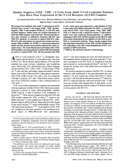
From www.bloodjournal.org by guest on February 6, 2015. For personal use only.
CD3+, CD56+ Aggressive Variant of Large
Granular Lymphocyte Leukemia
By Teresa C. Gentile, Aysegul H. Uner, Robert E. Hutchison, Jonathan Wright, Jonathan Ben-Ezra,
E. Clifton Russell, and Thomas P. Loughran, Jr
Clonal expansions of CD3+ large granular lymphocytes (LGL)
have been classified as T-LGL leukemia. The majority ofpatients with T-LGL leukemia have a chronic disease (years)
manifested often bysevere neutropenia, rheumatoid arthritis, and mild-to-moderate splenomegaly. The characteristic
phenotype of theleukemic LGL is CD3'. CD8+, CD16'. CD57+,
and CD56-. In this report wedescribe an aggressive variant
of T-LGL leukemia in which leukemic LGL also expressed
CD56, as identified by two-color flow-cytometry
analysis. In
contrast to the chronic nature typical of T-LGL leukemia,
these patients presented with a severesystemic illness that
was rapidlyprogressive and resistant t o treatment. Atypical
clinical features included rapidly increasing spleen size t o
massive proportions, extensive lymphadenopathy, and the
presence of B symptoms (fever, nightsweats, weight loss).
Hematologic and pathologic features were also unusual for
T-LGL leukemia. These patients had very high LGL counts
at diagnosis (range 11,692 t o 26,312 pL), which increased
rapidly despite treatment. Histopathologic examination of
splenic sections showed extensive infiltration of red pulp
cords and sinuses by leukemic cells with atrophy of the
white pulp, These clinicopathologic features are similar t o
those described for patients with natural killercell (NKI-LGL
leukemia, whose cells are also CD56+. However, unlike NKLGL leukemia, we could not show a direct pathogenic role
for Epstein-Barr virus (EBV), asSouthern-blot analyses using
an EBV-joinedtermini probe were negative in these patients.
Our findings suggest that CD3+, CD56+ LGL leukemia is a
distinctclinicopathologic entity separate from the usual
CD3+, CD56- T-LGL leukemia. The expression on leukemic
LGL of CD56, an adhesion molecule, may determine the aggressive biologic nature of this newly described disease.
0 1994 by The American Society of Hematology.
C
clinicopathologic features of these patients were similar to
those of NK-LGL leukemia.
LONAL DISORDERS of large granular lymphocytes
(LGL) may have either a CD3+ or a CD3- phenotype,"3 and have been designated as T-cell (T)-LGL leukemia or natural killer cell (NK)-LGL leukemia respectively:
T-LGL leukemic cells have a characteristic phenotype,
CD3+, CD8+, CD16+,
CD57', and show clonal T-cell receptor (TCR) gene rearrangement. NK-LGL leukemic cells are
indistinguishable by light microscopy from T-LGL leukemic
cells and show the following phenotype: CD3-, CD4-,
CD8-, CD16+, CD56+,CD57-. It is more difficult to prove
clonality in CD3- LGL because these cells do not rearrange
TCR genes. However, clonal cytogenetic abnormalities have
been shown in a number of patients and support the diagnosis
of NK-LGL leukemia?-8
The clinical presentation of T-LGL leukemia is quite different from that of NK-LGL leukemia. The majority of patients with T-LGL leukemia have a chronic disease with
symptoms primarily resulting from the consequences of neutropenia, which can often be severe. Recurrent bacterial infections are a hallmark of the disease and often the primary
indication for therapy. Rheumatoid arthritis also occurs frequently in patients with T-LGL leukemia, 9-11 as well as
numerous serologic abnormalities including polyclonal hypergammaglobulinemia, circulating immune complexes, and
positive tests for rheumatoid factor, antinuclear antibodies,
antineutrophil antibodies, and antiplatelet antibodies4 In
contrast, manifestations of systemic disease are often the
initial clinical finding in NK-LGL l e ~ k e m i a l ~ fever
" ~ ; without signs of infection, night sweats, and weight loss are
common. Most patients with NK-LGL leukemia die of disseminated disease within a few months of presentation, despite therapy. Rheumatoid arthritis has not been observed
in any patient with NK-LGL leukemia, and other serologic
abnormalities have for the most part not been evaluated.
In this report we describe three patients with clonal CD3'
LGL leukemia whose leukemic cells also co-expressed
CD56, a marker usually found in CD3- LGL leukemia. Despite expressing CD3 and showing clonal TCR gene rearrangements typical of patients with T-LGL leukemia, the
Blood, Vol 84, No 7 (October l),
1994:
pp 2315-2321
CASE REPORTS
Patient no. 1. A 14-year-old girl, originally from Jordan, presented in May 1992 with massive splenomegaly, adenopathy (most
notably in the cervical and axillary areas), and fever. Workup for
infection was unrevealing and she subsequently underwent excisional biopsy of a lymph node for diagnosis and elective splenectomy
for severe abdominal pain. The lymph node showed mostly matureappearing lymphocytes and a provisional diagnosis of lymphoma
was made. DNA flow cytometry of the spleen showed a diploid cell
population, and cell cycle analysis showed 15% S phase. Bone marrow examination at that time showed about 50% lymphocytes similar
to those seen in the lymph node. Unstimulated cultures of bone
marrow showed a normal karyotype. Approximately 3 weeks after
splenectomy, she developed rapidly enlarging cervical lymph nodes,
a peripheral white blood cell ( W C ) count of 64,300 with 79%
lymphocytes, and worsening anemia and thrombocytopenia. Repeat
bone marrow showed 65% small, mature-appearing lymphocytes.
Immunophenotyping and TCR gene rearrangement studies were subsequently performed on her peripheral blood mononuclear cells and
led to the diagnosis of LGL leukemia. Rheumatoid factor (RF) and
antinuclear antibody (ANA) were negative. Quantitative immuno-
From the Departments of Medicine, Microbiology and Immunology, and Pathology, Health Science Center, State University of New
York at Syracuse; Department of Veterans Affairs Medical Center,
Syracuse, NY; and the Departments of Pediatrics and Pathology,
Medical College of Virginia, Richmond.
Submitted March ii, 1994; accepted June 9, 1994.
Supported by Grants No. CA46903 and CA54552 awarded by the
National Cancer institute, National Institutes of Health.
Address reprint requests to Teresa C. Gentile, MD, PhD, Veterans
Affairs Medical Center, 800 Irving Ave, Syracuse, NY 13210.
The publication costs of this article were defrayed in part by page
charge payment. This article must therefore be hereby marked
"advertisement" in accordance with 18 U.S.C. section 1734 solely to
indicate this fact.
0 1994 by The American Society of Hematology.
0006-497i/94/8407-0032$3.00/0
2315
From www.bloodjournal.org by guest on February 6, 2015. For personal use only.
2316
GENTILE ET AL
globulins showed normal IgG, IgM, and IgA concentrations. Serum
protein electrophoresis (SPEP) was normal. Over the next 7 to 8
months she was treated with varying doses of prednisone, vincristine,
methotrexate, and 6-mercaptopurine, with temporary disease control.
In January 1993 she again had rapid progression of her disease as
evidenced by dramatic increases in the size of axillary and cervical
lymph nodes. She was treated with induction chemotherapy similar
tothat administered for childhood acute lymphoblastic leukemia
(vincristine, prednisone, adriamycin, L-asparaginase) followed by
five cycles of consolidation therapy with intermediate-dose methotrexate and cytosine arabinoside. Her disease control was again temporary and she has recently had increasing adenopathy. She is currently undergoing conditioning for allogeneic bone
marrow
transplantation.
Patient no. 2. A 52-year-old whiteman presented to his physician in July 1993 complaining of a cough and postnasal drip. Examinationat that time showed an enlarged spleen (palpated 1I to12
cm below the left costal margin) and an elevated WBC count. On
further questioning the patient reported a 20-pound weight loss over
the prior 2 months and some mild sweating episodes. Bone marrow
examination showed a hypercellular marrow (70% to 80%) with
markedly increased numbers of lymphoid cells. Unstimulated cultures of bone marrow showed a normal karyotype. Immunophenotyping and T-cell gene rearrangement studies led to the diagnosis of
LGL leukemia. RF and ANA tests were negative. Serum immunoglobulin levels were mildly decreased (IgG: 551 mg/dL, normal 591
to 1965 mg/dL; IgM: 60 mg/dL, normal 77 to 400 mg/dL; IgA: 44
mg/dL, normal 50 to 31 I mg/dL). SPEP showed an increased alpha
1 fraction at 6.0% (normal 1.9% to 4.5%). Over the next month the
patient continued to lose weight and complained of progressively
increasing severe left upper quadrant and left shoulder pain. Because
of his worsening symptoms, therapy was instituted and he subsequently received 3 courses of cytoxan 1,400 mg, adriamycin 95 mg,
vincristine 2 mg, and prednisone 100 mg (CHOP) every day for 5
days, resulting in a decrease in his peripheral WBC count hutno
significant improvement in his splenomegaly. In November 1993 he
underwent elective splenectomy. Cell-cycle analysis showed an S
phase of 5.5%. Markedly increased numbers of LGL (in the range
of 70,000 to 100,OOO/pL) with unusually prominent nucleoli, were
noted postsplenectomy. Thrombocytopenia did not improve. Highdose cytoxan (4.5 gm/m2) was administered, and he is being evaluated for HLA-identical allogeneic bone marrow transplantation.
Patientno. 3. A 41-year-old Brazilian man was noted to have
an enlarged spleen (palpated 15 cm below the left costal margin)
during a routine physical exam in September 1992. Subsequent laboratory evaluation showed an elevated WBC count with 80% lymphocytes, the majority of which had the morphology of LGL. Irnmunophenotyping of peripheral blood and TCR gene rearrangement
studies confirmed the diagnosis of LGL leukemia. The RF test was
positive at a titer of 1:320, andANAwas negative. Quantitative
immunoglobulins and SPEP were within normal limits. Over the
next 3 months he developed systemic symptoms of low-grade fever
and night sweats as well as increasing abdominal painfrom an
enlarging spleen and splenic infarctions. In January he was treated
with daily oral cytoxan which decreased his peripheral WBC count
(from 32,000 to 18,000/pL) but did not much improve his splenomegaly. Subsequently he was treated with chlorodeoxyadenosine (2CdA), again with little improvement in the symptomatology caused
by enlarging spleen. In March 1993 he underwent splenectomy,
which was complicated by sepsis, and led to his eventual death.
MATERIALS AND METHODS
Cell phenotyping and separation. Peripheral blood mononuclear
cells (PBMCs) were first isolated from whole blood using FicollHypaque density gradient centrifugation (Pharmacia, Piscataway,
NJ). These cells were subsequently analyzed for the presence of cell
surface antigens using FACScan (Becton-Diclunson, Mountainview,
CA) and a panel of monoclonal antibodies (MoAbs) directly conjugated with either fluorescein isothiocyanate or phycoerythrin. These
MoAbs included PE-leu 4 (CD3 specific; Becton-Dickinson), FITCT3 (CD3 specific; Olympus, Lake Success, NY), FITC-leu 3 (CD4
specific; Becton-Dickinson), FITC-leu 2 (CD8 specific; BectonDickinson), FITC-leu 1l (CD16 specific; Becton-Dickinson), FITCleu 7 (CD57 specific; Becton-Dickinson) and PE-leu 19 (CD56 specific; Becton-Dickinson). Data were collected and analyzed using
Consort 30 software (Becton-Dickinson), gating on leukocytes using
Leukogate (Becton-Dickinson). Results are reported as percentage
positive based on threshold values of fluorescence determined using
suitable isotype-specific control antibodies (Becton-Dickinson or
Olympus).
HistopathologyandImmunopathology.
Four micron sections
were cut andmounted on Probe-on Plus (Fisher Scientific, Pittsburg,
PA) slides for immunohistochemical staining. One slide was stained
with hematoxylin and eosin for histopathology. The antibody panel
used was a modification of a panel shown to identify lymphocyte
lineage with approximately 95%accuracy.14 Antibodies included
leukocyte common antigen (LCA, CD45; DAKO, Carpenteria,CA),
L26 (CD20; DAKO), 4KB5 (CD45Ra; DAKO), MB2 (Biotest,
Swedesboro, NJ), UCHL-I (CD45Ro; DAKO), CD3 (DAKO),"
MTI (CD43; Biotest), Ber-H2 (CD30; Biotest)," Leu-MI (CD15;
Becton-Dickinson), and KP-1 (CD68; DAKO)." Additional antibodies including CD5 (Becton-Dickinson), CD8 (Becton-Dickinson),
CD4 (Becton-Dickinson), CD2 (Coulter, Marietta, Ga), CD7 (Becton-Dickinson), CD1 (Becton-Dickinson), and CD22 (DAKO) were
applied to frozen sections. Each antibody was applied at optimal
dilutions followed by application of a biotinylated secondary antibody. Alkaline phosphatase-conjugated streptavidin wasthen reacted withthe biotinylated secondary antibody andthe complex
detected using fast red TR. Counterstaining was with hematoxylin
followed by a polymeric covering. Each reagent was applied in
uniform volume by capillary action to paired slides on a code-on
immunostainer (Fisher Scientific, Rochester, NY). Appropriate
blocking agents, washes, and enzyme treatments were integrated.
Reactions were scored by light microscopy.
Clonalanalyses. High molecular weightDNAwas isolated by
subjecting cells to lysis and digestion in I % sodium dodecyl sulfate
and 200 ug/mL of Proteinase K (BRL Life Technologies, Gaithersburg, MD) and incubating overnight at37°C.DNAwas extracted
with Tris-saturated phenol (0.5 molL Tris, pH 7.5) followed by
chloroform-isoamyl alcohol (24: 1) and then precipitated in 0.3 mol/
L sodium acetate and 2 v01 of 95% ethanol. DNA was next digested
withthe restriction enzymes BamHI (Oncor, Gaithersburg, MD),
Table 1. Clinical Features of Patients With
CD3'. CD56' LGL Leukemia
Patient No.
Age at diagnosis (yrs)
52
Sex
Disease duration (mos)
Recurrent infections
Rheumatoid arthritis
Systemic B symptoms
Massive splenomegaly (g)*
Lymphadenopathy
1
2
3
14
F
19
M
41
M
5
8*
+
+
+
+(2,926)
+(1,700)
+(2.900)
* Deceased.
t Normal spleen weight <250 g.
+
+
+
From www.bloodjournal.org by guest on February 6, 2015. For personal use only.
2317
CD3+, CD56+ LGL LEUKEMIA
Table 2. Hematologic Features of Patients With
CD+, CD%' LGL Leukemia
Patient No.
3
1
2
14,800
11,692
2,368
30.8
97
29,900
26,312
2,691
47.8
101
19.000
64,300
50,797
99,400
90,454
62,800
55,264
At presentation:
Leukocytes/pL
LGUpL
NeutrophiWpL
Hematocrit %
Platelets (1 X 10³/pL)
Within 3 mos of presentation:
Leukocytes/pL
LGUpL
15,200
3,800
35.1
208
EcoRI (Oncor) and Hind111 (Oncor) followed by Southern blot analysis using a 32P-labeled TCR p probe (Oncor).
Epstein-Barr virus ( E N ) analyses. Genomic DNA was tested
for an EBV-specific clonal marker using NJ6 E clone as probe
(provided by Dr E. Kieff, Department of Medicine, Harvard University) in Southern blot analysis, as de~cribed.".'~This clone contains
the 5.2-kb BamHI-EcoRI fragment that includes the 500-base pair
terminal fragment of EBV.I8.l9 PCR amplification of EBV genome
was performed as described19using oligonucleotide primers TC 70
and TC 72 (Research Genetics, Huntsville, AL). These primers are
designed to amplify a 265-bp fragment of BMLF1, a single-copy
gene of the EBV genome. Positive control DNA for both Southern
blot and polymerase chain reaction (PCR) assays was DNA extracted
from Raji cell line. The sensitivity of the PCR assay in these experiments enables the detection of as few as 10 EBV-infected Raji cells.
Strict precautions were taken to guard against false-positive results
cased by PCR contamination, as described."
RESULTS
Table 1 summarizes the clinical features of the patients.
The acute, aggressive nature of the disease is evidenced by
the presentation with systemic B symptoms, massive splenomegaly, extensive lymphadenopathy, and resistance to therapy. Features also atypical for T-LGL leukemia include absence of recurrent infections or rheumatoid arthritis.
The hematologic profiles of the patients are outlined in
Table 2. An absolute lymphocytosis was present in all pa-
tients at presentation, with relatively high numbers of LGL
compared with values typically observed in patients with TLGL leukemia (median 4 2 0 0 / ~ L )Of
. ~ note, there were rapid
increases (within 3 months) in circulating LGL to high levels
in all patients. Such a finding is not observed in T-LGL
leukemia but is typical for NK-LGL leukemia. None of the
patients was neutropenic. All patients had moderate degrees
of thrombocytopenia, either at presentation or with disease
progression.
Leukemic cells in all three patients coexpressed CD3 and
CD56 (Table 3). In patient no. 1, the percentage of doublelabeled CD3+CD56+cells was lower; however, this analysis
was not done on presentation but after extensive treatment
with multiple chemotherapeutic agents. It is reasonable to
assume that this value may have been higher at presentation
because decreases in circulating LGL were seen, at least
transiently, as a consequence of treatment. Leukemic cells
were also CD8+. Variable expression of CD16 and CD57
were noted on leukemic LGL.
Characteristic histopathologic features of CD3+, CD56+
T-LGL leukemia are shown in Fig 1. Massive splenomegaly
was aprominent feature of the disease in each patient. Spleen
weights ranged from 1,700 to 2,900 g, with normal being
less than 250 to 300 g. Histologically, each spleen specimen
showed extensive, diffuse infiltration of the splenic red pulp
cords and sinuses by lymphoid cells (Fig IC and d). These
findings are similar to those seen in typical T-LGL leukemia." Variable degrees of atrophy of the white pulp were
also noted in these patients, in contrast to follicular hyperplasia usually noted in T-LGL leukemia." Immunoperoxidase
staining of frozen section from each spleen confirmed that
infiltrating lymphoid cells had the same phenotype as circulating LGL (not shown).
DNA hybridization studies showed clonal rearrangement
of TCR p gene in each patient (Fig 2). There was no hybridization with the NJSE probe (not shown), indicating that the
clonal expansion of LGL was not related to Epstein-Barr
virus (EBV) infection. Using PCR analysis we were able to
detect EBV sequences in PBMC from only one of these
patients, at a low copy number. Subsequent testing byin
situ hybridization showed only occasional positive cells consistent with a background of previous EBV infection.
Table 3. One- and Two-Color Flow Cytometry Analysis of PBMC
% Positive
Absolute No./pL
Patient No.
1
2
3
Normals
Antigens
CD3
CD4
CD8
CD16
CD57
CD3TDI6'
CD3+CD56+
CD3TD57'
94
8
77
3
6
ND
45'
ND
95
1
93
66
0
66
94
0
99
0
85
88
40
89
95
41
74 + 6
47 + 4
25 + 5
9+4
13 + 6
+
Normals values are mean SD of 10 normal subjects.
Abbreviation: ND, not done.
Value obtained after treatment.
I f 1
2+1
10 4
+-
1
2
3
10,990
935
9,003
351
701
ND
1,439'
ND
24,996
263
24,970
13,638
0
13,638
19,521
0
10,169
0
8.731
5,650
4,109
9,142
9,758
4,212
Normals
1.448
910
485
174
263
25
32
204
+ 325
+ 188
+ 149
+ 94
+ 131
+
+
+
28
16
99
From www.bloodjournal.org by guest on February 6, 2015. For personal use only.
GENTILE
2318
AL
Fig 1. Characteristic
histopathologic features of CD3'.
CD56' T-LGL leukemia. (A) Peripheral blood showing typical
LGL (hematoxylin-eosin
stain,
original magnificationx 250). (B)
Anormal lymphoidcells admixed
diffusely among normalhematopoietic elements inbone marrow (hematoxylin-eosin
stain,
original magnification x 2501.
Serologic reactivity to human T-cell leukemia virus VI1
antigens was tested in all three patients. Serum from each
patient was enzyme-linked immunosorbent assay negative
and Western-blot indeterminant, similiar to previously published observations in patients with T-LGL leukemia?'
DISCUSSION
We describe a new disease, CD3+, CD56' LGL leukemia.
which is anaggressive variant of T-LGL leukemia. The characteristic features of this disorder include clonally expanded
CD3+,CD56+ LGLthat increase rapidly after diagnosis,
systemic B symptoms (fever, nightsweats, weight loss), and
massive splenomegaly. Moderate thrombocytopenia and
lymphadenopathy are also observed. Common features of TLGL leukemia such as neutropenia, recurrent bacterial infections, and rheumatoid arthritis did not occur in the CD3+,
CD56+ variant. Indeed, the clinical presentation of this disease is unlike the usual clinical course of T-LGL leukemia,
but remarkably similar to features of NK-LGL leukemia.
The cellular origin of the malignant LGL clone in our
From www.bloodjournal.org by guest on February 6, 2015. For personal use only.
CD3'.CD56'
LGLLEUKEMIA
2319
Fig 1. ICont'd) (C) Infiltration
of splenic red pulp cords and sinuses by LGL (hematoxylin-eosinstain, original magnification
x 251. ID) Infiltration ofsplenic
red pulp cords and sinuses by
LGL fhematoxylin-eosin stain,
original magnification x 250).
patients is not known. CD56 is expressed on approximately
15% of normal PBMC, which have LGL morphology and
mediate non-major histocompatibility complex-restricted
cytotoxicity. The majority of these cells are NK cells, i.e.
CD3-, CD56+. However, a small percentage ofnormal
PBMC coexpress CD3 and CD56.22Itis likely that these
LGL represent the normal counterparts of the leukemic LGL.
Alternatively, it isconceivable that leukemic cells arose from
normal T cells (CD3', CD56-), with upregulation of CD56
after activation. Supporting this hypothesis, marked expansion of CD3', CD56' cells can be generated in vitro from
normal CD3+,CD56- T-cell precursors after activation with
interleukin (1L)-l, IL-2, and interferon
Our data suggest that expression of CD56 is an important
biological marker in LGL leukemia. LGL leukemias expressing CD56, whether of NK (CD3-) or T-cell (CD3+) origin,
have similar aggressive clinical features. In contrast, T-LGL
leukemias not expressing CD56 have different manifestations and a chronic clinical course. A relationship between
NK immunophenotype and biological behavior has been previously reported. Sheibani et a124observed that lymphoblastic lymphomas expressing NK-associated antigens showed
From www.bloodjournal.org by guest on February 6, 2015. For personal use only.
2320
GENTILE ET AL
mw
24
-
episomes have a uniform configuration.'" These data support
a clonal process that implicates EBV directly in the pathogenesis of both these diseases. In contrast, such analyses
were negative in our patients. Furthermore, after PCR amplification we detected EBV sequences in only one patient, at
a low copy number. These results do not support a role
for EBV infection in CD3', CD56' LGL leukemia. Further
studies are needed to elucidate the pathogenesis of this newly
delineated disease.
I
-:
REFERENCES
I . Loughran TP Jr, Starkebaum G: Large granular lymphocyte
leukemia: Report of 38 cases and review of the literature. Medicine
-
7.7
66397, 1987
2. Chan WC, Link S, Mawle A, Check I, Brynes RK, Winton
E F Heterogeneity of large granular lymphocyte proliferations: Delineation of two major subtypes. Blood 68:1142, 1986
3. Semenzato G, Pandolfi F, Chisesi T, DeRossi G , Pizzolo G,
Zambello R, Trentin L, Agostini C, Dini E, Vespignani M, Cafaro
A, Pasqualetti D, Giubellino MC, Migone N. Foa R: The lymphoproliferative disease of granular lymphocytes. A heterogeneous disorder ranging from indolent to aggressive conditions. Cancer
60297 1, 1987
4. Loughran TP Jr: Clonal diseases of large granular lymphocytes.
Blood 82: I , 1993
5. Taniwaki M, Tagawa S, Nishigaki H. Horiike S, Misawa S,
-
3.7
I
2
3
4
Fig2.Southern-blotanalysisusing32-P-labeled
TCR /3 probe.
Lane 1: placental DNA digested with B a d 1 restriction enzyme. Lane
2 DNA from patient no. 2 digested with B a d 1 restriction enzyme.
Lane 3 placental DNA digested with Hindll restriction enzyme. Lane
4 DNA from patient no. 2 digested with Hindll restriction enzyme.
Similiar results were obtained for patients no.1 and 3 (not shown).
a more aggressive clinical course. However, CD56 was not
included in their panel of NK-associated antigens. CD56
(leu-19, NKH-1) is a 220,000-d glycoprotein corresponding
to neural cell adhesion molecule (NCAM). NCAM isa member of the immunoglobulin superfamily of cell adhesion molecules, and is found on cells in a variety of tissues including
brain, nerve, muscle, gastrointestinal, and nasopharynx.2s''"
NCAM mediates cell adhesion by exhibiting homophilic
(like to like) binding, and as such may lead to cell targeting
to tissues which also express NCAM. For example, nonHodgkin's lymphomas of T-cell lineage and coexpressing
CD56 characteristically involve the nasopharynx and other
tissues that normally express NCAM.3"O'3' Such CD56'
lymphomas are part of a clinical spectrum of angiocentric
immunoproliferative lesions (AIL), including lethal midline
granuloma and lymphomatoid granulomatosis, as reviewed
by Peiper.32
Molecular analyses of EBV-fused termini in CD56 expressing NK-LGL leukemias and AIL have shown that viral
Shimazaki C, Maekawa T, Fujii H, Kitani T, Abe T Chromosomal
abnormalities define clonal proliferation in CD3- large granular lymphocyte leukemia. Am J Hematol 33:32, 1990
6. Koizumi S, Seki H, Tachinami T, Taniguchi M, Matsuda A,
Taga K, Nakarai T, Kat0 E, Taniguchi N. Nakamura H: Malignant
clonal expansion of large granular lymphocytes with a Leu I I +,
Leu 7-surface phenotype: In vitro responsiveness of malignant cells
to recombinant human interleukin 2. Blood 68:1065. 1986
7. Ohno T, Kanoh T, Anta Y,Fujii H, Kuribayashi K. Masuda
T, Horiguchi Y,Taniwaki M, Nosaka T, Hatanaka M, Uchino H:
Fulmanent clonal expansion of large granular lymphocytes. Characterization of their morphology, phenotype, genotype, and function.
Cancer 64:1918, 1988
8. Ohno Y,Amakawa R, Fukuhara S, Huang C-R, Kamesaki H,
Amano H, Imanaka T, Takahashi Y,Anta Y,Uchiyama T, Kita K,
Miwa H: Acute transformation of chronic large granular lymphocyte
leukemia associated with additional chromosome abnormalities.
Cancer 64:63, 1989
9. Wallis WJ. Loughran TP Jr. Kadin ME, Clark EA, Starkebaum
GA: Polyarthritis and neutrapenia associated with circulating large
granular lymphycytes. Ann Intern Med 103:357,1985
IO. Loughran TP Jr. Starkebaum GA, Kidd P, Neiman P Clonal
proliferation of large granular lymphocytes in rheumatoid arthritis.
Arthritis Rheum 31:31, 1988
1 I . Gonzales-Chambers R, Przepiorka D, Winkelstein A, Agarwal A, S t a n T
W
, Kline WE, Hawk H: Lymphocyte subsets associated with T cell receptor beta chain rearrangement in patients with
rheumatiod arthritis and neutropenia. Arthritis Rheum 35:516, 1992
12. Fernandez LA, Pope B, Lee C, Zayed E Aggressive natural
killer cell leukemia in an adult with establishment of an NK cell
Line. Blood 67:925, 1986
13. Sheridan W, Winton EF, Chan WC, Gordon DS, Vogler WR,
Phillips C, Boniovanni KF, Waldman T A Leukemia of non-T lineage natural killer cells. Blood 72:1701. 1988
14. Elghetany MT. Kurec AS, Schueler K. Forbes BA, Duggan
DB, Davey FR: lmmunophenotyping of non-Hodgkin's lymphomas
in paraffin-embedded tissue sections. A comparison with genotypic
analysis. Am J Clin Path01 95:517, 1991
From www.bloodjournal.org by guest on February 6, 2015. For personal use only.
CD3+, CD56+ LGL LEUKEMIA
15. Mason DY, Cordell J, Brown M, Pallesen G, Ralfkiaser E,
Rothbard J, Crumpton M, Gatter KC: Detection of T-cells in paraffin
wax embedded tissue using antibodies against a peptide sequence
from the CD3 antigen. J Clin Pathol 42:1194, 1989
16. Schwarting R, Gerdes J, Durkopp H, Falini B, Pileri S, Stein
H: BER-H2: A new anti-Ki-l (CD30) monoclonal antibody directed
at a formol resistant epitope. Blood 74:1678, 1989
17. Pulford KA, Rigney EM, Micklem KJ, Jones M, Stross WP,
Gatter WP, Mason DY: KP1: A new monoclonal antibody that detects a monocytelmacrophage associated antigen in routinely processed tissue sections. J Clin Path01 42:414, 1989
18. Loughran TP Jr, Zambello R, Ashley R, Guderian J, Pellenz
M, Semenzato G, Starkebaum G: Failure to detect Epstein-Barr virus
DNA in peripheral blood mononuclear cells of most patients with
large granular lymphocyte leukemia. Blood 81:2723, 1993
19. Raab-Traub N, Flynn K: The structure of the termini of the
Epstein-Barr virus as a marker of cellular clonal proliferation. Cell
47:883, 1986
20. Agnarsson BA, Loughran TP Jr, Starkebaum G, Kadin ME:
The pathology of large granular lymphocyte leukemia. Hum Pathol
20:643, 1989
21. Starkebaum G, Loughran TP,Jr, Kalyanaraman VS, Kadin
ME, Kidd PG, Singer JW, Ruscetti F W : Antibodies to human T cell
leukemidymphoma virus type I in sera of patients with leukemia
of large granular lymphocytes. Lancet 1:596, 1987
22. Lanier L, Le AM, Civin C, Loken M, Phillips J: The Relationship of CD16 (Leu-11) and Leu-l9 (NKH-1) antigen expression on
human peripheral blood NK cells and cytotoxic T lymphocytes. J
Immunol 136:4480, 1986
23. Schmidt-Wolf IGH, Lefterova P, Mehta BA, Fernandez LP,
Huhn D,, Blume KG, Weissman IL, Negrin RS: Phenotypic characterization and identification of effector cells involved in tumor cell
recognition of cytokine induced killer cells. Exp Hematol 21: 1673,
1993
2321
24. Sheibani K, Nathwani BN, Winberg CD, Burke JS, Swartz
WG, Blayney D, Van De Veld S, Hill LR, Rappaport H: Antigenically defined subgroups of lymphoblastic lymphoma. Relationship
to clinical presentation and biologic behavior. Cancer 60: 183, 1987
25. Lanier L, Testi R, Bind1 J, Phillips J: Identity of Leu-l9
(CD56) leukocyte differentiation antigen and neural cell adhesion
molecule. J Exp Med 169:2233, 1989
26. Lanier L, Chang C, Azuma M, Ruitenberg J, Hemperly J,
Phillips J: Molecular and functional analysis of human natural killer
cell associated neural cell adhesion molecule (N-CAWCD56). J
Immunol 146:4421, 1991
27. McClain D, Edelman G: A neural cell adhesion molecule
from human brain. Proc Natl Acad Sci USA 79:6380, 1982
28. Cunningham B, Hemperly J, Murray B, Prediger E, Brackenbury R, Edelman G: Neural cell adhesion molecule: Structure,
immunoglobulin like domains, cell surface modulation and alternative RNA splicing. Science 236:799, 1987
29. Thomas P, Pietrangeli C, Hayashi S-I, Schachner M, Goridis
C, Low M, Kincade P: Demonstration of neural cell adhesion molecules on stromal cells that support lymphopoeisis. Leukemia 2: 171,
1988
30. Kern W, Spier E, Hanneman T, Miller M, Matzner M, Grogan
T: Neural cell adhesion molecule positive peripheral T-cell
lymphoma: A rare varient with a propensity for unusual sites of
involvement. Blood 79:2432, 1992
31. Ng CS, Chan JKC, Lo STH: Expression of natural killer cell
markers in non-Hodgkin's lymphomas. Hum Pathol 18: 1257, 1987
32. Peiper SC: Angiocentric lymphoproliferative disorders of the
respiratory system: Incrimination of Epstein-Barr virus inpathogenesis. Blood 82:687, 1993
33. Ho FCS, Srivastava G, Loke SL, Fu KH, Leung BPY, Liang
R, Choy D:Presence of Epstein-Barr virus DNA innasal lymphomas
of B and T cell type. Hematol Oncol 8:271, 1990.
From www.bloodjournal.org by guest on February 6, 2015. For personal use only.
1994 84: 2315-2321
CD3+, CD56+ aggressive variant of large granular lymphocyte
leukemia [see comments]
TC Gentile, AH Uner, RE Hutchison, J Wright, J Ben-Ezra, EC Russell and TP Jr Loughran
Updated information and services can be found at:
http://www.bloodjournal.org/content/84/7/2315.full.html
Articles on similar topics can be found in the following Blood collections
Information about reproducing this article in parts or in its entirety may be found online at:
http://www.bloodjournal.org/site/misc/rights.xhtml#repub_requests
Information about ordering reprints may be found online at:
http://www.bloodjournal.org/site/misc/rights.xhtml#reprints
Information about subscriptions and ASH membership may be found online at:
http://www.bloodjournal.org/site/subscriptions/index.xhtml
Blood (print ISSN 0006-4971, online ISSN 1528-0020), is published weekly by the American
Society of Hematology, 2021 L St, NW, Suite 900, Washington DC 20036.
Copyright 2011 by The American Society of Hematology; all rights reserved.


© Copyright 2026