
Chemokine Regulation of Human Megakaryocytopoiesis
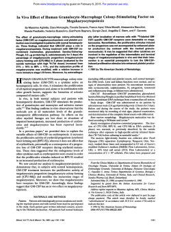
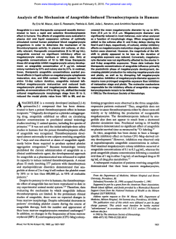
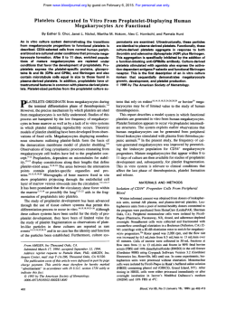
Chemokine Regulation of Human Megakaryocytopoiesis
By Alan M. Gewirtz, Jin Zhang, Janina Ratajczak, Mariusz Ratajczak, Kwang Sook Park,
Changqing Li, Zhanqing Yan, and Mortirner Poncz
We have previously shown that platelet factor 4 (PF4). a
platelet-specific CXC chemokine, can directly andspecifically
inhibit human megakaryocyte colony formation. We therefore hypothesized that PF4 might functionas anegative autocrine regulator of megakaryocytopoiesis. Herein we present additional studies characterizing the inhibitory effect
of CXC chemokines on humanmegakaryocyte development.
We first corroborated our initial studies by showing that
recombinant human (rH1 PF4, like the native protein, inhibited megakaryocytopoiesis. We then examined the inhibitory properties of other CXC family members. Neutrophil
activating peptide-2 (NAP-2). a naturally occurring N-terminally cleaved PTG peptide, was foundt o inhibit megakaryocytopoiesis with two t o three orders of magnitude greater
potency thanPF4. Structure functionstudies showed thatan
N-terminal mutation, which eliminated NAP-2's neutrophil
activating properties (NAP-2mA), also abrogated its ability
t o inhibit megakaryocyte development. Further investigations of this type
demonstrated that a chimeric PF4 protein
(AELR/PF4) in which PF4's N-terminus wasreplaced with the
first four amino
acids of NAP-P was also a potent inhibitorof
megakaryocytopoiesis. lnterleukin (IL)-8, another CXC
chemokine, and three CC chemokines (macrophage inhibitory protein-l& [MIP-la], MIP-1/3, and C101 also specifically
inhibited megakaryocyte colonyformation atNAP-2 equivalent doses. CXC and CC chemokine inhibition was additive
suggesting that the effects might be mediated through a
common pathway. The inhibitory effects of NAP-2 and MIPl a could not overcome
be
by adding physiologically relevant
amounts ofrecombinant human megakaryocyte growth and
development factor (MGDR) (50 ng/mL) t o t h ecultures. UsingNorthernblotand
reverse transcriptase-polymerase
chainreaction (RT-PCR) based analyses, we documented
mRNA expression of IL-8 receptor isoforms a and /3 in total
platelet RNA and in normal humanmegakaryocytes, respectively. Based on these results, we hypothesize that chemokines play a physiologic role in regulating megakaryocytopoiesis. Because chemokines are elaborated by ancillary
marrow cells, both autocrine and paracrine growth control
exerted, in part,
is suggested, the effects of which might be
through a and /3 IL-8 receptors.
0 7995 by The American Society of Hematology.
H
for example factor V mRNA expression, could be inhibited
by nanogram quantities of PF4, colony inhibition occurs in
the range of -25 pg/mL.I6 Though such concentrations are
routinely observed in serum, whether marrowconcentrations
of this magnitude can really be achieved in vivo is debatable.
Accordingly, we entertained the possibility that other members of the chemokine family to which PF4 and PTG belonged might possess similar, or perhaps even more potent,
inhibitory activity against the megakaryocyte lineage.I9-*'
Chemokines are 70 to 100 amino acids long and contain
four cysteine r e s i d ~ e s . l The
~ - ~ family
~
has diverged into two
main subgroups. PF4 and PTG are members of the CXC or
a chemokine family subgroup. In this group the first two of
the four conserved cysteine residues are separated from each
other by one additional amino acid residue. This subfamily
also includes interleukin-8 (IL-8), a potent neutrophil activating agent. PF4 and PTG are poor neutrophil activators,
but a cleavage product of PTG, neutrophil activating peptide-
UMAN MEGAKARYOCYTOPOIESIS and platelet
production are complex processes whose regulation
remains incompletely understood. The identification and
cloning of the c-mpl ligand, now known as thrombopoietin
(TPO) or MGDR, represents an important advance in this
area because this protein appears to be the major regulator
of megakaryocyte development. MGDR has been shown,
for example, to increase the number, size, andploidy of
megakaryocytes, and in vivo administration markedly increases platelet count in recipient anirnal~."~
Nevertheless,
MGDR is clearly not the sole regulator of the megakaryocytelplatelet axis, as innumerable cytokine experiments5"'
and knockout experiments of the MGDR receptor, c-Mpl,"
have shown.
Besides positive effectors, it is possible that megakaryocytopoiesis is regulated by inhibitory influences as well. In
this regard, it has been observed that megakaryocyte colony
growth is inferior in serum than in platelet-poor plasma suggesting that a platelet released product inhibits megakaryocyte g r o ~ t h . ' ~Megakaryocytes
"~
also appear to release products that inhibit their own development. We,and others,
have shown that one of the platelet-specific a-granule chemokines, platelet factor 4 (PF4), can inhibit megakaryocyte
colony formation in a lineage-specific fashion.I6"' Inhibition
is manifested by a decrease in the number and size of megakaryocyte colonies. In our hands,16 the related platelet-specific a-granule chemokine, PTG, does not exert similar effects on cultured cells, though conflicting findings have been
reported by others." In aggregate, these studies suggest that
megakaryocytopoiesis and thrombopoiesis maybe
controlled to an as yet undetermined extent by a negative autocrine growth loop.
One caveat in attributing a physiologic role to PF4 in
the regulation of human megakaryocyte development is the
amount of material required to observe the negative growth
effect. Though some markers of megakaryocyte maturation,
Blood, Vol 86, No 7 (October l), 1995: pp 2559-2567
Fromthe Departments of Pathology, Medicine and Pediatrics,
University of Pennsylvania School of Medicine, Philadelphia; The
Children's Hospital of Philadelphia, Philadelphia, PA; and the Department of Microbiology, Korea University School of Medicine,
Seoul, Korea.
Submitted January 9, 1995; accepted June 2, 1995.
Supported by Grants No. CA36896 to A.M.G. and HL.37419 to
M.P. from the National Institutes of Health and the Tobacco Research Council Grant No. 3152 to M.P.
Address reprint requests to Mortimer Poncz, MD, The Children 'S
Hospital of Philadelphia, 34th St and Civic Center Blvd, Philadelphia, PA 19104.
The publication costs of this article were defrayed in part by page
charge payment. This article must therefore be hereby marked
"advertisement" in accordance with I 8 U.S.C. section 1734 solely to
indicate this fact.
0 1995 by The American Society of Hematology.
0006-4971~.5/8607-0019$3.00/0
2559
2560
GEWIRTZ ET AL
2 (NAP-2), is a potent neutrophil a c t i v a t ~ r . ~Of
~ ”note,
~
two
different IL-8 receptors (IL-8R) are expressedbyneutrophils. One is designated the
a or type I IL-8R and binds
IL-8 efficiently, but NAP-2 poorly. A second /? or type 2
IL-8R has also been described, which binds IL-8 well, and
NAP-2 with moderate affinity.20.26,27The C C o r p chemokines, in which the first two cysteine residues are immediately adjacent to each other, represent the other branch of
this family. CC chemokines such as macrophage inhibitory
protein (MIP)-la**appear to be important monocyte stimulating agents andmay also selectively suppress human
megakaryocyte development. A CC chemokine receptor has been
defined.*’
The PF4 and PTG studies described above suggested that
neutrophil activating potency and megakaryocyte colony inhibitory activity paralleled each other. To test this hypothesis, we examined the effect of the neutrophil activatorNAP2 on human megakaryocyte development in vitro. We also
sought to determine the megakaryocytecolonyinhibitory
activity of other members of this chemokine family. Herein,
we present data showing that
a number of CXC and CC
chemokines are potent inhibitors of human megakaryocytopoiesis and that megakaryocytes
express a number of chemokine receptors. These data suggest that chemokines, perhaps
elaborated by ancillary cells in the marrow microenvironment, may play a physiologically significant role in regulating human megakaryocyte development by both autoctine
and paracrine mechanisms.
MATERIALS AND METHODS
Cells
Light-density bone marrow mononuclear cells (MNC) were obtained from normal remunerated consenting donors and depleted of
adherent cells and T lymphocytes (A-T-MNC) as described.3o
CD34’ were enriched from the A-T-MNC population by incubation
with anti-human progenitor cell antigen (HPCA)-1 murine monoclonal antibodies (Becton Dickinson, San Jose, CA) and subsequent
immunoselection of antibody-labeled cells with magnetic beads according tothe manufacturer’s protocol (Dynal, Oslo, Norway) as
described.” Purity of CD34’ cells selected in this manner exceeded
85%.
Hematopoietic Colony Assays
All assays were performed with either peripheral blood or bone
marrow obtained from normal consenting volunteers. Hematopoietic
In
colonies were grown and identified as previously
brief, colony assays were performed with either unseparated lightdensity marrownuclear cells (MNC) or with MNC depleted of
monocyte-macrophages and T-lymphocytes as previously described.” Final plated cell concentrations were 2 X 10’lmL. Cultures
were supplemented with 30% vollvol of aplastic anemia serum, except when supplemented with recombinant human MGDF (generously provided by Dr Pamela Hunt, Amgen Corp, Thousand Oaks,
CA).4 Megakaryocyte and granulocyte colonies were supplemented
with rH IL-3 (-29 U/mL) and rH granulocyte-macrophage colonystimulating factor (GM-CSF) (-5 ng/mL) (Genetics Institute, Cambridge, MA), except for the megakaryocyte studies that tested MGDF
effect. Erythroid colonies were supplemented with recombinant
erythropoietin (5 U/mL) (Amgen Corp). Recombinant human chemokines were added just before plating.
Megakaryocyte colonies were enumerated after 12 days in culture
by indirect immunofluorescence using either a highly-specific rabbit
antihuman platelet membrane glycoprotein antiserum’h~3”.31
or :I
monoclonal anti-GPIIbflIIa complex antibody A2AY.” Binding of
the probe antibody was detected with a species appropriate Ruorescein-conjugated secondary antibody (Meloy, Springfield, VA). A
cluster of three or more intensely fluorescent cells were defined as
one colony. Plates were read by two separate individuals, including
one blinded toplate designation. Results were in agreement between
both readers within 5% to 10%. Results are reported as the mean L
standard error (SE) of colonies enumerated.
Colony forming units-erythroid (CFU-E) were cultured for 7 days
and thenenumerated after staining with I % benzidine and hematoxylin as previously described.’” Colony-forming unit granulocyte-macrophage (CFU-GM) were cultured and identified as previously described.3”
In Vitro Synthesis of Recombinant Human Chemokines
The construction of the T,-promoter expression vectors for PF4,
NAP-2, AELRPF4, in which the N-terminal amino acid sequence
of PF4 preceding the first cysteine residue is replaced with the amino
acids A-E-L-R, and NAP-2“2’A in whichthe second amino acid
residue ofNAP-2is
mutated from glutamic acidto alanine has
been previously described.” These vectors wereusedto
express
BL21(DE3)pLys S (Novarecombinant proteins in Escherichia coli
gen. Madison, WI), which were allowed to grow to an optical density
(OD),,of
-0.9, which was followed byan additional 3 hours of
growth in 1 mmol/L isopropyl-thiogalactopyranoside that induced
recombinant protein expression.
deThe recombinant proteins were processed aspreviously
~ c r i b e d . ’Bacteria
~ . ~ ~ were centrifuged at 3,OOO rpm in a Sorvall GSA
rotor for 10 minutes andthen resuspended in %,,th the volume of
TED (50 mmollL Tris HCI, pH 8.0; I mmol/L EDTA; and I mmotl
L dithiothreitol [D=]). The cells were recentrifuged andresuspended in the same volume of TED with 0.1 mg/mL of lysozyme
added for 30 minutes. The lysate was sonicated at 4°C three times
with a Branson Soniiier (Branson Ultrasonics, Danbury, CT) using
a microtip at a power of 6 for 1 minute each cycle. The samples
were centrifuged for 10 minutes in a Sorvall GSA rotor for l 0
minutes at 10,000 rpm, andthe supernatants were collected and
stored.
Recombinant proteins were purified from the supernatants of bacterial lysates described above by a two-step procedure. Initially, they
were applied to a heparin agarose column (Sigma Chemical CO, St
Louis, MO), washed with TE buffer (50 mmoln Tris HCl, pH 8.0.
and 1 m m o a EDTA) containing 0.15 m o l n NaCl and eluted with
TE buffer containing 0.5 mollL NaCl for all the NAP-2 proteins and
1.5 moULNaCl for all the PF4 proteins. The eluted proteins were
concentrated usingYM3 Amico ultrafiltration filters (W.R. Grace
C O ,Beverly, MA), and the buffer was switched to 0.1% trifluoroacetic acid. The concentrated samples were further fractionated on reverse phase high performance liquid chromatography (HPLC).
All purified proteins wererun on 20% precast sodium dodecyl
sulfate-polyacrylamide gel electrophoresis (SDS-PAGE) underreduced condition followed by Coomassie blue staining. Further identification was done by enzyme-linked immunosorbent assay (ELBA1
as previously described.” Recombinant protein concentrations were
determined using a Coomassie Protein Assay Reagent Kit with bovine serum albumin as a standard (Pierce, Rockford, IL).
Recombinant 72 amino acid IL-8 protein was purchased (R & D
Inc, Minneapolis, MN), and MIP- la and 0 were generous gifts from
Drs Barbara Sherry and Anthony Cerami (Rockefeller University,
New York, NY).” Medium containing the CC chemokine C-l0 and
control medium were generously provided by Dr Mark Berger (University of Pennsylvania, Philadelphia).
2561
CHEMOKINEREGULATION OF HUMAN MEGAKARYOCYTOPOIESIS
similar to those we previously reported with highly purified,
serum-derived material. Mature rH PF4 protein was syntheThe IL-8Ra and IL-8RP cDNAs were kindly provided by Dr
sized in Escherichia coli as described in Materials and MethIngrid U. SchraufsWer (Scripps Institute, La Jolla, CA) in the exods. Its amino acid sequence was identical to the native
pression vector ~ s F F V . n e oThe
. ~ ~ 1.2-kb coding regions for both
protein, except that the recombinant material retained an
the IL-8Ra and IL-8RP cDNAs, the 3.3-kb platelet glycoprotein IIb
initiating methionine residue.34Biologic integrity of the engi(GPIlb), and the 0.4-kb P G cDNA inserts”-38 were released from
their respective vectors by EcoRI digestion. All of the above cDNA
neeredprotein was demonstrated in chemotactic studies
inserts were purified from an agarose gel using GENECLEAN
where the rH PF4 was as effective as serum-derived PF4.34
(BiolOl, Vista, CA). These inserts were then labeled using ( Y - ~ ~ P - In a plasma clot assay, both recombinant and native material
dCTP, random primers, and Klenow to -lo* CPM/pg DNA.
inhibited megakaryocyte colony formation in an identical
Total platelet RNA was prepared from platelet rich plasma (PRP)
manner throughout the dose range tested (Fig 1A). In
obtained from 1 0 0 mL of sodium citrate anticoagulated blood. Only
agreement with our previously reported results, inhibition
the top two-thirds of the PRP was used. After spinning down the
was dose-dependent and most significant at concentrations
platelets, platelet RNA was prepared using the guanidinium thiocya2 10 pg/mL.I6 Also in agreement with our previous results,
The amount
natdacid phenol single-step RNA isolation te~hnique.’~
the numbers of cells/colony (Fig 1B) and the size of cells
of RNA applied per lane was equivalent to -25% of the total yield.
The RNA was fractionated on gels containing formaldehyde folcomprising the colonies (Fig 1C) were also diminished in the
lowed by transfer to Genescreen Plus membrane (DuPont CO, Wilpresence of exogenous PF4. Finally, inhibition was megamington, DE) and hybridization to the labeled probe as previously
karyocyte lineage specific (data not shown).
IL-8Ra and IL-8RP Platelet Northern Blot
-
described.40
Reverse Transcriptase-Polymerase Chain Reaction of
Purijied Megakaryocyte mRNA
Human megakaryocytes were isolated from the marrow of remunerated, normal addt volunteers. The isolation of greater than 99%
pure megakaryocytes was accomplished as previously described using counterflow centrifugal elutriation (Beckman J2-21M; Standard
elutriation motor; Beckman Instruments; Mountainview, CA) to obtain an initial megakaryocyte enrichment:’ followed by isolation of
morphologically recognizable mature megakaryocytes with a micromanipulator. RNA was isolated from -50 megakaryocytes in less
than 3 hours’ time.
mRNA was isolated from these cells using the Quick-Pre mRNA
Purification Kit (Pharmacia, Piscataway, NJ). The final mRNApellet
was washed with 75% ethanol and resuspended in 10 pL of triple
distilled autoclaved water. Reverse transcription of the megakaryocyte mRNA was performed using 4 pL of the original sample heated
to 65°C for 10 minutes and then cooled on ice for 3 minutes. A total
of 100 U of Moloney murine leukemia reverse transcriptase (RT)
(GIBCO BRL, Gaithersburg, MD), 50 ng of random primers (Boehringer Mannheim, Indianapolis, IN), 40 U of RNAzin (Promega,
Madison, WI), and dNTPs (50 pmoVL each) were added to the tube
and incubated for 1 hour at 37°C. Specific oligo primer pairs used for
the polymerase chain reaction (PCR) amplification with the megakaryocyte random cDNA were as follows: II-8Ra: 5“ATGTCAAATATTACAGATCC-3’ (sense oligonucleotide; 1 to 20 base, beginning at the transcriptional start site) and 5”AGATTCATAGACAGTCCCCA-3’ (antisense, 500 to 481 base);and IL-8Rp: 5’GAGGACCCAGGTGATCCAGG-3’ (sense, 816 to 835 base) and
5‘-GAGAGTAGTGGAAGTGTGCC-3‘ (antisense, 1065 to 1046
The anticipated PCR products are, therefore, 500 bp and
249 bp for IL-8Ra and P, respectively. Ten microliters of the reverse
transcription reaction or 100 ng of the appropriate IL-8R cDNA or
water was used in a 100-pL PCR reaction using 30 ng of both sense
and antisense primers for each receptor and 2.5 U Taq polymerase
(Promega) using manufacturer’s supplied buffer and 2.5 pmoVL
MgClz. PCR conditions were melting at 95°C for 1 minute, annealing
at 60°C, and extension at 72°C for 30 rounds. A total of 10 pL of
each final PCR product was size-fractionated on a 1.2% agarose gel.
RESULTS
Hematopoietic Effects of Recombinant PF4
We firstdetermined whether rH PF4 made in a prokaryotic
system would manifest effects on megakaryocytopoiesis
NAP-2, AELWPF4 and Mutation Proteins
We have previously shown that PTG does not inhibit in
vitro megakaryocytopoiesis.’6However, it is now known that
a neutrophil cathepsin G N-terminal cleavage product of
PTG, a 70-amino acid protein termed NAP-2, is a biologically active form of this protein. NAP-2 is a potent activator
of neutrophils, binding to the IL-8RP on neutrophils.26To
extend our structure/function studies and to begin to understand the mechanismby which chemokines inhibit megakaryocyte development, we tested whether NAP-2, as opposed
to PTG, would inhibit megakaryocytopoiesis. As shown in
Fig 2A, megakaryocyte colony formation wasmuchmore
sensitive to the inhibitory effects of NAP-2 than PF4. Where
PF4 inhibited in the pg/rnL range, half maximal inhibitory
concentrations of NAP-2 were in the 10 to 100 ng/mL range.
These concentrations were comparable to those needed for
NAP-2 to activate neutrophil^.^^ As was the case with PF4,
NAP-2’s inhibitory effect was lineage-specific. It has previously been reported that at PF4 concentrations 2 10 pg/mL
inhibition of CFU-E and C m - G M might be ~bserved.”.’~
However, we did not observe such effects in our cultures
and no definitive changes in CFU-E or CFU-GM colony
formation were observed with increasing doses of NAP-2.
We also sought to determine if NAP-2 inhibited megakaryocyte development directly, or indirectly via a secondary
effect on accessory marrow cells. To address this question,
marrow mononuclear cells were depleted of monocyte-macrophages and T-lymphocytes and then enriched for CD34+
cells using immunomagnetic beads before exposure to NAP2. Purity of the CD34+ cells in this population consistently
exceeded 85%. At 50 pg/mL, PF4 decreased the number of
megakaryocyte colonies to 8% of the control, and at 750 ngl
mL, NAP-2 decreased expression to 10% of the control.
These results support the hypothesis that the chemokines
tested exerted a direct inhibitory effect on megakaryocyte
progenitor cells.
Because NAP-2 is thought to activate neutrophils through
IL-8R0, we tested other NAP-2 and PF4 mutant constructs
whose interactions with the neutrophil IL-8 receptors have
been previously defined.33For example, alanine substitutions
GEWIRTZ ET AL
2562
I
I
3 5 3 00 2 5 2 5
0 15 10
I
PF4
I
I
I
I
I
40
@g/ml)
no PF4 supplement
0
3 5 53 0 2 51 02 0 1 5
PF4
40
(pg/ml)
PF4 supplement
Fig 1. Inhibition of megakaryocytopoiesis by rPF4. (A) Percent decrease in the number of colonies seen per lo5 marrow cells plated at
increasing concentrations of PF4. (01,rH PF4; (0).native PF4. (B)The total numbers of cells seen per colony with increasing concentrations
of rPF4 are shown. (C) Immunofluorescence of typical megakaryocyte colonies with (bottom) and without rH PF4 (top), demonstrating the
decrease in the size of the colony andof the megakaryocytes within thecolony. Data in (A) and (B) represent analysis of two or moreseparate
experiments with four separate plates for each data point. The number of megakaryocyte colonies seen without added chemokine ranged
from 56 t o 107 per plate.
at the N-terminus of NAP-2 markedly inhibit its ability to
activate neutrophils. Interestingly, NAP-2""*, in
which the
second amino acid is mutated from glutamic acid to alanine
(Table l), also loses its ability to inhibit megakaryocytopoiesis (Fig 2B). In contrast, AELRPF4, in which PF4's
first eight amino acids proximal to the first cysteine residue
are replaced with the four NAP-2 N-amino acid residues
preceding its first cysteine residue, is equivalent to NAP-2
in terms of its ability to activate neutrophils and to inhibit
megakaryocyte colony formation (Fig 2B).
Inhibitory Effects of Other Chemokines
We tested additional chemokines to see whether they too
could inhibit megakaryocytopoiesis. IL-8 was almost as effective as NAP-2 in inhibiting megakaryocytopoiesis in the
5 to 500 ng/mL concentration range (Fig 3A). Surprisingly,
the CC chemokine MIP-la was equally effective (Fig 3A).
Inhibition with both of these chemokines was again noted
to be lineage specific (data not shown). Two additional CC
chemokines were tested and also inhibited megakaryocytopoiesis. MIP- 10 decreased megakaryocyte colony formation
to 52% of the untreated control value at 500 ng/mL, and a
culture supernatant containing C-IO, another member of the
CC chemokine family, decreased megakaryocyte colonies to
35% of control values at a concentration of 2% (vol/vol).
We thensought to determine whether the inhibitory effects
of the CXC and CC chemokines might be additive or synergistic. Progenitor cells were therefore cultured with the individual chemokines at varying doses, or with the two added
together. As shown in Fig 3B, the combination of NAP-2 and
MIP-la had greater inhibitory effect than either chemokine
individually. There does not appear to be a synergistic effect.
REGULATIONCHEMOKINE
OF HUMAN MEGAKARYOCYTOPOIESIS
chemokine dose, megakaryocyte colony formation was not
completely extinguished. When compared with an untreated
control group, -30% of maximal colony formation was still
observed even atthe highest combined dose level. The nature
of the difference@)between this resistant subpopulation and
those progenitor cells that were sensitive to the chemokine
inhibitory effects remains to be determined.
Finally, we also determined whether thechemokine inhibitory effect could be abrogated by MGDF stimulation. We
first established that, in our culture system, megakaryocyte
colony growth was maximal at MGDF doses -20 ng/mL.
At such doses, the number ofcolonies observed in the culture
dishes was comparable to the number observed when optimal
concentrations of aplastic anemia serum plus L - 3 and E-6
were used as stimulators. We then cultured progenitor cells
in the presence of 50 ng/mL of MGDF and varying doses
of either NAP-2 or MIP-la. As shown inFig 4,in comparison to the numbers of colonies obtained in the presence of
MGDF alone, NAP-2 at 2 5 ng/mL and MIF"1a at >50 ng/
mL significantly inhibited colony formation. Therefore, even
in the presence of a significant physiologic stimulator, used
at maximally effective concentrations, these chemokines appeared capable of blunting megakaryocyte colony growth.
" - 1
B
2563
'"7
Demonstration of the Expression of IL-8Ra and IL-8Rp by
Megakaryocytes
0 '
d
;OO
1'01
1'02
NAP-2
1'0s
1'04
1'0s
1'06
1'07
(ng/ml)
Fig 2. inhibition of m.gakaryocytopoi.rh by NAP-2 and related
constructs. (A) Percant decr.rw in the number of megakaryocyte
colonies aeon par I C marrow caih plated at incroasing concentrations of NAP-2. (01,CFU-Meg colonies; (m), CFU-GM colonies; and
(AI, CFU-E colonin. PM studlrat 25 pglmL were also done. (0).
CFU-Mag colonies; (U), CFU-GM colonies; and (A), CFU-E colonies.
Error bar nfm to OM standard devlatlon for r m p l n studied four
or mora times. IBI Analyois of CFU-Meg colony n u m h forPF4
(01. NAP-2 (01,NAP-2(X), and AEWPF4 (t). Data In (AI and (B)
analysis of two or mora .rpernte exporimento wlth four
soparota plates for r c h doto point. The numbor of megakaryocyte
colonin m n without added chemokine rangedfrom 51 to 128 per
pinta.
Rather, the combination of NAP-2 and MIP-la better approximates an additive effect. If the inhibition observed is
additive, CXC and CC chemokines may inhibit megakaryocyte development by signaling through a common pathway.
It should also be noted that even at the highest combined
The above studies suggested that chemokines exert a direct effect on megakaryocyte development. If our interpretation ofthe experiments was correct, it follows that megakaryocytes should express the receptors that bind these ligands.
To provide these important data, we pursued two independent, but complementary approaches. First, we isolated total
platelet RNA and looked for E-8Ra and IL-8R@-A.
The presence of these messages in the total RNA pool would
provide evidence that these receptors might, in fact, be expressed by megakaryocytes. We
also performed RT-PCRfor
these same messages on an essentially pure population of
normal human megakaryocytes.
Platelet RNA was extracted from the top two-thirds of
platelet-rich plasma of low-speed centrifuged blood. White
cell contamination was approximately 1 cell per 5,000 platelets. Duplicate lanes of total RNA were hybridizedto cDNAs
for the L-8Ra and IL-8RP and platelet GPIIb and P G as
platelet-specific positive controls. As seen in Fig 5A, there
were single detectable bands of the expected size of -3 kb
for both of the L - 8 receptors and 3.3 kb for GPIIb.38.42 An
intense signal was detected with pTG at 0.8 kb with additional bands seen at 1.2 and 1.8 kb.Total peripheral blood
neutrophils detect two major L-8R bands of 2.4 and 3.0 kb,
-
Table 1. Recombinant PF4 and NAP-2 Chemoklnes
Name
Amino Acid Sequence
NAP-2 (wild type)
NAP-2EW
PF4 (wild type)
AELWPF4
AELRCMC
THCN
DGRKICLDPDAPRIKKIVQKLAGDESAD
+RCMC.
THCN. . DGRKICLDPDAPRIKKIVQKLAGDESAD
EAEEDGDLQCLC
PHCP . NGRKICLDLQAPLYKKIIKKLLES
e C L C . . PHCP
.NGRKICLDLQAPLYKKIIKLLES
Amino acid differences from wild type are underlined.
...
..
...
...
.
.
. .
..
GEWIRTZ ET AL
2564
2
3
o
7
0-
-\ a
0
2
;
0
0
O
3
m
.
a
m
a
\ 2 ag\
- 5
"
0
O
Ya
d
MIP-1 a
IL-8
a
g
m
=
z
Y
g
0
z
2.5
NAP-2
while HL60 cell lines only express the 3.0 kb band, similar
to the single band we detected in platelet RNA.4'
Even though the blots depicted in Fig 5 were hybridized
under high stringency conditions, it is possible that we were
only detecting a single IL-8R species because of the -70%
homology in the coding regions of the IL-8R cDNAs. Therefore, to provide additional proof that we were, in fact, detecting both IL-8Ra and IL-gRP, weperformedRT-PCR
reactions on mRNA isolated from a small number of 100%
pure
normal
human megakaryocytes!3 Primers chosen were
unique for the two IL-8 receptors, as shown by the fact that
the primers do not yield cross-amplify using IL-8 receptor
cDNAs (data not shown). These RT-PCR studies demonstrate that
both
IL-8 receptors are expressed in megakaryocytes (Fig 5B).
DISCUSSION
o
k
T
L
X
O
Chemokine
added
(ng/ml)
Fig 3. Inhibitory effects of CXC and CC chemokines on megakaryocytopoiesis. (AI The inhibitory effect of 11-8 and MIP-la onmegakaryocyte formation relative t o an untreated control. NAP2 data
done simultaneously are also included. Data represent the average
of two experiments with four plates readeach
for point in each experiment. Errorbars refer t o one SD. At every concentration shown, the
inhibitory effect was significant atP c .003. (B)Effect of combined
treatment withNAP-2 and MIP-la.Megakaryocyte colony formation
was tested withNAP-2 ( W , MIP-la (01,or both (XI. Error bar refers
t o 1 SD for samples studied four or more times.Data in (A) and (El
represent analysis of two or more separate experiments with four
separate plates for each data point.
100461
0.04
0
T
T 0.009
S
NAP-2
We have previously shown that PF4, a platelet-specific
CXC chemokine, can directly inhibit in vitro megakaryocytopoiesis. We now show that other CXC chemokines, NAP2, IL-8, and even the more distantly related CC chemokines,
MIP-la and P, and C10, have a similar direct inhibitory
effect on in vitro megakaryocyte development as manifested
by the appearance of fewer colonies composed of smaller
numbers of less mature cells. Inhibition is observed when
chemokines are added to progenitor cell cultures at concentrations equivalent to those at which theyactivate neutrophils
and monocytes,-" suggesting that the inhibitory signals they
generate maybeof
physiologic significance. Studies performed with mutated NAP-2 and PF4 proteins also support
this hypothesis as the ability to activate neutrophils and to
inhibit megakaryocytopoiesis closely parallel each other.
Another highly suggestive finding is that megakaryocytes
express both a and isoforms of the IL-8 receptor. Nevertheless, although the presence of these receptors provides a
mechanism for the observed inhibitory effects of some of
the CXC chemokines, further studies will be needed to define
the full repertoire of megakaryocyte chemokine receptors
and, in particular, whether CC chemokine receptors also
exist on these cells.
.L8
10
0.y02
5'0 5 0 0
(ng/ml)
5
5'0 560
M I P - l a (ng/rnl)
Fig 4. Interaction of MGDF and chemokines. Inhibitory studies with NAP-2 and MIP-la were performed in the presence of 50 ng/mL of rH MGDF.
Data represent the average of two experiments with
four plates read for each point in each experiment.
Error barsrefer t o one SD. The Pvalue foreach study
is shownabove the error bar.
CHEMOKINE REGULATION OF HUMAN MEGAKARYOCYTOPOIESIS
2565
Although many stimulatory cytokines have been described,
relativelyfewinhibitoryproteinshavebeenelucidated.The
best-studied examples of these include the interferons (INFs),
W
tumor necrosis factor-a (TNF-a),transforming growth factorlp (TGF-p), and MP-la.""' Thephysiologicroleofthese
c?!
cytokines in the regulation ofhuman hematopoiesis remains
speculative.44 This is, in part, because the biologic effects of
kb
thesecytokinesarecontextdependent,
a situationthathas
tendedtogenerateapparentlycontradictoryreportsontheir
activity. For example, M P - l a appears to specifically inhibit
4.4
the growth of cycling hematopoietic cells,46 but when admixed
with IL-3 or GM-CSF appears to promote colony growth in a
synergistic manner:'In
addition, TNF-a and the INFs appear
to be general suppressers of hematopoietic cell growth, while
2.4
the activity of TGF-p is similar to MIP-la in that it appears
to block proliferation of growth factor stimulated progenitor
cells, but alone may stimulate CFU-GM and CFU-E.These
observationshavepromptedspeculationthatsomeofthese
inhibitors, in particular TGF-p and MP-la, may play a role
in maintaining stem cells in a Go state when hematopoiesis is
otherwise stimulated." In support of this hypothesis,it has been
reportedthatinhibitionof
TGF-p expression with antisense
DNA increases the apparent numberof assayable multilineage
progenitor cells in cord blood?R These apparently discordant
results may also be explained by the observation that the inhibitory effects of M P - l a and TGF-p depend on the maturation
of theprogenitorthathasbeenexposedtothischemokine.4".49.%1 Many investigatorshavereportedthat
MP-la
either has no effect on, or actually stimulates the growth of
morematureprogenitors,whileitsuppressesthegrowthof
more primitive C ~ I I The
S ~ fact
~ ~ that
~ ~ the chemokines we have
investigated have no apparent effect on CFU-GM derived colony formation is in accord with these results. Why the apparently mature CFU-Meg
are inhibited by these chemokines when
CFU-GM are not, remains unknown.
Whether the findings we report are of physiologic significance remains unclear. The fact that members of this family
can inhibit megakaryocyte development at concentrations
three orders of magnitude lower than PF4 is suggestive of
physiologic relevance because concentrations in this range
are likely achievable in vivo. Further, the ability to have
significant inhibition despite maximal levels of TPO also
supports a potential in vivo role. Nevertheless, establishing
whether there is a correlation between megakaryocyte/platelet mass and chemokine concentrations, either in plasma or
in the marrow, will have to be investigated. This willbe
particularly important in patients with inflammatory states
who maybe expected to have relatively high circulating
"
chemokine levels, and perhaps paradoxically, reactive
thrombocytosis. Measurement of MGDF levels will be of
Ra
equal importance then, as we have already speculated that
Fig 5. Expression of IL-8Ra and f3 RNA in megakaryocytes. (A)
stimulatory growth factors may overdrive the effects of apNorthern blot analysis of totalplateletRNAprobed
with the
parently
weaker negative regulators.'" Indeed, we have also
cDNAs for pTG, GPllb, IL-8Rrr, and IL-8Rf3. (B) Agarose gel of the
hypothesized that negative regulatory loops, whether autoRT-PCR products of IL-8Ra and p. Lane 1, mRNA from isolated
megakaryocytes, Lane 2, the cDNA for appropriate receptor, and
crine or paracrine in nature, are more likely to play a role
Lane 3, water control. Size markers are indicated at the side of
in regulating basal, or nonstimulated, megakaryocyte proboth (A) and (B).
duction." If shown to be of physiologic significance, these
studies may represent a significant advance in the development of new pharmacologic strategies to regulate disordered
or inappropriate thrombopoiesis.
A
7.5
m
I
t
B
M 1 2 3 1 2 3
I L-8
IL-8RR
2566
GEWIRTZ ET AL
N, Izaquirre C: The growth of large
14.MessnerHA,Jamal
megakaryocyte colonies from human bone marrow. J Cell Physiol
We thank Dr Pamela Hunt at AmgenCorp (Thousand Oaks,CA)
1:45, 1994
for the MGDF, Ingrid U. Schraufstlitter at the Scripps Institute (La
JS, Harker
15.Kimura HS, BursteinSA,ThorningD,Powell
Jolla, CA) for the E-8R cDNAs, and Dr Mark Berger at the UniverLA, Fialkow PJ, AdamsonJ W : Human megakaryocytic progenitors
sity of Pennsylvania (Philadelphia, PA) for the C10-containing me(CFU-M) assayed in methylcellulose: Physical characteristics
and
dium.
requirements for growth. J Cell Physiol 118:87, 1984
16. Gewirtz AM, Calabretta B, Rucinski B, Niewiarowski S, Xu
REFERENCES
WY: Inhibition of human megakaryocytopoiesis in vitroby platelet
1. De SauvageFJ,HassPE,SpencerSD,MalloyBE,Gurney
factor 4 (PF4) and a synthetic COOH-terminal PF4 peptide. J Clin
Invest 83:1477, 1989
AL, SpencerSA,DarbonneWC,
Hemel WJ, Wong SC, Kuang
W-J, Oles KJ, Hultgren B, Solberg LA, Jr., Goeddel DV, Eaton DL:
17.HanZC,SenskbeL,AbgrallJF,
Bribe J: Platelet factor 4
Stimulation of megakaryocytopoiesis and thrombopoiesis by the cinhibits human megakaryocytopoiesis in vitro. Blood751234, 1990
Mpl ligand. Nature 369:533, 1994
18. Han ZC, Bellucci S,Tenza D, Caen JP: Negative regulation
2. Luk S, Kaushansky K, HollyRD, Kuijper JL, Lofton-Day CE,
of human rnegakaryocytopoiesisby human platelet factor 4 and beta
Oort PJ, Grant FJ, Heipel MD, BurkheadSK, Kramer JM, Bell LA,
thromboglobulin:Comparativeanalysisinbone
m o w cultures
Sprecher CA, Blumberg H, Johnson R, Prunkard D, Ching
AFT,
from normal individuals and patients with essential thrombocythaeMathewes SL, Bailey MC, Forstrom JW, Buddle MM, Osbom SG,
mia and immune thrombocytopenic purpura. Br J Haematol 74:395,
Evans SJ, Sheppard PO, F’resnell SR Cloning and expression of
1990
murine thrombopoietin cDNA and stimulation
of platelet production
19. Baggiolini M, Dewald B, Moser B: Interleukin-8 and related
in vivo. Nature 369565, 1994
chemotactic cytokines-CXC and CC chemokines. Adv Immunol
3. Kaushansky K, Lok S. Holly R D , Broudy VC, Lin N, Bailey
55:97, 1994
MC, ForstromJ W , Buddle MM, Oort PJ, HagenFS,Roth GI, Papa20.Horuk R: The interleukin-8-receptorfamily:Fromchemoyannopoulou T, Foster DC: Promotion of megakaryocyte progenitor
kines to malaria. Immunol Today 15:169, 1994
expansion and differentiation by the c-Mpl ligand thrombopoietin.
21. Mantovani A, Sozzani S : Chemokines. Lancet 343:923, 1994
Nature 369:568, 1994
22. Walz A, Baggiolini M: A novel cleavage product
of 0-throm4. Bartley T D , Bogenberger J, Hunt P, Li Y-S, Lu HS, Martin
boglobulin formed in cultures of stimulated mononuclear cellsactiF, Chang M-S, Samal B, Nichol L,Swift S , JohnsonMJ,Hsu
vates human neutrophils. Biochem Biophys Res Commun 159:969,
R-Y, Parker VP, Suggs S , Skrine JD, Merewether LA, Clogston C,
1989
Hsu E, Hokom MM, Hornkohl A, Choi E, Pangelinan M, Sun Y,
23. Walz A, Dewald B, Tscharner V, Baggiolini M: Effect of
Mar V: Identification and cloning of a megakaryocyte growth and
neutrophil-activating peptide NAP-2,platelet basic protein, connecdevelopment factor that is a ligand for the cytokine receptor Mpl.
tivetissue-activatingpeptide m,andplatelet factor 4onhuman
Cell 77:1117, 1994
neutrophils. J Exp Med 170:1745, 1989
5. Leary AG, Yang YC, ClarkSC, Gasson JC, GoldeDW, Ogawa
24. Walz A, Baggiolini M: Generation of the neutrophil-activatM: Recombinant gibbon Interleukin 3 supports formation of human
ing peptide NAP-2 from platelet basic protein or connective tissue
multilineage colonies and blast cell colonies in culture: Comparison
peptide 111 through monocyte protease. J Exp Med 171:449, 1990
with recombinant human granulocyte-macrophage colony stimulat25. Holt JC, Yan Z, Lu W, Stewart GJ, Niewiarowski
S: Isolation,
ing factor. Blood 701343, 1987
characterization, and immunological detection of neutrophil activat6. WarrenMK,ConroyLB.Rose
JS: Therole of interleukin
ing peptide 2: A proteolytic degradation product of platelet basic
6 and interleukin 1 in megakaryocyte development. Exp Hematol
protein. Proc Soc Exp Biology Med 199:171, 1991
17:1095, 1989
26. Leonard El, Yoshimura T, Rot A, Noer K, Walz A, Baggiolini
7. Williams N, De Giorgio T, Banu N, Withy R, Hirano T, KishiM: Chemotactic activity and receptor binding of neutrophil attractmot0T:Recornbinantinterleukin
6 stimulatesimmaturemurine
ant/activation protein-l (NAP-l) and structurally related host demegakaryocytes. Exp Hematol 18:69, 1990
fense cytokines: Interaction of NAP-2 with the NAP-l receptor. J
8. Quesenberry PJ, McGrath HE, Williams M E , Robinson BE,
Leukoc Biol49:258, 1991
Deacon DH,Clark S, Urdal D, McNiece I K Multifactor stimulation
27. Murphy PM, Tiffany HL: Cloning of complementary DNA
of megakaryocytopoiesis: Effects of interleukin 6. Exp Hematol
encoding
functional
a
human
interleukin-8 receptor.
Science
19:35,1991
253:1280, 1991
9. Takahashi T,Tsuyuoka R, Ueda Y, Suzuki A, IchibaS, Okuno
28. Koch AE, Kunkel SL, Harlow LA, Mazarakis DD, Haines
Y, Nakamura K,Imura H: Megakaryocyte potentiating activity of
GK, BurdickMD, Pope RM, Strieter RM: Macrophage inflammatory
IL-l, IL-6 and GM-CSF as evaluated by their action on in vitro
protein-la. A novel chemotactic cytokinefor macrophages in rheuhuman megakaryocytic colonies. Br J Haematol78:480, 1991
matoid arthritis. J Clin Invest 93:921, 1994
10. Kaushansky K, O’Hara P, Bemer K, Segal GM, Hagen FS,
29. Neote K, Digregorio D, Mak JY, Honk R, Schall TJ: MolecuAdamson W. Genomic cloning, characterization and multilineage
lar-cloning, functional expression, and signaling characteristics of a
growth-promoting activity of human granulocyte macrophage colC-C chemokine receptor. J Immunol72415, 1994
ony-stimulating factor. Proc Natl Acad Sci USA 83:3101, 1986
30. Ratajczak M Z , Kuczynski W,Onodera K, Moore J, Ratajc11. Briddell RA, Bruno E, Cooper RJ, Brandt J E , Hoffman R
zak
J, Kregenow DA, DeRiel K, Gewirtz AM. A reappraisal of the
Effect ofc-kit ligand on in vitro human megakaryocytopoiesis. Blood
role of the insulin like growth factor-I (IGF-I) in the regulation of
78:2854, 1991
the human hematopoiesis. J Clin Invest 94:320, 1994
MW:
12.Gurney AL, Carvermoore K, DesavageFJ,Moore
31. Gewirtz A M , Xu WY, Mangan KF: Role of natural killer
Thrombocytopenia in c-Mpl-deficient mice. Science 265:1445, 1994
cells, in comparisonwith T lymphocytes and monocytes in the regu13. Vainchenker W, ChapmanJ, Deschamps JF, Vinci G, Bouget
lation of normal human megakaryocytopoiesis.J Immunol 139:2915,
J, Titeux M, Breton-Gorius J Normal human serum contains a fac1987
tor(s) capable of inhibiting megakaryocyte colony formation. Exp
32.BennettJS, Hoxie JA,Leitman SF, Vilaire G, CinesDB:
Hematol 10650, 1982
ACKNOWLEDGMENT
CHEMOKINEREGULATION OF HUMAN MEGAKARYOCYTOPOIESIS
Inhibition of fibrinogen binding to stimulated human platelets by a
monoclonal antibody. Proc Natl Acad Sci USA 80:2417, 1983
33. Yan Z, Zhang J, Holt JC, Stewart GJ, Niewiarowski S, Poncz
M: Structural requirements of platelet chemokines for neutrophil
activation. Blood 84:2334, 1994
34. Park KS, Rifat S, Eck H, Adachi K, Surrey S, Poncz M:
Biologic and biochemic properties of recombinant platelet factor 4
demonstrate identity with the native protein. Blood 75:1290, 1990
35. Wang JM, Sherry B, Fivash MJ, Kelvin DJ, Oppenheim JJ:
Human recombinant macrophage inflammatory protein-l alpha and
-beta and monocyte chemotactic and activating factor utilize common and unique receptors on human monocytes. J Immunol
150:3022, 1993
36. Schraufsttitter IU, Barritt DS, Ma M, Oades ZG, Cochrane
CG: Multiple sites on IL-8responsible for binding to Q and p IL8 receptors. J Immunol 151:6418, 1993
37. Majumdar S, Gonder D, Koutsis B, Poncz M: Characterization of the human P-thromboglobulin gene. Comparison with the
gene for platelet factor 4. J Biol Chem 2665785, 1991
38. Poncz M, Eisman R, Heidenreich R, Silver SM, Vilaire G,
Surrey S, Schwartz E, Bennett JS: Structure of the platelet membrane
glycoprotein IIb: Homology to the alpha subunits of the vitronectin
and fibronectin receptors. J Biol Chem 262:8476, 1987
39. Newman PJ,Gorski J, White GCII, Gidwitz S, Cretney CJ,
Aster RH: Enzymatic amplification of platelet-specific messenger
RNA using the polymerase chain reaction. J Clin Invest 82:739,
1988
40. Alwine JC: Hybridization analysis of specific RNAs by electrophoretic separation in agarose gels and transfer to diazobenzyloxymethyl paper. Gene Amplif Anal 2565, 1981
41. Gewirtz AM, Keefer M, Doshi A, Annamali HC, Chiu C,
Colman RW: Biology of human megakaryocyte factor V. Blood
67:1639, 1986
42. Moser B, Barella L, Mattei S, Schumacher C, Boulay F,
Colombo MP, Baggiolini M: Expression of transcripts for two in-
2567
terleukin 8 receptors in human phagocytes, lymphocytes and melanoma cells. Biochem J 294285, 1993
43. Gewirtz A, Boghosian-Sell L, Catani L, Ratajczak M, Shen
YM, Schreiber AD. Expression of FcyRII and CD4 receptors by
normal human megakaryocytes. Exp Hematol20:512, 1992
44. Ogawa M. Differentiation and proliferation of hematopoietic
stem cells. Blood 81:2844, 1993
45. Keller JR, Bartelmez SH, Sitnicka E, Ruscetti F W , Ortiz M,
Gooya JM, Jacobsen SEW Distinct and overlapping direct effects
of macrophage inflammatory protein-la and transforming growth
factor p on hematopoietic progenitor/stem cell growth. Blood
84:2175, 1994
46. Jacobsen SEW, Ruscetti F W , Oniz M, Gooya JM, Keller JR:
The growth response of Lin-Thy-l+ hematopoietic progenitors to
cytokines is determined by the balance between synergy of multiple
stimulators and negative cooperation of multipIe inhibitors. Exp
Hematot 22:985, 1994
47. Mantel C, Kim YJ, Cooper S, Dwon B, Broxmeyer HE. Polymerization of murine macrophage inflammatory protein l a inactivates its myelosuppressive effects in vitro: The active form is a
monomer. Proc Natl Acad Sci USA 90:2232, 1993
48. Cardoso AA, Li ML, Batard P, Hatzfeld A, Brown EL, Levesque JP, Sookdeo H, Panterne B, Sansilvestri P, Clark SC, Hatzfeld
J. Release from quiescence of CD34TD38- human umbilical cord
blood cells reveals their potentiality to engraft adults. Proc Natl
Acad Sci USA 908707, 1993
49. Broxmeyer HE, Sherry B, Lu L, Cooper S, Oh K-0, TekampOlson P, Kwon BS, Cerami A. Enhancing and suppressing effects
of recombinant murine macrophage inflammatory protein la, an
inhibitor of primitive hematopoietic cells. Roc Natl Acad Sci USA
90:12015, 1990
50. Mayani H, Little MT, Dragowska W, Thornbury G, Lansdorp
PM. Differential effects of the hematopoietic inhibitors MP-la,
TGF-p, and TNF-a on cytokine-induced proliferation of subpopulations of CD34+ cells purified from cord blood and fetal liver. Exp
Hematol 23:422, 1995
© Copyright 2026