
Red Blood Cell Glycophorins
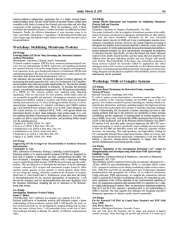
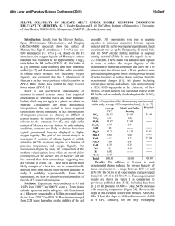
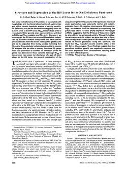
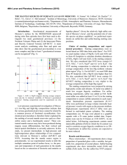
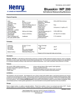
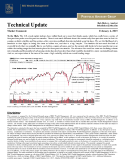
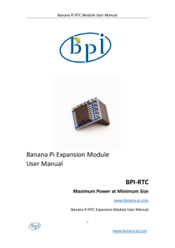
From www.bloodjournal.org by guest on February 6, 2015. For personal use only.
REMEW ARTICLE
Red Blood Cell Glycophorins
By Joel Anne Chasis and Narla Mohandas
G
r e ~ e a t s . 2 ~Although
3~~
the genomic structure of GPE is
LYCOPHORIN-A (GPA), GPB, GPC, and GPD
well-characterized, search for the protein encoded by this
constitute a group of red blood cell (RBC) transmemgene has yielded negative results. All the expressed glycophbrane proteins that, although perhaps not widely appreciorins are 0-glycosylated proteins with their amino terminal
ated in clinical hematology, have been important players in
domains exterior to the lipid bilayer and a single membranethe fields of membrane biochemistry and cellular biology
spanning domain. GPA and GPB share extensive sequence
for several decades. GPA was the first membrane protein to
homology with one another, while GPC and GPD are
be sequenced1,2and has subsequently served as a model for
closely related proteins-with no structural homology to
topology of receptors and other transmembrane glycoproGPA and GPB.
teins in both erythroid and nonerythroid cells. Initially,
In this review we will discuss the genomic organization
hematologic interest in the glycophorins was limited to
and
primary structure of cDNA and protein for each of the
blood bank serologists and the characterization of blood
glycophorins, with particular emphasis on the relationship
group antigens located on these sialoglycoproteins. Howof these biochemical characteristics to the functional role of
ever, with emerging data from functional studies, it is
the proteins in the cell membrane. Recent molecular
becoming apparent that certain glycophorins play imporbiologic
analyses have also provided a detailed characterizatant, but differing, roles in regulating RBC membrane
tion
of
the
primary structure of naturally occurring mutant
mechanical properties and in maintaining RBC shape.
forms of these molecules. This information, in association
Because several of these glycophorins are also expressed in
with biophysical studies of normal and mutant RBCs, has
various nonerythroid tissues, the functional importance of
enabled us to begin to understand the contributions of
their interactions with the membrane skeleton may have a
glycophorins
to the regulation of membrane material behavwidespread biologic significance.
ior.
In
the
sections
that follow we will describe these recent
The presence of glycophorins in the RBC membrane was
studies and the new insights that they have stimulated.
initially detected by Fairbanks et aL3 The four varieties of
glycophorin comprise approximately 2% of the total RBC
GPA AND GPB
membrane protein, with GPA as the major component
Structural characterization of GPA. Because of extensive
present at 5 to 9 x lo5 copies per cell, while the less
similarities in genomic organization, it appears that the
abundant GPB, GPC, and GPD are present at 0.8 to 3 x
genes for GPA and GPB arose from a common ancestral
lo5, 0.5 to 1 X 10⁵, and 0.2 x lo5 copies per cell,
gene through homologous recombinant events involving
re~pectively.4-~
Because of their high sialic acid content,
A h sequences. The GPA gene, located on chromosome
these molecules account for approximately 60% of the
4q28-q31,25,26
contains 7
The first exon, as well as
RBC’s negative surface charge. As such, they play a pivotal
part
of
the
second,
encode
a
cleavable
leader peptide. The
role in modulating RBC-RBC interactions, as well as RBC
exoplasmic domain of GPA, composed of 70 amino acid
interactions with the vascular endothelium and other circuresidues (Fig l), is encoded by the second through fourth
lating blood cells. Over the past 2 decades, an unfortunate
exons with the codons for M- and N-blood group antigens
confusion was created in the glycophorin field by the
contained in the NHz-terminal26 residues encoded by exon
appearance of four different nomenclatures (Table l).3,6,s-10
2. M- and N-phenotypes differ from one another at amino
Fortunately for us, a consensus has recently been reached
acid residues one and five, with the N-phenotype containing
among the investigators in the field, who have agreed to
leucine at residue 1 and glutamic acid at residue 5, while the
designate the various glycophorins as GPA, GPB, GPC, and
M-phenotype is characterized by serine at residue 1 and
GPD.
A membrane-spanning domain of
glycine at residue 5.9,z9,30
Protein, cDNA and genomic sequence analysis have
22 amino acids and a cytoplasmic domain of 39 residues are
provided a detailed characterization of the primary strucencoded by exons 5 and 6, respectively. With its cleavable
ture of GPA, GPB, and GPC.1,2J1-1s
The primary structure
signal peptide and single membrane-spanning domain,
of GPD is currently under study, but immunochemical and
biochemical data imply that this is a protein closely related
to GPC. Although these four sialoglycoproteins share the
From the Division of Cell and Molecular Biology, Lawrence
“glycophorin” name, suggesting a common genetic origin,
Berkeley Laboratory, University of Califomia, Berkeley; and the
Department of Medicine, University of California, San Francisco.
this is partially a misnomer, because recent molecular
Submitted June 2, 1992; accepted June 15, 1992.
biologic studies have firmly established that three of these
glycophorins constitute different gene p r o d u ~ t s . *GPA,
~ , ~ ~ , ~ ~ Supported by National Institutes of Health Grants No. DK26263
and DK32094 and by the Director, office of Health and EnvironmenGPB, and GPC are encoded by three different genes on two
tal Research Division of the US Department of Energy, under contract
different chromosomes. GPC and GPD do, however, apno. DE-ACO3-76SF00098.
pear to be closely related, arising from the same gene
Address reprint requests to Joel Anne Chasis, MD, Lawrence
through use of alternative translation initiation sites.21,Z2 Berkeley Laboratoiy, MS 74-157, I Cyclotron Rd, Berkeley, CA 94720.
Recently, a novel GPE gene was isolated that might have
This is a USgovemment work. There are no restrictions on its use.
evolved from GPA by homologous recombination at Alu
0006-497119218008-0008$0.00/0
Blood, Vol80, N o 8 (October 15). 1992: pp 1869-1879
1869
From www.bloodjournal.org by guest on February 6, 2015. For personal use only.
1870
CHASIS AND MOHANDAS
Table 1. RBC Membrane Glycophorins
Alternative nomenclature
Chromosomal location
Copies per cell ( x 10-1)
Apparent molecular mass
(Kd)
Amino acid residues
Oligosaccharide side chains
0-linked
N-linked
Blood group antigens
RBC specific
GPA
GPB
GPu
PAS-1
GP6
PAS-3
GPC
GPD
GPB
PAS-2
4
4
2
500-900 80-300 50-100
36
131
20
72
32
128
15
1
11
0
MN
Yes
Yes
12
1
Ge:3
No
ss
2
20
-
23
107
-6
0
Ge:2,3
Unknown
GPA is characterized as a class I transmembrane protein.
Analysis of the secondary structural organization of GPA
based on circular dichroism spectra and conformational
prediction from primary structure suggests that the molecule is about 20% p sheet.3I." One short stretch of p sheet,
composed of residues 90 through 93, is of particular
interest, as it may play a role in the formation of GPA
dimers, which are the predominant species in the native
membrane. This segment of p sheet lies closely adjacent to
the helical region within the bilayer; formation of intermolecular parallel p sheets between the two monomers could
then control alignment and packing of the helical regions of
the two molecules in the bilayer.32
Structural characterization of GPB. Five exons of the
GPB gene27.2slocalized to chromosome 4 encode a 70
amino acid class I transmembrane protein that possesses
significant structural similarity to GPA (Figs 1 and 2). The
nucleotide sequences of exons 1 through 5 of the GPB gene
are very similar to exons 1 through 5 of the GPA gene;
however, the 3' proximal sequences differ. The NH2terminal 26 amino acids of GPB, encoded by exon 2, are
homologous to those of GPA molecules of the blood group
antigen N-phenotype. Although GPB genomic DNA conI
I
I31
GPA NH2
COW
I
I
I
I
GPB
I
GPD
361
Iy)
107
COW
N%
I
I
Fig 1. Schematic representation of extracellular, intramembranous and cytoplasmic domains of GPA, GPB, GPC, and GPD. GPA and
GPBamino acids 1-26, homologous (solidareas); GPA71-101 and GPB
34-72, strikingly similar (very heavily shaded areas); GPA 58-71 and
GPB 26-34, some homology (heavily shaded areas); GPA 26-58 and
101-131. no homology (open areas). GPC amino acids 1-128 and GPD
1-107. homologous (lightly shaded areas). Extracellular domains are
on the left side of the bilayer and cytoplasmic domains on the right
side of the bilayer.
tains nucleotide sequences quite similar to GPA exon 3 and
its flanking introns, these sequences are not expressed in
GPB messenger RNA (mRNA). The cDNA of GPB thus
lacks nucleotides that encode residues 27 through 55 of
GPA, which includes the site at which GPA is N-glycoslated. The extraccllular domain of GPB, encoded by exon 4,
expresses the Ss-blood group polymorphism at amino acid
residue 29, with mcthionine and threonine imparting S-and
s-phenotypes, respectively.I2 Thc transmembrane domain
of GPB encoded by exon 5,like that of GPA, contains about
20 hydrophobic amino acids. In both sialoglycoproteins, the
cytoplasmic-transmembranejunction contains 3 to 4 basic
amino acids, which function as stop transfer signaP and, in
addition, may interact with the phospholipids to anchor the
proteins in the bilayer.34 Of potential functional significance is the fact that the cytoplasmic domain of GPB is
shorter than that of GPA, containing only 3 residues in
addition to the 3 membrane-anchoring amino acids.
Structural characterization of GPE gene. A new member
of the GPA and GPB gene family has very recently been
isolated and charactcrized.23.24.3.c
Initially called invariant
(inv) but renamed GPE, this gene may have evolved from
the GPA gene in a fashion analogous to that postulated for
the GPB gene. As both the GPE and GPB genes contain
similar 3' sequences and 3' Alu repeats, they may have
arisen from GPA by homologous recombination at A h
repeats.24Although this newly discovered gene is effectively
transcribed, there is to date no evidence of protein expression. cDNA sequencing studies show that the gene would
encode a 78 amino acid protein containing a 19 residue
leader ~ e p t i d e . 2The
~ ~ 29
~ ~ N-terminal amino acids are
identical to those of blood group M-type GPA, but residues
27 through 59 differ significantly from GPA and GPB.
Comparison of genomic and cDNA sequence shows that
the gene consists of 4 exons, with the nucleotide sequence
of exons 1 and 2 homologous to GPA and GPB. In contrast,
exon 3 differs from the GPB gene by several point mutations, a 24-bp insertion, and a stop codon that shortens the
reading frame. However, in the region 3' of exon 3, the
sequence of GPE is virtually identical to that of GPB.
Along with the genes for GPA and GPB, the gene for
GPE has been localized to chromosome 4 (Fig 2).24Based
on genomic analysis of glycophorin variants, an interesting
model has been proposed for the tandem organization of
the three genes along chromosome 4 with the order GPA,
GPB, and GPE.3s Deletion of structural genes within this
region could position the promoter of one glycophorin gene
upstream from the body of another glycophorin gene,
thereby generating hybrid gene structures.3sIn support of
this model are the observations that, except for a few point
mutations, the sequences of both the promoter region and
exon 1 of the GPA, GPB, and GPE genes are highly
homologou~.~~
Moreover, the rare point mutations do not
affect the potential cis-acting elements (CACC, NF-El, and
NF-E2) that are present in the promoter region.
Glycophorin variants. RBC membranes containing hybrid glycophorin variants and glycophorin deficiencies were
initially identified by serologic and immunochemical assays,
but have recently been characterized at the molecular level.
From www.bloodjournal.org by guest on February 6, 2015. For personal use only.
RED BLOOD CELL GLYCOPHORINS
-G
P
A
-
1871
-WE-
-0PB-
Normal
Mi V
5*4-j>,sY4-zzqf
- - - -
- -
- - - - pbe>
Y4-13
16
Fig 2. Alignmentof GPA, GPB, and OPE genes on chromosome 4 in normal and deletion variants. The first exon of each gene is separatedfrom
the other exons by at least 15 kb. (- 4 Deletions. (Modified and reprinted with permission.=)
-
Among these variant phenotypes are several that have been
of particular interest because of the insight they provide
into the functional role of glycophorin in the membrane.
This group of mutants includes Miltenberger V, in which
the RBC membrane contains biochemically altered GPA
m o l e c ~ l e s , ~ .as
' ~ well
. ~ ~ as the En(a-) and MkMkphenotypes, in which the membrane is totally deficient in GPA.3X-40
The Miltenberger V gene, as a consequence of unequal
crossing over between GPA and GPB genes, is composed of
exons 1 through 3 of the GPA gene and exons 3 through 5 of
the GPB gene (Fig 2).2R.3s.37
As a result, the glycoprotein
encoded by the Miltenberger V gene is a hybrid molecule
composed of the cxoplasmic domain of GPA (residues 1
through 58) fused to the transmembrane and cytoplasmic
domains of GPB (residues 27 through 72).
The extremely rare En(a-) phenotype characterized by
membranes totally lacking in GPA results from several
different mutations. In what is categorized as the Finnish
En(a-) phenotype, En(Fin), the individual is homozygous
for a complete deletion of the GPA gene with normal gcnes
encoding GPB and GPE (Fig 2):' However, the English
variant of En(a-), En(UK), is a more complex gcnctic
story, in part, because the individual is assumed to be
heterozygous for En(UK) and Mk. The presence of the Mk
gene supresses the expression of GPA and GPB, due to
deletion of both the GPA and GPB genes (Fig 2).".42
Although molecular biologic studies are still incomplete,
conclusions from protein and DNA analysis suggest that the
En(UK) gene is a fusion product formed from GPA and
GPB genes, which encodes a hybrid glycophorin with the
N-terminal domain of blood group M-type GPA and the
C-terminal domain of GPB.4'-434sTogether, the Miltenberger V, En(Fin), and Mk mutations have been extremely
Fig 3. RBC-RBC interactions.
Rouleau formation (right hand
panel), which occurs physiologically, resuits from weak cell-cell
interactions, which temporarily
overcome the repulsive negative
surface charges but are readily
dissociated. RBC aggregation
(left hand panel) results from
strong associations, such as
those produced by Ig binding,
and require much greater forces
for cell dissociation.
useful in studies defining the biologic functions of GPA, as
described below.
Biologic function of GPA. GPA expression is uniquely
erythroid. Studies of multiple nonerythroid tissues and cell
lines by both Northern analysis of RNA preparations and
immunocytochemical analysis of cell surface expression
have confirmed that GPA expression is restricted to the
erythroid li11eage.4"~ During normal erythropoiesis, GPA
can be detected on the surface of the proerythroblast, but
not on the surface of the earlier burst-forming uniterythroid (BFU-E) and colony-forming unit-erythroid
(CFU-E) p r 0 g e n i t o r s . 4 ~Because
~ ~ ~ ~ the biologic function
of GPA during terminal erythroid differentiation is entirely
unknown, this review will focus on functional studies in the
mature, circulating RBC.
GPA, with its high sialic acid content, is the major
contributor to the net negative surface charge of the mature
RBC membrane. This underappreciated biologic role is
critical for minimizing RBC-RBC interactions and preventing RBC aggregation (Fig 3). In circulation, the net
aggregation energy between adjacent RBCs is determined
by the balance between the aggregatingenergy from macromolecules bridging RBCs and the disaggregating energy
produced by the mechanical shear stress and the electrostatic repulsive energy (as recently reviewed by Chien and
Sung53).The net negative electrical charges contributed by
sialic acid residues on glycophorin produce the electrostatic
repulsive energy. Biochemical studies of two mutant phenotypes, En(a-) Finnish type and MkMk, suggest that a
mechanism may exist in the RBC to maintain the surface
content of sialic acid within a certain range. In both En(a-)
and MkMkcells, which completely lack GPA,3xv39
an intriguing, associated surface change is an increased glycosylation
From www.bloodjournal.org by guest on February 6, 2015. For personal use only.
1872
CHASIS AND MOHANDAS
06-
05-
=
d
04-
t
9
03-
5
2
02-
01O
00-
I
5
I 1 l I I
10
I
20
I
40
1
I
I
60
1 1 1 1
80 1W
I
150
Shear stress (dynes/cm')
'1'
00
0
I
I
50
I
I
I
100
I
150
I
1
200
Time (seconds)
Fig 4. Membrane deformabilityand mechanicalstability of RBCs deficient in various glycophorins.(Left) RBC membranes deficient in GPC and
GPD (-) required significantly higher values of applied shear stress as compared with normal membranes (shaded area) to reach equivalent
deformation. The lines are parallel; thus, GPC- and GPD-deficient membranes required 2.5-fold higher shear stress than did normal membranes to
reach equivalent deformation at all points along the curve, indicating that the membranes had 0.4 times normal deformability. In contrast,
GPA-deficient I--) and GPB-deficient (- -) membranes required the same shear stress as normal membranes to reach equivalent deformation,
implying normal membrane deformability. (Right) The deformability index of RBC membranes deficient in GPC and GPD I-)
decreased more
rapidly with time than normal RBC membranes (shaded area) when exposed to constant shear stress, implying decreased mechanical stability. In
contrast, the rate of decline of deformability index of RBC membranes deficient in GPA (--) and GPB (- -) was normal, implying that these
membranes had normal membrane mechanicalstability. (Reprinted with permission.w)
-
-
of band 3 resulting from the addition of sialic acid
moitie~.~
This
~ , posttranslational
~~,~~
modification of band 3
partially compensates for the loss of sialic acid residues on
GPA. As a consequence, these mutant RBCs maintain a net
negative surface charge that is only 20% less than normal,
rather than a 60% decrease, which would be expected in a
GPA-deficient RBC. Individuals with En(a-) and MkMk
RBCs are clinically well. Thus, increased glycosylation of
band 3 in En(a-) and MkMkcells functionally substitutes
for the loss of GPA and accounts for the normal RBC
behavior observed in these individuals. Mutations that
result in GPA deficiency are extremely rare and we speculate that without concomitant increase in glycosylation of
other membrane proteins these mutations may be incompatible with survival. Further support for the thesis that the
erythrocyte harbors a mechanism for preserving surface
glycosylation is provided by Dantu-positive erythrocytes.
Although these cells lack 57% of their normal GPA, they
contain a GPB-A hybrid molecule in a hybrid:GPA ratio of
2.4:1.55 Compensating for the increased glycosylation contributed by the glycophorin hybrid is a decrease in the
glycosylated residues on band 3 . From this body of data, we
conclude that maintaining its negative surface charge is
crucial to the RBC and that the critical biologic function of
GPA is to minimize cell-cell interactions in circulation.
Characterization of the cellular consequences of En(a-)
and MkMkmutations have also provided crucial leads for
understanding the role of GPA in regulating membrane
biophysical properties. Individuals with these two phenotypes have normally discocytic RBC morphology and no
clinically significant anemia. When membrane material
properties of En(a-) RBCs were characterized by ektacyt ~ m e t r y both
, ~ ~ membrane deformability and membrane
mechanical stability were found to be normal (Fig 4). These
studies unequivocally show that GPA plays no role in
regulating cell shape, membrane deformability, or membrane mechanical stability.
Although the findings outlined above suggest that in its
native state GPA does not regulate either cell shape or
membrane material properties, a number of recent studies
have shown that, following binding of a ligand specific for
this protein, profound changes occur in membrane material
behavior. Such an inducible change in membrane behavior
initiated by ligand-receptor interaction is a form of signal
transduction akin to ligand-induced changes in granulocyte
and lymphocyte function. The study of this form of signal
transduction in the RBC began with the initial observation
that binding of the lectin, wheat germ agglutinin, to the
RBC surface inhibited chemically induced echinocytic transformatiod7 and also markedly reduced RBC membrane
def~rmability.~~
Subsequent studies showed that monoclonal antibodies (MoAbs) specific for the exoplasmic
domain of GPA, as well as their monovalent Fab fragments,
also decreased membrane deformability, confirming that
this process was mediated by GPA and suggesting that the
process involved a transmembrane communication rather
than the formation of an external lattice of GPA molecules
cross-linked by lectin or divalent IgG.5s The importance of
the cytoplasmic domain in this transmembrane process was
underscored by the finding that ligand binding decreased
the lateral mobility of normal GPA molecules within the
but did not change the lateral mobility of the
Miltenberger V hybrid glycophorin A,6owhich has a significantly truncated cytoplasmic domain of only six amino acids
(Fig 5, left and middle panel^).*^,^^,^^ Based on these
findings, the following model has been proposed. In the
native state, the cytoplasmic domain of GPA has little or no
interaction with the skeletal network. However, ligand
binding induces a conformational change in the cytoplasmic
domain that results in its increased association with the
From www.bloodjournal.org by guest on February 6, 2015. For personal use only.
1873
RED BLOOD CELL GLYCOPHORINS
A
iB
Outside
UPlD
LIPID
inside
Normal
BILAYER
COOH
Inside
Mi V
+
R10
+
10F7
+
+
R10
10F7
Fig 5. Schematic diagram of normal and variant Miltenberger V GPA and the effect of R-10 and lOF7 on the relative rigidity of normal and
Miltenberger V RBCs. (Left) Normal GPA with cytoplasmic domain containing 39 amino acid residues. (Middle) Hybrid Miltenberger V molecule
composed of the exoplasmic domain of GPAfused t o the transmembrane and cytoplasmic domains of GPB and containing 6 amino acid residues
in its cytoplasmic tail. (Right) The deformability of nonliganded normal cells (lane 1) and Miltenberger V cells (lane 4) were normal with a relative
rigidity of 1. The relative rigidity of normal cells after R10 (lane 2) and lOF7 (lane 3) binding was 13.1 and 13.9, respectively. The rigidity of
Miltenberger RBCs after R-10 (lane 5) and 10F7 (lane 6) binding was only minimally increased. These results imply that marked increases in
antibody-induced rigidity require the presence of the cytoplasmic domain of GPA. (Reprinted with permission.62)
membrane skeleton and a decrease in lateral mobility. As a
consequence of this increased association, membrane deformability decreases (Fig 5, right panel). In the RBC, this
form of ligand-induced signal transduction appears to
involve only GPA molecules because binding of ligands
specific for other RBC surface components, including band
3 and blood group antigens A, B, Rh, and Kell, does not
alter membrane pr~perties.~*-~l
An important feature of this ligand-induced membrane
rigidity is that the extent of rigidity can be modulated by
varying the site on the exoplasmic domain to which the
ligand binds (Fig 6).62For example, binding of an MoAb to
an epitope close to the N-terminus of GPA increased
membrane rigidity 5.8-fold, binding to an epitope in the
midregion of the exoplasmic domain resulted in a 10.8-fold
increase in membrane rigidity, while binding to an epitope
9/43
20
-1
T
15-
10
t
.-..
0,
c
a,
m
$
5
Control
9A3
10F7
R-10
B14
Fig 6. Schematic diagram of antibody binding sites on GPA and the maximum relative rigidity induced by antibody binding. (Left) The peptide
and 1 N-linked (A)tetrasaccharide is shown traversing the lipid bilayer. The blood group antigens M and N are
backbone with its 15 0-linked (0)
determined by variations within the first five amino acids in the amino terminal end of the molecule. The MoAb 9A3 has anti-M specificity and
binds t o the amino terminus. Antibodies R-10 and 1 0 R bind in the midregion of the extracellular portion of GPA distal t o the trypsin cleavage site.
814 detects an epitope closely adjacent t o the lipid bilayer, between residues 56 and 67. (Right) Antibody binding t o different regions of GPA
induces different degrees of increase in membrane rigidity. Control M M RBCs with no bound antibody (lane 1) have normal deformability and
relative rigidity of 1. Binding of 9A3 (lane 2) produces cells with a relative rigidity of 5.8 f 1.5; 10F7 (lane 3) cells with a relative rigidity of 10.8 f
1.4; R-10 (lane 4) cells with a relative rigidity of 10.8 f 2.1; and 814 (lane 5) cells with a relative rigidity of 18.2 f 2.7. (Reprinted with permission.6Z)
From www.bloodjournal.org by guest on February 6, 2015. For personal use only.
1874
between residues 56 and 67 close to the bilayer increased
rigidity 18-fold (Fig 6). While the mechanism for this
modulatable process has not yet been characterized, we
speculate that the site of ligand binding determines the
extent of conformational change in the cytoplasmic domain.
This modulatable process represents an intriguing mechanism whereby a single receptor could communicate across
the membrane in multiple ways.
Although these studies clearly establish a ligand-induced
increased association of the cytoplasmic domain of GPA
with the membrane skeleton, the specific nature of this
association has not yet been characterized. What remains to
be elucidated is whether the changes in membrane material
properties result from a specific protein-protein interaction
or whether the ligand-induced conformational change in
the GPA cytoplasmic domain causes this region to become
physically entangled with the dense underlying skeletal
lattice. Two membrane proteins that GPA might specifically interact with after ligand binding are protein 4.1 and
band 3. Anderson and Marchesi have reported that an
association occurs between protein 4.1 and GPA that is
modulated by phosphoin~sitides.~~
However, it is doubtful
that this proposed interaction plays any role in the observed
membrane changes because RBCs totally deficient in protein 4.1 become rigid after ligand binding.58Parenthetically,
it is difficult to assign a physiologically relevant role for a
GPA and protein 4.1 interaction in light of the observed
normal membrane behavior of En(a-) cells completely
deficient in GPA. A second candidate protein for interaction with GPA is the other major RBC transmembrane
protein, band 3. Accumulated data suggest that at least
some subpopulations of band 3 and GPA molecules are in
close proximity within the bilayer. For example, antibodyinduced cross-linking of GPA molecules has been shown to
affect the rotational mobility of band 3.64Moreover, studies
on Wrb blood antigen expression showed that MoAbs
specific for Wrb immunoprecipitated both GPA and band 3
and reacted by radioimmunoassay only with cells in which
both of these proteins were e ~ p r e s s e d A
. ~ novel
~
concept
that emerges from these observations is that interaction
between these two major integral proteins may contribute
significantly to the biologic function of the RBC membrane.
A number of bacterial antigens bind to carbohydrate
residues on GPA.66,67An intriguing, but as yet untested
hypothesis, is that such binding induces membrane rigidity
that then stimulates increased entrapment and phagocytosis. In this scenario, GPA binding would provide a mechanism for enhanced antigen clearance. While much remains
to be learned regarding ligand-induced signal transduction
and GPA-band 3 associations in the membrane, it can be
stated with certainty that a major physiologic function of
GPA is to bestow negative surface charge to the membrane,
thus minimizing cell-cell interactions.
We have limited our discussion in this section to GPA
because, to date, no biologic function has been ascribed to
GPB other than that of carrying blood group antigens Ss
and U.
CHASE AND MOHANDAS
GPC AND GPD
Structural characterization of GPC and GPD. Unlike
GPA and GPB, which are encoded by two distinct genes,
GPC and GPD are encoded by a single gene located on
chromosome 2q14-q21.1y,21
Although GPD contains a truncated amino terminal domain, the remaining polypeptide
(residues approximately 21 to 128) is identical to that of
GPC (Fig l).18,68,69
While the mechanism of production of
these two proteins from the same gene is currently under
active investigation, evidence gathered to date suggests that
these polypeptides arise from the use of two different
translation initiation sites (AUGs) within the same reading
frame through a leaky scanning mechanism. Translation
initiated at the first AUG generates GPC, while initiation at
the second AUG gives rise to GPD.4y
Structurally, the GPC gene is organized (over 13.5 kb)
into four exons with exons 1 through 3 encoding the
extracellular domain and exon 4 encoding both the membrane spanning and carboxy-terminal domains.70Interestingly, exons 2 and 3 are within a 3.4-kb DNA fragment
containing two repeated domains with less than 5% nucleotide sequence d i ~ e r g e n c e These
. ~ ~ two exons vary from
one another only in that exon 3 contains 27 additional
nucleotides, which encode residues 42 through 50 of the
exoplasmic domain. Therefore, it appears likely that these
tandem repeated domains result from duplication of a
region of an ancestral gene. In contrast to GPA, GPC does
not express a cleavable signal sequence and thus belongs to
the type 111 class of membrane protein^.^^,^^ cDNA analysis
suggests a protein 128 amino acids in length. The extracellular amino terminal domain, composed of 57 hydrophilic
residues, contains 12 0-glycosylation sites and one N-glycosylation site,13 as well as the Gerbich (Ge:3) blood group
antigens (residues 41 through 50).71The membrane spanning domain contains 24 nonpolar residues (58 through 81),
while the cytoplasmic domain is composed of 47 residues
(82 through 128). It may be functionally significant that the
cytoplasmic domains of GPC and GPD are the longest of
the four glycophorins.
Several variant forms of GPC initially identified by
immunochemical and serologic
have recently
been characterized on a molecular leve1.21,22,75
These variant phenotypes include Gerbich, Yus, and Webb, in which
the RBC membrane contains normal amounts of GPC, but
with altered biochemical composition, as well as the Leach
phenotype, in which the membrane is totally deficient in
GPC. Polymerase chain reaction (PCR) amplification of
Yus mRNA and sequencing of the mutant fragment showed
a 57-bp deletion corresponding to exon 2 that resulted in a
deletion of N-terminal amino acids 17 to 35 (Fig 7).75
Similar analysis of the Gerbich phenotype showed a deletion of the 84-bp exon 3, which produced a deletion of
N-terminal amino acids 36 to 63 (Fig 7).75It is postulated
that the Yus and Gerbich deletions within the GPC gene
were produced by an unequal crossover between the
homologous 3.4-kb repeat sequences. If a crossover occurs
5’ to misaligned exons 2 and 3 in the two chromosomes, an
altered gene exhibiting an exon 2 deletion would be
produced (Yus-type). Alternatively, crossover 3’ to mis-
From www.bloodjournal.org by guest on February 6, 2015. For personal use only.
1875
RED BLOOD CELL GLYCOPHORINS
A
Fig 7. PCR analysis of GPC mRNA from indhriduals with normal and abnormal GPC proteins. (A) A
model of GPC mRNA with the coding region shaded
and exons 1 through 4 indicated. Arrows show location of 5’ (sense strand) and 3’ (antisense strand)
oligonucleotidesusedto amplify a 395-bp band including the entire coding regionand few flanking bases of
untranslated sequence. (B) Products obtained after
amplification of cDNA from individuals with the phenotypes indicated above each lane. Molecular weight
standards are in the left lane (0 x 174 DNA cut with
Hae 111). Deduced structure of each GPC mRNA is
shown at the right. (Reprintedwith permission.”)
B
PCR products
aligned exons 2 and 3 would yield a gene containing an exon
3 deletion (Gerbich phenotype). In contrast to the exon
deletions seen in the Yus and Gerbich phenotypes, the
Webb phenotype is the result of a substitution of serine for
asparagine at position 8.’5
To date, two different mutations have been characterized
that result in a total deficienty of GPC in the membrane,
known as the Leach phenotype. Southern blot analysis of
genomic DNA suggests that the most common genetic basis
for this deficienty involves a deletion of exons 3 and 4 of the
GPC gene.22.76.n
The second mutation to be identified is a
single nucleotide deletion in codon 45 within exon 3, which
causes a frameshift mutation in the mRNA, resulting in a
premature stop codon.77 Proteins translated from this
mRNA would presumably be truncated and not inserted
into the lipid bilayer. Characterization of the Leach, Yus,
and Gerbich mutations has provided important insights
into the biologic function of GPC, as we will discuss in the
following section.
Biologic function of GPC. It is clearly apparent that
GPC, unlike GPA and GPB, has a pattern of expression
that is not limited to the erythroid lineage. Immunocytochemical analysis of cell surface expression as well as
Northern analysis of RNA preparations confirm the presence of GPC in multiple nonerythroid tissues, including
breast, liver, and kidne~.*~.~l
Interestingly, the level of
mRNA expression is lower in nonerythroid tissues. This
difference may be the consequence of the use of different
transcription start sites in different tissues; for example, in a
lymphoid cell line transcription begins 11 nucleotides upstream from the site used in erythroid cells, while in a
megakaryocytic cell line the transcription start site is 57
nucleotides upstream.” Similar differences in the level of
expression of other genes such as c-myc and band 3 have
been attributed to variable transcription sites.m*w
Two murine MoAbs, one glycosylation dependent (MR4130) and the other sialic acid independent (AP03), have
been useful probes in examining the developmentallyregulated expression of GPC during terminal erythroid differentiati~n.~.~*.*l
While CFU-E-derived erythroblasts react with
both antibodies, erythroblasts derived from BFU-E are
APOfpositive but MR4-130-negative, indicating that a
desialated form of GPC is inserted into the membranes of
earlier progenitors. With this pattern of expression, GPC,
deduced mRNA structures
like carbonic anhydrase I and blood group antigen A, can
be used as a marker for identifying very early erythroid
precursors during normal and leukemic erythroid differentiation?‘
In the mature RBC, GPC, in contrast to GPA, appears to
play a critical role in regulating cell shape, membrane
deformability, and membrane mechanical stability. A number of mutations involving the GPC gene (which have been
described above), result in RBCs that are either completely
deficient in GPC or else contain a variant form of the
gly~oprotein.4.~~’.~~
Characterization of the cellular consequences of these mutations56.n2
have provided crucial leads
for understanding the function of GPC. Although a complete deficiency of GPC, as seen in the Leach phenotype,
does not cause clinically significant anemia, it does result in
elliptocytosis,4as well as a decrease in membrane deformability and a marked reduction in membrane mechanical
stability (Fig 4).56These abnormalities in cell shape and
membrane properties are in dramatic contrast to the
normal discocytic morphology and normal membrane properties observed in the En(a-) GPA-deficient RBCs. The
data obtained with Leach RBCs thus imply that GPC plays
a critical role in regulating both RBC shape and membrane
material properties.
Yus, Gerbich, and Webb mutations, as described above,
result in qualitative changes in the GPC rather than
quantitative changes of the Leach mutation^.^^.^^-^^ Interestingly, these three mutations are all limited to the region of
the gene encoding the exoplasmic domain, leaving the
primary structure of the cytoplasmic domain
By characterizing the cellular properties of these mutant
phenotypes, we have been able to define the role of various
domains in regulating membrane function. In contrast to
RBCs of the Leach phenotype, Gerbich, Yus, and Webb
RBCs have normal discocytic morphology and exhibit
normal membrane deformability and mechanical stability
(Fig 8).R2The normal membrane properties of these phenotypes, in the context of a structurally normal cytoplasmic
domain, implies that the cytoplasmic domain of GPC is the
critical region of the molecule for maintaining normal
shape and for regulating membrane properties.
An abundance of information is currently available
regarding the role of band 3 in anchoring the spectrin-based
membrane skeleton to the lipid bilayer through interaction
From www.bloodjournal.org by guest on February 6, 2015. For personal use only.
CHASIS AND MOHANDAS
1876
vant interaction does indeed occur between the cytoplasmic
domain of GPC and protein 4.1.
Although the molecular nature of the interaction between the two RBC membrane constituents, GPC and
protein 4.1, remains to be elucidated, it seems clear that
GPC plays a functionally important role in regulating RBC
shape and membrane properties, and that protein 4.1
serves as a membrane anchor for this sialoglycoprotein.
I
CONCLUSIONS
/Leach
O0.1
'*I
GPA and GPB, having arisen from a common ancestral
gene through homologous recombinant events involving
A h sequences, share extensive sequence homology with
one another. GPC and GPD, which have no structural
homology to GPA and GPB, are encoded by a single gene,
probably through the use of different translation initiation
sites via a leaky scanning mechanism. Translation initiated
at the first AUG generates GPC, while initiation at the
second AUG gives rise to GPD, which contains 21 fewer
amino acid residues in its amino terminal domain than does
GPC. The structural data accumulated on the GPA gene
family serves as a springboard for detailed analysis of
multiple GPA variants. It has already been shown that
certain of these variants are encoded by genes produced by
a variety of recombinations and deletions within and
between the glycophorin genes. With their extensive diversity, the glycophorins could, therefore, serve as an impor-
I
0.0 0
50
100
150
200
Time (seconds)
Fig 8. Membrane mechanical stability of RBCs with various mutant GPC polypeptides. The deformability index of resealed membranes prepared from RBCs of the Leach phenotype decreased more
rapidly with time when compared with normal membranes (shaded
area), implying decreased mechanical stability. In contrast, the rate of
decline of deformability index of membranes of the Gerbich and Yus
phenotypes was normal, implying these membranes had normal
mechanical stability. (Reprinted with permission.=)
of its cytoplasmic domain with ankyrin. Less well-appreciated, but strongly substantiated by the observations outlined above, is a similarly important anchoring function for
the cytoplasmic domain of GPC. Evidence to date implies
that protein 4.1 is the membrane skeletal component with
which GPC interacts. Mueller and Morrison originally
suggested this specific interaction based on the observation
that nonionic detergents do not extract GPC from normal
membranes, but do extract this sialoglycoprotein from
membranes deficient in protein 4.1.x3 Subsequent studies
substantiating this hypothesis include the observation that
GPC and protein 4.1 copurify after extraction from the
membranew and that GPC content of the membrane is
related to protein 4.1 content.""' For example, flow cytometric analysis showed that the GPC content of membranes
with 50% deficiency of protein 4.1 was 44% of normal,
while it was only 9% of normal in membranes totally
deficient in protein 4.1. In these individuals, molecular
analysis of both GPC and protein 4.1 genes documented
only an abnormality in the protein 4.1 gene, implying that
GPC deficiency in these membranes is secondary to protein
4.1 deficiency rather than the result of a primary defect in
the glycoprotein itself.% Convincing evidence that protein
4.1 anchors GPC in the bilayer has recently been provided
by a series of experiments in which detergent extractability
of GPC was examined in protein 4.1-deficient RBCs in their
native state and after reconstitution with exogenously
purified protein 4.1.M While GPC was readily extractable
from protein 4.1-deficient membranes, the glycoprotein was
retained with the skeletal proteins after reconstitution of
the 4.1-deficient membranes with purified protein 4.1 (Fig
9). These data, considered with that obtained with the
mutant RBCs, strongly suggest that a physiologically rele-
- Glycophorin C
dimer
- Glycophorin C
monomer
a a ' b
b ' c
c'
Fig 9. Western blot analysis using anti-GPC antibody on membranes and detergent extracted membrane skeletons from protein
4.1-deficient RBCs before and after reconstitution with purified protein 4.1. The first two lanes show the presence of GPC In membranes
(lane a) and detergent extracts (lane a') from normal RBCs. The third
lane shows that GPC is present in membranes totally deficient in
protein 4.1 (lane b), but absent from detergent extracts preparedfrom
these membranes (lane b'). The last two lanes show the results of
tests on protein 4.1-deficient membranes that have been reconstituted with exogenous protein 4.1. GPC is present in both the
membrane (lane c) and the detergent extracted membrane skeletons
(lane c'). (Reprinted with permission.-)
From www.bloodjournal.org by guest on February 6, 2015. For personal use only.
1877
RED BLOOD CELL GLYCOPHORINS
tant model for future study of polymorphisms in other
human genes.
Certain important and previously unsuspected functions
for various RBC sialoglycoproteins have recently been
identified. GPA, with its abundant sialic acid content, is the
major contributor to the net negative surface charge of the
mature RBC membrane. The electrostatic repulsive energy
produced by these negative charges plays a critical role in
minimizing cell-cell interactions in the circulation. An
intriguing hypothesis is that the erythrocyte harbors a
mechanism for preserving negative surface charge. This
thesis is suggested by the apparent compensatory changes
in glycosylation of band 3 observed in mutant phenotypes
[En(a-), MkMk,Dantu-positive] in which GPA molecules
are either deficient or present in an overly glycosylated
variant form.
While GPA, in its native state, does not appear to
regulate either cell shape or membrane material properties,
ligand binding induces both a profound increase in membrane rigidity and a decrease in the lateral mobility of GPA
molecules. This ligand-induced change in membrane properties results from increased association of the cytoplasmic
domain of GPA with the skeletal network and can be
modulated by varying the site on the exoplasmic domain to
which the ligand binds. This inducible change in membrane
behavior, mediated by the cytoplasmic domain, can be
considered a form of signal transduction akin to ligandinduced changes in cellular functions described in granulocytes and platelets. Moreover, recent studies show that
different VLA integrin a subunit cytoplasmic domains
mediate distinct cellular functions.R8Molecular character-
ization of the mechanism involved in GPA signal transduction may, therefore, provide insights into novel cell communication pathways present also in nonerythroid cells.
GPC in the native state, in contrast to GPA, appears to
play a pivotal role in regulating cell shape, membrane
deformability, and membrane mechanical stability. The
GPC-related regulation of these membrane functions appears to be through the interaction of the cytoplasmic
domain of the sialoglycoprotein with protein 4.1. A deficiency in the anchoring protein 4.1 results in GPC-deficient
RBC membranes. The coexistence of GPC and protein 4.1
in a wide variety of tissues raises the possibility that the
interaction between these two proteins may have important
but as yet undefined functions in these nonerythroid cells.
Because the level of GPC mRNA transcription is higher in
erythroid than in nonerythroid tissues, this sialoglycoprotein will be a relevant molecule for future study of differential tissue specificity and factors regulating gene expression.
It is now abundantly clear that the glycophorins function
as more than just blood group antigens. Indeed, the
glycophorins can continue to serve as models for exploring
mechanisms of human gene polymorphisms, differential
tissue expression, signal transduction, and surface component glycosylation, thereby contributing to our understanding of the biology of both erythroid and nonerythroid cells.
ACKNOWLEDGMENT
We thank Dr Ross Coppel for assistance with the computerdrawn figures, Dr Marion Reid for advice, Ricky Winardi for useful
discussions on secondary structure, and Cynthia Long for assistance in preparation of the manuscript.
REFERENCES
1. Tomita M, Marchesi VT: Amino-acid sequence and oligosac-
charide attachment sites of human erythrocyte glycophorin. Proc
Natl Acad Sci USA 72:2964, 1975
2. Tomita M, Furthmayr H, Marchesi VT: Primary structure of
human erythrocyte glycophorin-A. Isolation and characterization
of peptides and complete amino acid sequence. Biochemistry
17:4756, 1978
3. Fairbanks G, Steck TL, Wallach DFH: Electrophoretic analysis of the major polypeptides of the human erythrocyte membrane.
Biochemistry 10:2602, 1971
4. Anstee DJ, Parsons SF, Ridgwell K, Tanner MJA, Merry AH,
Thomson EE, Judson PA, Johnson P, Bates S, Fraser ID: Two
individuals with elliptocytic red cells lack three minor erythrocyte
membrane sialoglycoproteins. Biochem J 218:615,1984
5. Merry AH, Thomson EE, Anstee DJ, Stratton F: The
quantification of erythrocyte antigen sites with monoclonal antibodies. Immunology 51:793, 1984
6. Dahr W: Immunochemistry of sialoglycoproteins in human
red blood cell membranes, in Vengelen-Tyler V, Judd WJ (eds):
Recent Advances in Blood Group Biochemistry. Arlington, VA,
American Association of Blood Banks, 1986, p 23
7. Merry AHJ, Hodson C, Thomson E, Maallinson G, Anstee
DJ: The use of monoclonal antibodies to quantify the levels of
sialoglycoproteins (Y and 6 variant sialoglycoproteins in human
erythrocyte membranes. Biochem J 233:93, 1986
8. Dahr W, Uhlenbruck G, Knott H: Immunochemical aspects
of the MNSs blood group system. J Immunogenet 2:87,1975
9. Furthmayr H: Glycophorins A, B, C: A family of sialoglycoproteins. Isolation and preliminary characterization of trypsin-derived
peptides. J Supramol Struct 9:79,1978
10. Anstee DJ: The blood group MNSs-active sialoglycoproteins. Semin Hematol18:13,1981
11. Furthmayr H: Structural comparison of glycophorins and
immunochemical analysis of genetic variants. Nature 271519, 1978
12. Dahr W, Beyreuther K, Steinbach H, Gielen W, Kruger J:
Structure of the Ss blood group antigens. 11. A methionine/
threonine polymorphism within the N-terminal sequence of the Ss
glycoprotein. Hoppe Seyler’s Z Physiol Chem 3612395, 1980
13. Dahr W, Beyreuther K, Kordowicz M, Kriiger J: N-terminal
amino acid sequence of sialoglycoprotein D (glycophorin C) from
human erythrocyte membranes. Eur J Biochem 12557,1982
14. Siebert PD, Fukuda M: Isolation and characterization of
human glycophorin A cDNA clones by a synthetic oligonucleotide
approach: Nucleotide sequence and mRNA structure. Proc Natl
Acad Sci USA 83:1665,1986
15. Siebert PD, Fukuda M: Molecular cloning of human glycophorin-B cDNA: Nucleotide sequence and relationship to glycophorin-A. Proc Natl Acad Sci USA 85:421,1987
16. Colin Y, Rahuel C, London J, Romeo PH, d’Auriol L,
Galibert F, Cartron JP: Isolation of cDNA clones and complete
amino acid sequence of human erythrocyte glycophorin C. J Biol
Chem 261:229,1986
17. Blanchard D, Dahr W, Hummel M, Latron F, Beyreuther K,
Cartron JP: Glycophorin-B and C from human erythrocyte mem-
From www.bloodjournal.org by guest on February 6, 2015. For personal use only.
1878
branes: Purification and sequence analysis. J Biol Chem 262:5808,
1987
18. Blanchard D, El-Maliki B, Hermand P, Dahr W, Cartron JP:
Structural homology between glycophorins C and D, two minor
glycoproteins of the human erythrocyte membrane carrying blood
group Gerbich antigen, in Proceedings of the IXth International
Symposium on Glycoconjugates, p F54, July 6-11, 1987, Lille,
France
19. Mattei MG, Colin Y, LeVan Kim C, Mattei JF, Cartron JP:
Localization of the gene for human erythrocyte glycophorin-C to
chromosome 2, q14-q21. Hum Genet 74:420,1986
20. Tate CG, Tanner MJA: Isolation of cDNA clones for human
erythrocyte membrane sialoglycoproteins a and 6. Biochem J
254:743,1988
21. LeVan Kim C, Colin Y, Blanchard D, Dahr W, London J,
Cartron JP: Gerbich group deficiency of the Ge:1,-2,-3 and
Ge:1,-2,-3 types. Eur J Biochem 165:571,1987
22. Tanner MJA, High S, Martin PG, Anstee DJ, Judson PA,
Jones TJ: Genetic variants of human red-cell membrane sialoglycoprotein p. Study of the alterations occurring in the sialoglycoprotein-p gene. Biochem J 250:407, 1988
23. Kudo S, Fukuda M: Identification of a novel human glycophorin, glycophorin E, by isolation of genomic clones and complementary DNA clones utilizing polymerase chain reaction. J Biol
Chem 265:1102,1990
24. Vignal A, Rahuel C, London J, Cherif-Zahar B, Schaff S,
Hattab C, Okubo Y, Carton J-P: A novel member of the glycophorin A and B family. Molecular cloning and expression. Eur J
Biochem 191:619,1990
25. Cook PJL, Lindenbaum RH, Salonen R, De La Chapelle A,
Daker MG, Buckton KE, Noades JE, Tippett P: The MNSs blood
group of families with chromosome 4 rearrangements. Ann Hum
Genet 45:39,1981
26. Rahuel C, London J, d’Auriol L, Mattei MG, Tournamille
C, Skrzynia C, Lebouc Y, Galibert F, Cartron JP: Characterization
of cDNA clones for human glycophorin A. Use for gene localization and for analysis of normal and glycophorin A deficient
(Finnish type) genomic DNA. Eur J Biochem 172:147,1988
27. Kudo S, Fukuda M: Structural organization of glycophorin
A and B genes: Glycophorin B gene evolved by homologous
recombination at A h repeat sequences. Proc Natl Acad Sci USA
86:4619, 1989
28. Vignal A, Rahuel C, El Maliki B, LeVan Kim C, London J,
Blanchard D, d’Auriol L, Galibert F, Blajchman MA, Cartron JP:
Molecular analysis of glycophorin A and B gene structure and
expression in homozygotes Milterberger class V (Mi”) human
erythrocytes. Eur J Biochem 184:337,1989
29. Dahr W, Uhlenbruck G, Janssen E, Schmalisch R: Different
N-terminal amino acids in the M,N glycoprotein from MN and NN
erythrocytes. Hum Genet 35:335,1977
30. Wasniowska K, Drzeniek Z, Lisowoska E: The amino acids
of M and N blood group glycopeptides are different. Biochem
Biophys Res Commun 76:385,1977
31. Verpoorte JA: Purification and characterization of glycoprotein from human erythrocyte membranes. Int J Biochem 6:855,
1975
32. Welsh ET,Thom D: Molecular organization of glycophorin
A Implications for molecular interactions. Biopolymers 24:2301,
1985
33. Von Heijne G: Transcending the impenetrable: How proteins come to terms with membranes. Biochim Biophys Acta
947:307, 1988
34. Segrest JP, Jackson RL, Marchesi VT, Guyer RB, Terry W:
Red cell membrane glycoprotein: Amino acid sequence of an
CHASIS AND MOHANDAS
intramembranous region. Biochem Biophys Res Commun 49:964,
1972
35. Vignal A, London J, Rahuel C, Cartron J-P: Promoter
sequence and chromosomal organization of the genes encoding
glycophorins A, B and E. Gene 95:289,1990
36. Mawby WJ, Anstee DJ, Tanner MJA: Immunochemical
evidence for hybrid sialoglycoproteins of human erythrocytes.
Nature 291:161,1981
37. Huang CH, Blumenfeld 0: Identification of recombination
events resulting in three hybrid genes encoding human MiV, MiV
(J.L.) and St(a) glycophorins. Blood 77:1813,1991
38. Tanner MJA, Anstee DJ: The membrane change in En(a-)
human erythrocytes. Absence of the major erythrocyte sialoglycoprotein. Biochem J 153:271,1976
39. Tokunaga E, Sasakawa S, Tamaka K, Kawamata H, Giles
CM, Ikin EW, Poole J, Anstee DJ, Mawby W, Tanner MJA: Two
apparently healthy Japanese individuals of type MkMkhave erythrocytes which lack both the blood group MN and Ss-active
sialoglycoproteins. J Immunogenet 6:383, 1979
40. Okubo Y, Daniels GL, Parsons SF, Anstee DJ, Yamaguchi
H, Tomita T, Sen0 T: A Japanese familywith two sisters apparently
homozygous for Mk. Vox Sang 54:107,1988
41. Rahuel C, London J, Vignal A, Cherif-Zahar B, Coliin Y,
Siebert P, Fukuda M, Cartron JP: Alteration of the genes for
glycophorin A and B in glycophorin-A-deficient individuals. Eur J
Biochem 177:605,1988
42. Tate CG, Tanner MJA, Judson PA, Anstee DJ: Studies on
human red cell membrane glycophorin A and glycophorin B genes
in glycophorin deficient individuals. Biochem J 263:993, 1989
43. Dahr W, Uhlenbruck G, Leikola J, Wagstaff W: Studies on
the membrane glycoprotein defect of En(a-) erythrocytes. 111.
N-terminal amino acids of sialoglycoproteins from normal and
En(a-) erythrocytes. J Immunogenet 5:117,1978
44. Bigbee WL, Langlois RG, Vanderlaan M, Jensen RH:
Binding specificities of eight monoclonal antibodies to human
glycophorin A-Studies with MCM,and MkEN(UK) variant chimpanzee erythrocytes. J Immunol 133:3149,1984
45. Langlois RG, Bigbee WL, Jensen RH: Flow cytometric
characterization of normal and variant cells with monoclonal
antibodies specific for glycophorin-A. J Immunol34:4009, 1985
46. Gahmberg CG, Jokinen M, Anderson LC: Expression of
major sialoglycoprotein (glycophorin) on erythroid cells in human
bone marrow. Blood 52:379,1978
47. Yurchenco PD, Furthmayr H: Expression of red cell membrane proteins in erythroid precursor cells. J Supramol Struct
13:255,1980
48. Fukuda M: Tumor-promoting phorbol diester-induced specific changes in cell surface glycoprotein profile of K562 human
leukemic cells. Cancer Res 41:4621, 1981
49. Cartron JP, Colin Y, Kudo S, Fukuda M: Molecular genetics
of human erythrocyte sialoglycoproteins Glycophorins A, B, C and
D, in Harris JR (ed): Blood Cell Biochemistry, vol 1. New York,
NY, 1990, p 322
50. Robinson J, Sieff C, Delia D, Edwards PAW, Greaves M:
Expression of cell-surface HLA-DR, HLA (ABC) and glycophorin
during erythroid differentiation. Nature 289:68,1981
51. Sieff C, Bicknell D, Caine G, Robinson J, Lam G, Greaves
M: Changes in cell surface antigen expression during hematopoietic differentiation. Blood 60703,1982
52. Fukuda M: Cell Surface glycocojugates as onco-differentiation markers in hematopoietic cells. Biochim Biophys Acta 780:
119,1985
53. Chien S, Sung LA: Physiochemical basis and clinical implications of red cell aggregation. Clin Hemorheol7:71,1987
From www.bloodjournal.org by guest on February 6, 2015. For personal use only.
RED BLOOD CELL GLYCOPHORINS
54. Gahmberg CG, Myllyla G, Leikola J, Pirkola A, Nordling S:
Absence of the major sialoglycoprotein in the membrane of human
En(a-) erythrocytes and increased glycosylation of band 3. J Biol
Chem 251:6108,1976
55. Dahr W, Moulds J, Unger P, Kordowicz M: The Dantu
erythrocyte phenotype of the NE variety. Blut 55:19,1987
56. Reid ME, Chasis JA, Mohandas N Identification of a
functional role for human erythrocyte sialoglycoproteins p and y.
Blood 69:1068,1987
57. Lovrien RE, Anderson RA: Stoichiometry of wheat germ
agglutinin as a morphology controlling agent and as a morphology
protective agent for the human erythrocyte. J Cell Biol85:534,1980
58. Chasis JA, Mohandas N, Shohet SB: Erythrocyte membrane
rigidity induced by glycophorin-A-ligand interaction. Evidence for
a ligand-induced association between glycophorin-A and skeletal
proteins. J Clin Invest 75:1919,1985
59. Knowles D, Chasis JA, Evans E, Mohandas N: Decreased
lateral mobility of glycophorin-A following ligand binding: Implications for a signal transduction process involving the cytoplasmic
domain. Blood 76:10a, 1990 (abstr, suppl 1)
60. Chasis JA, Knowles D, Winardi R, George E, Mohandas N:
Conformational changes in cytoplasmic domains of band 3 and
glycophorin-A affect red cell membrane properties. Blood 78:252a,
1991 (abstr, suppl 1)
61. Pasvol G, Chasis JA, Mohandas N, Anstee DJ, Tanner MJA,
Merry AH: Inhibition of malaria parasite invasion by monoclonal
antibodies against glycophorin-A correlates with reduction in red
cell membrane deformability. Blood 74:1836, 1989
62. Chasis JA, Reid ME, Jensen RH, Mohandas N: Signal
transduction by glycophorin-A Role of extracellular and cytoplasmic domains in a modulatable process. J Cell Biol 1071351,1988
63. Anderson RA, Marchesi V T Regulation of the association
of membrane skeletal protein 4.1 with glycophorin-A by polyphosphoinositide. Nature 318:295,1985
64. Nigg EA, Bron C, Girardet M, Cherry RJ: Band 3-glycophorin-A association in erythrocyte membrane demonstrated by
combining protein diffusion measurements with antibody-induced
cross-linking. Biochemistry 19:1887, 1980
65. Telen M, Chasis JA: Relationship of the human erythrocyte
Wrb antigen to an interaction between glycophorin-A and band 3.
Blood 76:842,1990
66. Jokinen M, Ehnholm C, Vaisanen-Rhen V, Korhonen T,
Pipkorn R, Kalkkinen N, Gahmberg G: Identification of the major
human sialoglycoprotein from red cells, glycophorin AM, as the
receptor for Escherichia coli IH11165 and characterization of the
receptor site. Eur J Biochem 147:47,1985
67. Parkkinen J, Rogers GN, Korhonen T, Dahr W, Finne J:
Identification of the 0-linked sialyloligosaccharides of glycophorin
A as the erythrocyte receptors for S-fimbriated Escherichia coli.
Infect Immun 54:37,1986
68. Dahr W, Blanchard D, Kiedrowski S, Poschmann A, Cartron
JP, Moulds J: High frequency antigens of human erythrocyte
membrane sialoglycoproteins. VI. Monoclonal antibodies reacting
with the N-terminal domain of glycophorin-C. Biol Chem Hoppe
Seyler 3702349,1989
69. El-Maliki B, Blanchard D, Dahr W, Beyreuther K, Cartron
JP: Structural homology between glycophorins C and D of human
erythrocytes. Eur J Biochem 183:639,1989
70. Colin Y, LeVan Kim C, Tsapis A, Clerget M, d’Auriol L,
London J, Cartron JP: Human erythrocyte glycophorin-C. Gene
structure and rearrangements in genetic variants. J Biol Chem
264:3773,1989
71. High S, Tanner M J A Human erythrocyte membrane sialoglycoprotein-p. The cDNA sequence suggests the absence of a
cleaved N-terminal signal sequence. Biochem J 243:277, 1987
1879
72. Anstee DJ, Ridgwell K, Tanner MJA, Daniels GL, Parsons
SF: Individuals lacking the Gerbich blood-group antigen have
alterations in the human erythrocyte membrane sialoglycoproteins
p and y. Biochem J 221:97,1984
73. Dahr W, Kiedrowski S, Blanchard D, Hermand P, Moulds
JJ, Carton JP: High frequency of human erythrocyte membrane
sialoglycoproteins. V. Characterization of the Gerbich blood group
antigens. Ge2 and Ge3. Biol Chem Hoppe Seyler 368:1375,1987
74. Reid EM, Anstee DJ, Tanner MJA, Ridgwell K, Nurse CT:
Structural relationships between human erythrocyte sialoglycoproteins p and y and abnormal sialoglycoproteins found in certain rare
human erythrocyte variants lacking the Gerbich blood group
antigen(s). Biochem J 244:123,1987
75. Chang S, Reid ME, Conboy J, Kan YW, Mohandas N:
Molecular characterization of erythrocyte glycophorin-C variants.
Blood 77:644,1991
76. High S, Tanner MJA, Macdonald EB, Anstee DJ: Rearrangements of the red-cell membrane glycophorin-C (sialoglycoprotein
p) gene. A further study of the alterations in the glycophorin-C
gene. Biochem J 262:47,1989
77. Telen MJ, LeVan Kim C, Chung A, Cartron J-P, Colin Y:
Molecular basis for elliptocytosis associated with glycophorin C
and D deficiency in Leach phenotype. Blood 78:1603,1991
78. LeVan Kim C, Colin Y, Mitjavila MT, Clerget M, Dubart A,
Nakazawa M, Vainchenker W, Cartron JP: Structure of the
promoter region and tissue specificity of the human glycophorin C.
J Biol Chem 264:20407,1989
79. Battey J, Moulding C, Taub R, Murphy W, Stewart T, Potter
H, Lenoir G, Leder P: The human c-myc oncogene: Structural
consequences of translocation into the IgH locus in Burkitt
lymphoma. Cell 34:779, 1983
80. Kopita RR, Andersson MA, Lodish HF: Multiple tissuespecific sites of transcription initiation of the mouse antiport gene
in erythroid and renal cells. Proc Natl Acad Sci USA 84:7149, 1987
81. Villeval JL, LeVan Kim C, Bettaieb A, Debili N, Colin Y, El
Maliki B, Blanchard D, Vainchenker W, Cartron JP: Early expression of glycophorin-C during normal and leukemic human erythroid differentiation. Cancer Res 49:2626, 1989
82. Reid ME, Anstee DJ, Jensen RH, Mohandas N: Normal
membrane function of abnormal p-related erythrocyte sialoglycoproteins. Br J Haematol67:467, 1987
83. Mueller TJ, Morrison M: Glycoconnectin (PAS 2); a membrane attachment site for the human erythrocyte cytoskeleton, in
Brewer G , Eaton J (eds): Erythrocyte Membranes 11: Recent
Clinical and Experimental Advances. New York, NY,Liss, 1986, p
85
84. Elliot C, Ralston GB: Solubilization of human erythrocyte
band 4.1 protein in non-ionic detergent Tween-20. Biochim Biophys Acta 775:313,1984
85. Alloisio N, Morle L, Bachir D, Guetarni D, Colonna D,
Delaunay J: Red cell membrane sialoglycoprotein in homozygous
and heterozygous 4.1 (-) hereditary elliptocytosis. Biochim Biophys Acta 816:57,1985
86. Reid ME, Takakuwa Y, Conboy J, Tchernia G, Mohandas
N: Glycophorin-C content of human erythrocyte membrane is
regulated by protein 4.1. Blood 75:2229,1990
87. Sondag D, Alloisio NN, Blanchard D, Ducluzeau MT,
Colonna P, Bachir D, Bloy C , Cartron JP, Delaunay J: Gerbich
reactivity in 4.1( -) hereditary elliptocytosis and protein 4.1 level in
blood group Gerbich deficiency. Br J Haematol65:43,1987
88. Chan BMC, Kassner PD, Schiro JA, Byers HR, Kupper TS,
Hemler ME: Distinct cellular functions mediated by different VLA
integrin 01 subunit cytoplasmic domains. Cell 68:1051, 1992
From www.bloodjournal.org by guest on February 6, 2015. For personal use only.
1992 80: 1869-1879
Red blood cell glycophorins
JA Chasis and N Mohandas
Updated information and services can be found at:
http://www.bloodjournal.org/content/80/8/1869.citation.full.html
Articles on similar topics can be found in the following Blood collections
Information about reproducing this article in parts or in its entirety may be found online at:
http://www.bloodjournal.org/site/misc/rights.xhtml#repub_requests
Information about ordering reprints may be found online at:
http://www.bloodjournal.org/site/misc/rights.xhtml#reprints
Information about subscriptions and ASH membership may be found online at:
http://www.bloodjournal.org/site/subscriptions/index.xhtml
Blood (print ISSN 0006-4971, online ISSN 1528-0020), is published weekly by the American
Society of Hematology, 2021 L St, NW, Suite 900, Washington DC 20036.
Copyright 2011 by The American Society of Hematology; all rights reserved.

© Copyright 2026