
Cortisol and 17-Hydroxyprogesterone Levels in Saliva of
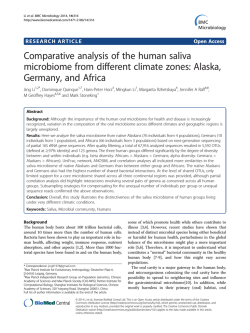
Original Paper
Biol Neonate 2000;78:22–26
Cortisol and 17-Hydroxyprogesterone
Levels in Saliva of Healthy Neonates
Normative Data and Relation to Body Mass Index, Arterial Cord Blood pH and
Time of Sampling after Birth
Ingo Klug a Regina Dressendörfer d Christian Strasburger d
Gonne Peter Kühl c Hans-Ludwig Reiter c Andrea Reich a Grit Müller a
Kerstin Meyer a Jürgen Kratzsch b Wieland Kiess a
a Children’s
Hospital, University of Leipzig, and b Institute of Clinical Chemistry and Pathobiochemistry, Leipzig,
Hospital, Justus Liebig University of Giessen, Giessen, and d Department of Endocrinology,
Klinikum Innenstadt, University of Munich, Munich, Germany
c Children’s
Key Words
Cortisol W 17-Hydroxyprogesterone W Neonate W Neonatal
stress W Saliva W Circadian rhythm
Abstract
The measurement of cortisol and 17-hydroxyprogesterone (17-OHP) in saliva has become a reliable tool for
both the scientist and the clinician for studying adrenal
cortical function in the adult and the older child. We have
now established in parallel normative data for salivary
cortisol and 17-OHP levels in healthy neonates. We have
asked whether or not there is a circadian rhythm of cortisol and 17-OHP saliva levels in neonates. Furthermore,
we have asked whether salivary hormone levels correlated with auxologic and clinical data and time of sampling. Cortisol and 17-OHP levels in saliva samples from
119 healthy neonates (55 girls, 64 boys) were measured
using in-house time-resolved fluorescent immunoassays. Saliva samples were obtained using a saliva collecting tube three times a day on the first or second day
of life. Gender and gestational age did not influence salivary cortisol and 17-OHP levels. No significant circadian
ABC
© 2000 S. Karger AG, Basel
0006–3126/00/0781–0022$17.50/0
Fax + 41 61 306 12 34
E-Mail [email protected]
www.karger.com
Accessible online at:
www.karger.com/journals/bon
rhythm of salivary hormone levels was detected in this
group of newborns. However, body mass index, arterial
cord blood pH and time of saliva sampling significantly
influenced salivary hormone levels. In conclusion, measurement of cortisol and 17-OHP in saliva is feasible in
healthy neonates. The existence of normative data forms
the basis for future studies on pathophysiologic states in
the newborn period.
Copyright © 2000 S. Karger AG, Basel
Introduction
Corticosteroids are essential for the development of
the fetus. In early neonatal life, cortisol is already of
importance for stress responses and metabolic homeostasis. The concentration of cortisol in amniotic fluid increases during gestation towards a maximum at 38–40
weeks of gestation with levels of 82 B 12 ng/ml [1, 2].
The 17-hydroxyprogesterone (17-OHP) levels in amniotic fluid in early gestation (1.63 B 0.21 ng/ml, mean B
SD) are also significantly lower than the levels found later
in gestation. From the 36th to the 38th week of gestational
Wieland Kiess, MD
Children’s Hospital, University of Leipzig
Oststrasse 21–25, D–04317 Leipzig (Germany)
Tel. +49 341 97 26000, Fax +49 341 97 26009
E-Mail [email protected]
age, 17-OHP levels reach a maximum of 3.8 B 0.74 ng/ml
[3]. High 17-OHP levels in amniotic fluid are found in
fetuses with congenital adrenal hyperplasia [4, 5].
The transplacental transfer of cortisol and synthetic
glucocorticoids has been demonstrated. In the middle of
gestation, maternal plasma cortisol levels are 8–10 times
higher than the fetal concentrations [6, 7]. The relative
biologic activity and metabolic rates of glucocorticoids
during gestation and at term have been studied previously
[8–11].
The measurement of cortisol and 17-OHP in saliva has
become a reliable tool for both the scientist and the clinician for studying adrenal cortical function in the adult
and the older child [12–15]. We have now established in
parallel normative data for salivary cortisol and 17-OHP
levels in healthy neonates. In addition, we have asked
whether or not there is a circadian rhythm of cortisol and
17-OHP saliva levels in neonates and whether salivary
hormone levels correlate with clinical data of the newborns and time of sampling. Cortisol and 17-OHP levels
in saliva samples from 119 healthy neonates were measured using in-house time-resolved fluorescent immunoassays. These normative data are expected to build the
basis for future studies on pathophysiologic states of adrenal function and stress responses in the newborn period.
Subjects and Methods
Subjects
Saliva cortisol and 17-OHP levels were determined in 119
healthy neonates (55 girls and 64 boys). Auxologic and clinical data
were recorded using a structured questionnaire (table 1). 25 neonates
were born by sectio caesarea, 94 vaginally. Duration of birth was 44–
54 min and the time from onset of labor until birth was 414 B 256
min in the 94 neonates with vaginal delivery. Saliva was collected
18–24 h (n = 21) or 25–55 h (n = 98) after birth.
Saliva Sampling
From each infant, three saliva samples were taken at 7:00, 13:00
and 19:00 h before meals. Saliva was obtained by using a saliva collecting tube with a cotton swab inside (Salivette®, Sarstedt, Nümbrecht, Germany). The cotton swab was left in the neonates’ mouth
for 5 min. This procedure did not cause any distress. Saliva samples
were centrifuged and stored frozen at –20 ° C until analysis.
Cortisol and 17-OHP Assays
Samples were analyzed using in-house time-resolved fluorescent
immunoassays as described before [12, 13]. In some cases, sample
volumes were too small for duplicate determination of the two hormones. Therefore, some measures contain smaller sample numbers
(!119). In brief, specific polyclonal antibodies against cortisol and
17-OHP respectively were used as first antibodies. Cortisol or 17OHP biotin conjugates were used as tracers. Cross-reactivities of the
Cortisol and 17-Hydroxyprogesterone
Levels in Saliva of Healthy Neonates
Table 1. Anthropometric and clinical data of the 119 healthy neonates investigated in respect to salivary cortisol and 17-OHP levels
Gestational age, weeks
Weight, g
Length, cm
Head circumference, cm
Body surface, m2
BMI, kg/m2
Cord blood pH
Median
Range
39
3,290
51
34
0.2
12.5
7.31
31–42
2,080–4,550
45–57
30–38
0.15–0.25
9.8–15.9
7.07–7.53
immunoassays were very low [12, 13]. Hence, the assays are considered to be ideal for measuring steroid levels in the neonatal period.
The interassay coefficient of variation was between 4.1 and 11.7%
for the cortisol assay and between 5.0 and 9.8% for the 17-OHP
assay. The intraassay coefficient of variation was between 5.3 and
8.8% for the cortisol assay and between 5.0 and 9.8% for the 17-OHP
assay.
Results
Anthropometric and Clinical Data
Anthropometric and clinical data of the neonates are
summarized in Subjects and Methods and in table 1. As
was expected, there was a correlation between gestational
age and auxologic parameters (data not shown).
Cortisol and 17-OHP Levels in 119 Healthy Neonates
Cortisol and 17-OHP levels were measurable in saliva
samples of all healthy neonates. Only on rare occasions
was there too small a saliva volume to measure both hormones in duplicate. There were no differences between
cortisol and 17-OHP levels in saliva from male and
female neonates (data not shown). Normative data for
salivary cortisol and 17-OHP levels in the neonatal period
were generated (table 2, 3). Interestingly, there was a significant difference in salivary hormone levels when time
of sampling was analyzed: significantly higher morning
cortisol levels (p = 0.015) were found in the group in
which samples had been taken 18–24 h after birth compared to the group where samples had been obtained 25–
55 h after birth (fig. 1a). 17-OHP levels were significantly
higher at all three times (morning p = 0.001, midday p =
0.017, evening p = 0.038) in the group where saliva samples had been taken 18–24 h after birth compared to the
group where samples had been taken 25–55 h after delivery (fig. 1b).
Biol Neonate 2000;78:22–26
23
a
b
Fig. 1. Salivary cortisol (a) and 17-OHP levels (b) (median, 1st and 3rd quartile) in 119 healthy neonates with
samples taken 18–24 (X) or 25–55 (!) h after birth as described in Methods. In the group in which saliva samples
were taken within 24 h after birth, morning, midday and evening 17-OHP and cortisol levels were significantly higher
(morning p = 0.001, midday p = 0.017, evening p = 0.04 for 17-OHP; morning p = 0.0015 for cortisol) compared to the
group in which samples were obtained later than 24 h after birth.
Table 2. Salivary cortisol levels (ng/ml) from healthy newborns
according to time of sampling
Mean B SD
Range
Median n
Samples taken 18–24 h after birth
Morning
14.25B11.82
Midday
12.97B11.26
Evening
10.71B6.58
2.21–49.46
2.62–46.55
3.42–23.45
11.19
10.18
8.15
21
21
19
Samples taken 25–55 h after birth
Morning
9.76B9.36
Midday
8.61B5.71
Evening
8.19B5.29
2.41–60.40
2.37–28.66
1.21–34.66
6.71
6.66
6.80
98
96
96
Lack of Circadian Rhythm of Cortisol and 17-OHP
Levels
Neither saliva cortisol levels nor 17-OHP levels
showed a circadian rhythm (fig. 1a, b). However, there
was a significant correlation between 17-OHP levels in
the morning, midday and evening (morning and midday
r = 0.7, midday and evening r = 0.67, morning and evening r = 0.63).
Salivary Cortisol Levels as an Indicator of Stress in the
Neonate
Arterial cord blood pH values were significantly and
inversely related to the duration of labor in the whole
group of neonates considered to be healthy and not
exposed to known unexpected stresses (r = 0.37, p 1
24
Biol Neonate 2000;78:22–26
Table 3. Salivary 17-OHP levels (pg/ml) from healthy newborns
according to time of sampling
Mean B SD
Range
Median n
Samples taken 18–24 h after birth
Morning
623.1B481.4
Midday
497.0B323.3
Evening
397.5B260.6
211.4–1723.3
183.4–1781.0
161.6–945.6
443.4
323.3
287.6
15
17
10
Samples taken 25–55 h after birth
Morning
305.6B222.4
Midday
295.4B175.8
Evening
247.1B131.4
81.8–1528.9
116.4–983.6
92.3–739.2
239.4
256.4
205.6
75
65
56
0.001; data not shown). When we asked whether or not
early cortisol levels were related to cord blood pH as an
indicator of stress to the baby, a significant inverse relation was found in the group of infants where saliva samples had been taken within 24 h after birth (r = –0.52).
Relation between Cortisol and 17-OHP Levels in
Saliva
Since both cortisol and 17-OHP can be derived from
the adrenal cortex of the infant, we have asked whether or
not cortisol and 17-OHP levels in saliva of healthy neonates correlated. In fact, when parallel measurements of
cortisol and 17-OHP in a total of 240 saliva samples were
analyzed, there was a significant correlation between cortisol and 17-OHP levels in saliva (r = 0.33, p ! 0.0001).
Klug et al.
Discussion
Significantly higher salivary cortisol levels (mean
14.25 ng/ml) were found in infants in which saliva had
been sampled within the first 24 h after birth compared to
the group of infants in which samples had been obtained
25–55 h after birth (mean 9.76 ng/ml; p ! 0.001). We
hypothesize that stress might contribute to high cortisol
production of the neonate around birth and during delivery [15, 16]. As a consequence, high cortisol saliva levels
are present at birth and shortly thereafter. Due to the
short half-life of cortisol, the high cortisol levels then
decline rapidly during the first days of life. This assumption is in agreement with the findings of Bertrand et al.
[14] who also found that cortisol levels declined after
birth. They also investigated plasma cortisol levels of
75 neonates born at term. Maximum cortisol levels (40.5
ng/ml) were found shortly after birth, while a minimum
(6.7 ng/ml) was observed between the 3rd and the 5th day
of life. These data are in accordance with our data in
respect to saliva cortisol levels. In addition, Bespalova et
al. [15] showed a decline in plasma cortisol levels within
the first day of life in healthy neonates. Kurihara et al.
[17] investigated saliva and plasma cortisol levels as an
indicator of stress in neonates. Saliva cortisol levels
showed a close correlation with plasma cortisol levels.
Cortisol levels were positively related to stress in this
group of neonates and infants.
In our study, low cord blood pH was related to longer
duration of labor (r = 0.26, p = 0.01). It has to be emphasized in this context that all infants studied by us were
healthy, not asphyxiated and undistressed. Therefore the
range of cord blood pH values is limited. It would be
expected that in a study also involving ill or distressed
neonates, a larger range of pH values would be found. Still
there was a striking inverse relation between morning cortisol levels and cord blood pH (r = –0.52, p = 0.017) in the
group in which samples were taken within 24 h after birth.
In the same group, there was a positive correlation
between morning cortisol levels and body mass index (r =
0.58, p = 0.006). A multiple regression analysis showed
that both correlations just exist because of two extremely
high morning cortisol levels, which belong to two participants with extremely high body mass indices. We hypothesize that elevated salivary cortisol levels – as an
expression of increased stress – might be related to high
body mass index and near asphyxiation. This hypothesis
should be tested in a prospective study involving distressed and asphyxiated as well as healthy neonates.
Cortisol and 17-Hydroxyprogesterone
Levels in Saliva of Healthy Neonates
It is unclear at what time of infancy the circadian
rhythmicity of cortisol release is first present. Some authors believe that rhythmicity starts as early as within the
2nd week of life [15, 18–23], while others state that the
circadian rhythm of cortisol plasma levels is only present
after 9 months of life [16]. In our study, no circadian
rhythmicity was present in the whole group of 119 infants
during the two days of the study period.
Reference values of plasma 17-OHP levels in neonates
and infants have been reported previously [24, 25]. We
now provide normative data on 17-OHP saliva levels in
neonates. In our study, 17-OHP salivary levels rapidly
decreased from the first to the second day of life. This
finding goes along with the data from Sippell et al. [20]
who showed that 17-OHP blood levels decrease rapidly
after birth [20, 25]. In contrast to salivary cortisol levels,
17-OHP morning, midday and evening levels show a
remarkable correlation: high morning levels are related to
high midday and high evening levels. As a result of this, it
could indeed be possible to take just one sample a day in
order to, e.g. screen for congenital adrenal hyperplasia
using saliva samples.
In conclusion, we have established normative data for
salivary cortisol and 17-OHP in healthy neonates (table 2, 3; fig. 1). The salivary concentrations of both hormones decline rapidly after birth. No gender difference of
salivary cortisol and 17-OHP levels was found. The determination of salivary cortisol levels in neonates might be a
helpful measure to control for neonatal stress and birth
asphyxia.
Acknowledgments
We wish to thank Christian Smit for helping collect samples. We
also wish to express our sincere gratitude to the parents who gave
permission to study their infants. We gratefully acknowledge the generous gift of saliva collecting devices (Salivette®) by Sarstedt, Nümbrecht, Germany.
Biol Neonate 2000;78:22–26
25
References
1 Sizonenko PC, Aubert ML: Hypothalamopituitary axis and adrenal cortex: Fetoplacental
unit; in Pediatric Endocrinology. Baltimore,
Williams & Wilkins, 1993, pp 127–128.
2 Ballard PL: Hormonal regulation of pulmonary
surfactant. Endocr Rev 1989;10:165–181.
3 Sippell WG, Muller-Holve W, Dörr HG, Bidlingmaier F, Knorr D: Concentrations of aldosterone, corticosterone, 11-deoxycorticosterone, progesterone, 17-hydroxyprogesterone,
11-deoxycortisol, cortisol, and cortisone determined simultaneously in human amniotic fluid
throughout gestation. J Clin Endocrinol Metab
1981;52:385–392.
4 Pang S, Levine LS, Cederqvist LL, Fuentes M,
Riccardi VM, Holcombe JH, Nitowsky HM,
Sachs G, Anderson CE, Duchon MA, Owens R,
Merkatz I, New MI: Amniotic fluid concentrations of delta4- and delta5-steroids in fetuses
with congenital adrenal hyperplasia due to 21hydroxylase deficiency and in anencephalic fetuses. J Clin Endocrinol Metab 1980;51:223–
229.
5 New MI: Present status of prenatal diagnosis of
congenital adrenal hyperplasia; in Lee PA, Kowarski AA, Migeon CJ (eds): Congenital Adrenal Hyperplasia. Baltimore, University Park
Press, 1977, pp 511–526.
6 Allen JP, Cook DM, Kendall JW, McGilvra R:
Maternal-fetal ACTH relationship in man. J
Clin Endocrinol Metab 1973;37:230–234.
7 Simmer HH, Frankland M, Greipel M: On the
regulation of fetal and maternal 16alpha-hydroxydehydroepiandrosterone and its sulfate
by cortisol and ACTH in human pregnancy at
term. Am J Obstet Gynecol 1975;121:646–
652.
8 Beitins IZ, Bayard F, Ances IG, Kowarski A,
Migeon CJ: The metabolic clearance rate,
blood production, interconversion, and transplacental passage of cortisol and cortisone in
pregnancy near term. Pediatr Res 1973;7:509–
519.
26
9 Murphy BEP: Cortisol and cortisone in human
fetal development. J Steroid Biochem 1979;11:
509–513.
10 Diczfalusy E: Endocrine functions of the human fetus and placenta. Am J Obstet Gynecol
1974;119:419–433.
11 Jaffe RB: Fetoplacental endocrine and metabolic physiology. Clin Perinatol 1983;10:669–
693.
12 Dressendörfer RA, Kiess W, Bidlingmaier F,
Smit C, Rascher W, Klug I, Kistner A, Strasburger CJ: A time resolved fluorescence immunoassay for the determination of salivary 17hydroxyprogesterone:
Reference
ranges
throughout childhood and adolescence. Pediatr
Res 1998;44:650–655.
13 Dressendörfer RA, Kirschbaum C, Rohde W,
Stahl F, Strasburger CJ: Synthesis of cortisolbiotinconjugate and evaluation as a tracer in an
immunoassay for cortisol measurement. J Steroid Biochem Mol Biol 1992;7:683–692.
14 Bertrand J, Bonnet H, Forest MG, Loras B,
Rokicki W: Free cortisol of human plasma in
the first three months of life. Biol Neonate
1990;57:21–29.
15 Kniazev IA, Sen’kevich OA, Bespalova VA,
Saliaeva MV: Course of the early neonatal period and hormonal indicators in newborn infants
from multiple pregnancies (in Russian). Pediatriia 1990;(10):24–29.
16 Rosendahl W, Schulz U, Teufel T, Irtel von
Brenndorf C, Gupta D: Surgical stress and neuroendocrine response in infants and children. J
Pediatr Endocrinol Metab 1995;8:187–194.
17 Kurihara H, Chiba H, Shimizu Y, Yanaihara
T, Takeda M, Kawakami K, Takai-Kawakami
K: Behavioral and adrenocortical responses to
stress in neonates and the stabilising effects of
maternal heartbeat on them. Early Hum Dev
1996;46:117–127.
Biol Neonate 2000;78:22–26
18 Santiago LB, Jorge SM, Moreira AC: Longitudinal evaluation of the development of salivary
cortisol circadian rhythm in infancy. Clin Endocrinol 1996;44:157–161.
19 Kiess W, Meidert A, Dressendörfer RA,
Schriever K, Kessler U, König A, Schwarz HP,
Strasburger CJ: Salivary cortisol levels
throughout childhood and adolescence: Relation with age, pubertal stage and weight. Pediatr Res 1995;40:502–506.
20 Sippell WG, Dörr HG, Bidlingmaier F, Knorr
D: Plasma levels of aldosterone, corticosterone,
11-deoxycorticosterone, progesterone, 17-hydroxyprogesterone, cortisol and cortisone during infancy and childhood. Pediatr Res 1980;
14:39–46.
21 Bettendorf M, Albers N, Bauer J, Heinrich UE,
Linderkamp O, Maser-Gluth C: Longitudinal
evaluation of salivary cortisol levels in fullterm and preterm neonates. Horm Res 1998;
50:303–308.
22 Walker RF, Hughes IA, Riad-Fahmy D: Salivary 17-OHP in congenital adrenal hyperplasia. Clin Endocrinol 1979;11:631–637.
23 Riad-Fahmy D, Read GF, Walker RF, Griffiths K: Steroids in saliva for assessing endocrine function. Endocr Rev 1982;3:367–395.
24 Otten BJ, Wellen JJ, Rijen JC, Stoelinga GB,
Benraad TJ: Salivary and plasma androstenedione and 17-hydroxyprogesterone levels in
congenital adrenal hyperplasia. J Clin Endocrinol Metab 1983;57:1150–1154.
25 Sippell WG, Becker H, Versmold HT, Bidlingmaier F, Knorr D: Longitudinal studies of plasma aldosterone, corticosterone, deoxycorticosterone, progesterone, 17-hydroxyprogesterone, cortisol, and cortisone determined simultaneously in mother and child at birth and during the early neonatal period. I. Spontaneous
delivery. J Clin Endocrinol Metab 1978;46:
971–985.
Klug et al.
© Copyright 2026