
Microstructural Evidence for the Condensation - USRA
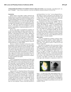
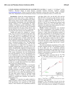
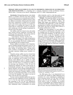
46th Lunar and Planetary Science Conference (2015)
2214.pdf
MICROSTRUCTURAL EVIDENCE FOR THE CONDENSATION ORIGIN OF HIBONITE-SPINEL
INCLUSIONS FROM ALH A77307 (CO3.0). Jangmi Han1,2, Lindsay P. Keller2, and Adrian J. Brearley3. 1Lunar
and Planetary Institute, Houston, TX 77058, USA ([email protected]); 2ARES, NASA/JSC, Houston, TX
77058, USA; 3Department of Earth and Planetary Sciences, MSC03-2040, University of New Mexico, Albuquerque,
NM 87131, USA.
Introduction: Hibonite is a highly refractory phase
occurring in many CAIs and Wark-Lovering rims from
different chondrite groups [1]. Hibonite is predicted to
be one of the earliest refractory phases to form by equilibrium condensation from a cooling gas of solar composition [2]. Therefore, hibonite can provide important
insights into the earliest stages of solar nebular evolution.
A previous FIB/TEM study of a hibonite-spinel
CAI in ALH A77307 (CO3.0) presented the crystallographic relationship between hibonite and spinel, suggesting that their structural similarity caused kinetic
inhibition of melilite condensation and instead favored
epitaxial nucleation and growth of spinel on the
hibonite surfaces [3]. Here, we present the microstructures of two hibonite-spinel CAIs in ALH A77307 using FIB/TEM techniques to better understand their
formation conditions and processes.
Methods: Each FIB section of two hibonite-spinel
CAIs in ALH A77307 was extracted from a region
consisting of subparallel hibonite laths intergrown with
spinel and minor perovskite. These FIB sections were
cut normal to the elongation direction of the hibonite
laths [3,4]. The FIB sections were investigated in detail
using a variety of TEM techniques (BF- and HR-TEM,
STEM, EDS, and electron diffraction).
Results: Two hibonite-spinel CAIs (03 and 08) in
ALH A77307 share characteristics common to this type
of CAI [3,5]: (1) they are irregularly-shaped, porous
objects with a small size of 70×40 µm for 03 and
55×50 µm for 08, (2) lath-shaped hibonite grains 3-40
µm long and 0.3-9 µm wide are embedded in spinel
and are subparallel to each other, and (3) fine-grained,
subrounded to elongated perovskite grains with a size
of 0.2-4 µm commonly occur as inclusions in spinel
and often in hibonite. In contrast, only CAI 03 contains
melilite, but CAI 08 is melilite free.
Both FIB sections (03-A and 08-A) consist mainly
of hibonite and spinel with minor perovskite inclusions.
Individual mineral constituents have similar microstructures. Hibonite grains are typically lath-shaped
and intergrown with each other. Hibonite grains show
various thicknesses and lengths; 0.2-5 μm long and 60
nm-2.5 μm wide in 03-A and 2-11 μm long and 0.5-6.5
μm wide in 08-A. Electron diffraction patterns of
hibonite grains reveal that the grains are consistently
elongated along the a axis, but have different orientations relative to one another. Spinel is usually irregular-
ly-shaped with a size of 0.5-3.5 µm in 03-A and 1-6.5
µm in 08-A. Perovskite inclusions (0.2-5 µm in size) in
hibonite and spinel are subrounded to elongated. In
addition, FIB 03-A contains elongated melilite (0.2-2
µm long) interstitial to hibonite laths.
While spinel and perovskite are relatively featureless and free of defects, most hibonite grains in both
FIB sections contain numerous planar defects; however, the concentration of planar defects varies among
and within the hibonite grains. Electron diffraction
patterns of hibonite show weak streaking along c*,
indicating stacking disorder. As shown in Figure 1,
HR-TEM images of hibonite grains show variations in
spacing of the lattice fringes, suggestive of stacking
disorder. In most cases, isolated layers that are 1.6 nm
wide are randomly intergrown with the prominent 1.1
nm wide (002) layers of stoichiometric hibonite. We
also observe more complex intergrowth combinations.
We observe the intergrowth of 7 nm wide layers with
the 2.2 nm wide layers of stoichiometric hibonite. The
most complex case is the intergrowth of layers of various spacings ranging in width from 4.8 to 11.4 nm with
the 2.2 nm wide layers of stoichiometric hibonite. Analytical electron microscopy shows that hibonite grains
are close to Ca(Al,Si,Ti,Mg)12O19 in composition, but
show Ca deficiencies by up to ~10 mol% where the
defects are concentrated.
Figure 1. HR-TEM image of hibonite from FIB 03-A. The
1.6 nm wide layers (yellow dashed lines) are randomly distributed in ordered regions with the 1.1 nm wide layers (red
dashed lines).
Discussion: Our overall petrologic and mineralogical observations from two hibonite-spinel CAIs in ALH
A77307 are generally consistent with the predictions of
mineral formation from equilibrium condensation calculations [2]. The irregular shapes and porous textures
46th Lunar and Planetary Science Conference (2015)
of these CAIs also support a condensation origin. It is
therefore very likely that hibonite formed by hightemperature condensation from a nebular gas, followed
by perovskite and spinel.
Textural relationships in the hibonite-spinel CAIs,
however, indicate that spinel, rather than melilite, condensed following hibonite condensation, inconsistent
with equilibrium condensation calculations [2]. This
suggests that conditions of condensation for these CAIs
departed from thermodynamic equilibrium. Melilite
condensation was extremely limited, demonstrated by
its rarity in both CAIs. Instead, the occurrence of spinel
in crystallographic continuity with hibonite demonstrates that spinel condensed directly onto hibonite.
Epitaxial nucleation and growth of spinel may have
occurred on the surfaces of hibonite, probably due to
the lower activation energy for spinel nucleation compared with that of melilite [3].
The presence of perovskite inclusions within
hibonite may be attributed to simultaneous condensation of these phases over a limited temperature interval
[2]. Early-condensed hibonite grains trapped adjacent
condensing fine-grained perovskite grains and continued to nucleate and grow into larger crystals. As nebular cooling proceeded, hibonite condensation eventually ceased, but perovskite kept condensing and was later
enclosed by spinel once spinel condensation commenced at lower temperatures [3].
The presence of planar defects in hibonite provide
useful insights into possible formation conditions of
these two CAIs. Ideal hibonite consists of a sequence
of four-closely-packed O layers and Al ions in a spinel
block (S) alternating with a Ca-containing layer (C),
resulting in a unit cell containing 2 (S+C) blocks
(S+C+S+C) [6]. The observed 1.1 nm wide lattice
fringes correspond to the c dimension of the ideal
hibonite structure consisting of one basic structural unit
representing the combination of a spinel block and a
Ca-containing layer (S+C) [6]. The local presence of
1.6 nm wide lattice fringes can be interpreted as the
result of modification of the stacking sequence due to
the presence of a thicker spinel block with six ccp O
layers (S+S+C) [6]. Therefore, the observed wide
range of spacings can be interpreted as different stacking combinations of the basic structural spinel block
versus the Ca-containing layer. This interpretation is
consistent with the observed Ca deficiency of hibonite.
There are three possible explanations for the Ca deficiency of hibonite; (1) reheating and partial evaporation of Ca from hibonite, (2) equilibrium condensation
of Al-rich spinel in hibonite, and (3) non-equilibrium
condensation of hibonite and (S+S+C) phase. Evaporative loss of Ca from hibonite may have occurred during
or after hibonite condensation as a result of later re-
2214.pdf
heating. Calcium loss requires extensive evaporation,
resulting in complete loss of moderately refractory elements (e.g., Mg). Therefore, Mg isotopic mass dependent fractionation is expected without Ca isotopic
mass dependent fractionation [7]. However, the absence of any Mg isotopic mass dependent fractionation
from similar hibonite-bearing CAIs in CO3 chondrites
[5,8] appears to rule out evaporation as an important
mechanism, although Mg isotopic data for the CAIs in
this study are lacking.
Second, Al-rich spinel may have condensed as isolated layers within hibonite. A thermodynamic mixing
model for the spinel MgAl2O4-Al8/3O4 solid solution
suggests that Al-rich spinel is stable between 1,9202,267 K [9]. However, equilibrium condensation calculations predict that, at these temperatures, Al-rich spinel fails to condense due to dominant Al and Ca condensation, instead forming Ca-aluminates or liquids in
dust-enriched systems [10].
However, the lack of melilite in these CAIs indicates that they did not form at equilibrium. Based on
the observations, we propose that the intergrowth of
hibonite and the (S+S+C) phase is the product of formation under non-equilibrium conditions, where the
formation of the defect-structured hibonite is energetically more favorable than corundum formation by direct condensation. Under non-equilibrium conditions,
corundum condensation may be inhibited. Excess Al in
the gas phase was incorporated into the hibonite as
extra spinel blocks, resulting in the planar defects with
the (S+S+C) stacking sequence.
Conclusions: Our TEM observations suggest that
two hibonite-spinel CAIs in ALH A77307 formed by
high-temperature condensation from the cooling solar
nebula under non-equilibrium conditions. The planar
defects in hibonite may have formed by the accommodation of some excess Al into the hibonite in the form
of the extra spinel blocks. Interestingly, similar microstructures are also observed in hibonites from WarkLovering rims [11], indicating the common formation
histories for the different occurrences of hibonite.
Acknowledgements: This research was supported by
NNX11AK51G to A.J. Brearley (PI) and 10-COS10-0049 to
L.P. Keller (PI).
References: [1] MacPherson G.J. (2014) Treatise on Geochemistry II vol.1 pp.139-179. [2] Ebel D.S. (2006) MESS2
pp.253-277. [3] Han J. & Brearley A.J. (2014) LPS XLV, Abstract #2125. [4] Han J. et al. (2014) MAPS, 49, A151. [5]
Russell S.S. et al. (1998) GCA, 62, 689-714. [6] Schmid H. &
De Jonghe L.C. (1983) Philos. Mag. A, 48, 287-297. [7] Simon J.I. & DePaolo D.J. (2010) EPSL, 289, 457-466. [8]
Fahey A.J. et al. (1994) GCA, 58, 4779-4793. [9] Sack R.O.
(2014) Am. J. Sci., 314, 858-877. [10] Ebel D.S. et al. (2014)
MAPS, 49, A101. [11] Keller L.P. (1991) AGU, 72, 141.
© Copyright 2026