
Graphite Morphologies in the Volcanic- Hosted Deposit at
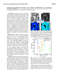
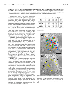
macla nº 9. septiembre ‘08
revista de la sociedad española de mineralogía
91
Graphite Morphologies in the VolcanicHosted Deposit at Borrowdale (NW England,
UK): Preliminary Raman and SIMS data
/ JOSÉ FERNÁNDEZ BARRENECHEA (1, *), JAVIER LUQUE (1), LORENA ORTEGA (1), MAGDALENA RODAS (1),
DAVID MILLWARD (2), OLIVIER BEYSSAC (3)
(1) Dpto. Cristalografía y Mineralogía, Facultad de Geología, Universidad Complutense de Madrid, 28040 Madrid ( Spain)
(2) British Geological Survey, Murchison House, West Mains Road, Edinburgh EH9 3LA (UK)
(3) Laboratoire de Geologie, CNRS-UMR 8538, Ecole Normale Supérieure, 24 rue Lhomond, 75231 Paris Cedex 5 (France)
INTRODUCTION.
Graphite structure consists of a
continuous bidimensional array of sixfold rings of carbon atoms stacked along
the c-axis. This determines that crystals
commonly show laminar (platy) habits.
In spite of this, other morphologies have
been reported, but the widest range of
morphologies known within a single
paragenesis is reported here from the
Borrowdale graphite deposit in Cumbria,
UK. There, graphite mineralization is
hosted within volcanic rocks, one of only
two examples of such an association
known
worldwide.
The
crystal
morphology of any mineral is controlled
by both its structure and the physicochemical conditions prevailing during
nucleation and growth. On the other
hand, some geochemical features are
also dependent upon the conditions of
crystal growth. Thus, the structural and
isotopic characterization of the graphite
morphologies in the volcanic-hosted
deposit at Borrowdale could help unravel
the evolution of the mineralizing process
in this particular environment.
GEOLOGICAL
SETTING
MINERALOGICAL FEATURES.
AND
The Borrowdale graphite deposit
consists of mineralized faults hosted by
andesite lavas and sills belonging to the
upper Ordovician (Caradoc) Borrowdale
Volcanic Group, the geochemical
characteristics of which show evidence
of assimilation of pelitic material from
the
underlying
Skiddaw
Group
(McConnell et al., 2002). The richest
graphite deposits are developed at the
intersections of the faults within steeply
inclined pipe-like bodies up to 1 x 3 m in
over 100 m in length. The pipe-like
bodies contain nodular masses and
patches of graphite, typically 1-2 cm
across, but ranging from a few
millimetres to 1 m or more. The mineral
association of the deposit includes
graphite, chlorite, epidote, and quartz.
The study of fluid inclusions within
quartz
associated
with
graphite
indicates that the mineralizing fluids
evolved from H2O-CO2-CH4 mixtures
(X H2O = 0.646; X CO2 = 0.244; X CH4 =
0.114; X NaCl = 0.006) to H2O-CH4 (X H2O
= 0.93; X CH4 = 0.02; X NaCl = 0.05) fluids.
The wide diversity of graphite
morphologies recognized within the
deposit can be grouped into three
categories: laminar, cryptocrystalline,
and spherulitic. Laminar graphite
crystals (up to 300 µm long and 50 µm
wide) are by far the most abundant (≈
90%). Along fault zones, “graphic-like”
intergrowth textures, consisting of thin
curved and tapering graphite flakes or
“vermiform” graphite crystals within
chlorite
have
been
observed.
Cryptocrystalline graphite may form: 1)
“composite nodules” consisting of both
flaky and cryptocrystalline graphite, 2)
rounded patches within flaky graphite,
and 3) “colloform” bands and globules
dispersed within the host rock.
Spherulites occur in four different
settings: 1) as individual forms, 5-40 µm
in diameter, within flaky graphite, 2) as
individual forms, 1-3 µm in diameter,
included in quartz fragments, 3) as
individual forms, 1-5 µm in diameter, or
aggregates disseminated within the
volcanic rock, and 4) as aggregates, 510 µm in diameter, associated with
chlorite along fault zones.
palabras clave: Grafito, Raman, SIMS, Borrowdale
resumen SEM/SEA 2008
RESULTS.
Raman spectra were collected with a
Renishaw INVIA spectrometer at the
Ecole Normale Superieure (Paris,
France) on the polished thin sections
used for the petrographic study. This
method allows for measurements in situ
and recognition of the textural
relationships between graphite and the
rest of the minerals in the assemblage.
All the measurements were done
focusing the laser beam beneath the
surface of the transparent minerals
associated with graphite to avoid the
mechanical disruption of the graphite
structure at the surface of the thin
section due to polishing (Pasteris, 1989;
Beyssac et al., 2003). The 514.5 nm
wavelength of a 20 mW spectra Argon
laser focused through a 100x objective
was used for the analyses. Under these
conditions the spatial resolution is ~1
µm and the spectral resolution is close
to 1 cm-1. The Raman parameters (peak
position, band area, and band intensity)
were determined with the computer
program PeakFit 3.0 using a Voigt
function.
According to the Raman data all
graphite
morphologies
from
the
Borrowdale deposit correspond to
structurally well ordered graphite. The
intensity and area ratios for the D and G
bands in the first-order spectra (R1 and
R2) are consistent with highly crystalline
graphite (R2 averages 0.06 for laminar
and spherulitic graphite and 0.05 for
cryptocrystalline
graphite
within
composite nodules). In addition, no
significant changes were observed in the
second order Raman region, all the
spectra showing well-defined shoulders
key words: Graphite, Raman, SIMS, Borrowdale
* corresponding author: [email protected]
92
at ≈2685 cm-1 on the S1-peak, which is
indicative of attaining tri-periodic order
in the structure (Lespade et al., 1982).
Cryptocrystalline
graphite
forming
colloform textures within the host rock,
however, shows a significantly lower
crystallinity (R2=0.25).
nodules has significantly lighter isotopic
signatures (average δ13C = -33.70 ‰)
with no apparent zoning across the
banded texture (Fig. 1). The lightest
carbon isotope ratios correspond to
vermicular graphite within chlorite
(average δ13C = -34.49 ‰).
Secondary ion mass spectrometry
(SIMS) analyses were performed at the
Department of Geology and Geophysics,
University of Edinburgh, using a Cameca
DISCUSSION.
According to classical theories of
nucleation, spherulites and colloform
-34.73
-32.87
-34.01
-34.29
-35.12
-34.01
140 µm
fig. 1. SIMS data (δ13C relative to the PDB standard) of colloform graphite around quartz. The quartz grain
contains small graphite spherulites. Note the narrow range of δ13C values.
ims 1270 ion microprobe. Gold-coated
polished thin sections were analysed
using negative secondary ions sputtered
with a positively charged Cs+ beam. The
spot size was 20 µm, and hence stable
carbon isotope data were obtained only
from those morphologies displaying
sizes larger than this. The results were
checked using international standards,
including graphite USGS24 (δ13C =
-16.05 ‰). Five measurements were
made on the standard at the start and
end of each 20 analyses – thus enabling
calibration of the sample measurements
against 10 standard measurements.
Under these conditions, the precision of
the point analysis is close to 0.2 ‰.
It is worth noting that the analyses of
each morphological type show very
homogeneous values, thus falling within
narrow ranges. Graphite flakes have one
of the heaviest isotopic signatures
(average δ13C = -30.26 ‰). Such values
are close to those found in graphite
spherulites within chlorite (average δ13C
= -30.15 ‰). On the other hand,
cryptocrystalline graphite in composite
textures
represent
heterogeneous
nucleation on pre-existing substrates
under high supersaturation conditions.
Laminar graphite should nucleate later,
as a result of precipitation from fluids
with significantly lower supersaturation.
In spite of this sequence of
crystallization of the different graphite
morphologies, Raman data indicate that
there is not a significant change in the
structural order of graphite.
The light isotopic signatures of the
different morphologies of graphite from
the Borrowdale deposit suggest that the
carbon was derived from a biogenic
source. This is in good agreement with
the evidence of assimilation of Skiddaw
Group metapelites by the volcanic host
rocks, and with the presence of zones
within the metapelites that have been
depleted in carbon and other elements
during hydrothermal alteration (Cooper
et al., 1988). Independent bulk carbon
isotopic
analyses
of
Skiddaw
metapelites yielded values close to -28
‰.
In the fractionation that occurs between
two carbon-bearing phases, the more
oxidized species of the pair becomes
relatively enriched in the heavier
isotope, i.e. 13C. Thus, graphite
precipitating from CO2-rich fluids would
be lighter than CO2 in equilibrium. This
agrees with the fact that the earlier
morphologies (colloform) crystallizing in
the pipe-like bodies display lighter
signatures than flaky graphite that
formed somewhat later. However,
graphite within chlorite from fault-fills
points to an inverse relationship: the
isotopic ratio of spherulitic graphite
within chlorite is heavier than in the
vermicular intergrowths. This suggests,
though does not prove, that graphitechlorite veins represent a slightly later
mineralizing event related to deposition
from lower temperature, CH4-rich fluids.
Since graphite is the oxidized phase with
respect to methane, the earlier
crystallizing
morphologies
(i.e.
spherulites), retained heavier isotopic
signatures than those precipitating later
(vermicular graphite).
Finally, the small differences in the δ13C
values between the different graphite
morphologies suggest that graphite
precipitated over a narrow temperature
interval.
REFERENCES.
Beyssac, O., Goffé, B., Petitet, J.P., Froigneux,
E., Moreau, M., Rouzaud, J.-N. (2003): On
the characterization of disordered and
heterogeneous carbonaceous materials
by Raman spectroscopy. Spectrochim.
Acta, Part A, 59, 2267-2276.
Cooper, D. C., Lee, M. K., Fortey, N. J., Cooper,
A. H., Rundle, C. C., Webb, B. C., Allen, P.
M. (1988): The Crummock Water aureole:
a zone of metasomatism and source of
ore metals in the English Lake District. J.
Geol. Soc., London, 145, 523-540.
Lespade, P., Al-Jishi, R., Dresselhaus, M.S.
(1982): Model for Raman scattering from
incompletely
graphitized
carbons.
Carbon, 20, 427-431.
McConnell, B.J., Menuge, J.F., Hertogen, J.
(2002): Andesite petrogenesis in the
Ordovician Borrowdale Volcanic Group of
the English Lake District by fractionation,
assimilation and mixing. J. Geol. Soc.,
London, 159, 417-424.
Pasteris, J.D. (1989): In situ analysis in
geological thin-sections by Laser Raman
Microprobe Spectroscopy: a cautionary
note. Appl. Spectroscopy, 43, 567-570.

© Copyright 2026