
Low Level Laser Therapy for Tendinopathy. Evidence of A Dose
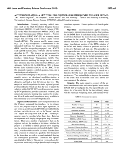
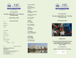
Low level laser therapy for tendinopathy. Evidence of a dose - response pattern
Bjordal , Jan Magnus, University of Bergen, Section of Physiotherapy Science, 5020 Bergen,
Norway
e-mail : [email protected], tel. + 47 55 585663, fax. +47 55 298364
Couppe, Christian, Willemoes gade 61, 4.sal, 2100 København Ø, Denmark
Ljunggren, Anne Elisabeth, University of Bergen, Section of Physiotherapy Science, 5020 Bergen,
Norway
To investigate if low level laser therapy (LLLT) can reduce pain from tendinopathy, we performed a
review of randomized placebo-controlled trials with LLLT for tendinopathy.
A literature search for trials published after 1980 using LLLT was conducted on Medline, Embase,
Cochrane Library and handsearch of physiotherapy journals in English and Scandinavian languages.
Validity assessment of each trial was done according to predefined criteria for location-specific dosage
and irradiation of the skin directly overlying the affected tendon..
The literature search identified 78 randomized controlled trials with LLLT, of which 20 included
tendinopathy. Seven trials were excluded for not meeting validity criteria on treatment procedure or
trial design. Twelve of the remaining thirteen trials investigated the effect of LLLT for patients with
subacute and chronic tendinopathy provided a pooled mean effect of 21 % [5.9-36.1, 95%CI]. If only
results from the nine trials adhering to assumed optimal treatment parameters were included, the mean
effect over placebo increased to 32 % [23.0-41.0, 95% CI].
Low level laser therapy can reduce pain in subacute and chronic tendinopathy if a valid treatment
procedure and location-specific dose is used.
Keywords: Low level laser therapy, dose – response pattern, tendinopathy, meta-analysis
BACKGROUND
Low level laser therapy (LLLT) was introduced in a clinical randomised controlled trial
(RCT) on musculoskeletal pain as early as in 1980 [1] . In the past two decades a number of
clinical RCTs have been performed with LLLT to treat a variety of musculoskeletal and
neurogenic pain conditions. Clinical applications of LLLT have been performed either by
direct exposure of the skin overlying the injury, exposure of trigger points or acupuncture
points, or of nerves inside or outside the painful area. A broad range of doses (0,0001 – 38
J/cm2) [2] has been reported to produce significant effects on musculoskeletal disorders in
about one third of the LLLT trials. Thus the rationale behind the selection of application
technique and treatment parameters like power density, size of exposure area, timing or
treatment frequency often remains unclear. However, the majority of LLLT-trials have failed
to provide successful results while employing doses within the same range as above. Recent
review articles [2-4] have concluded that there is little - if any - evidence in favour of LLLT for
the treatment of musculoskeletal pain. Several editorials in medical journals have supported
the criticism on the clinical use of LLLT [5-7]. Still the amount of RCTs with results in favour
of LLLT is by far too large to be explained by random chance alone. There is a missing link
between the increasing number of successful results from LLLT in the laboratory and the
mediocre results of clinical trials [2]. In an attempt to fill this gap, we decided to investigate if
there exists a dose- response pattern for a subgroup of patients from the clinical trials of
tendinopathy when the laser treatment procedure was similar to the successful laboratory
trials. Three validity criteria for clinical laser treatment procedure may be vital for
effectiveness. The first is that the tendinopathy is the target for irradiation. Secondly, power
density and dose at the target tendon should be similar to that of the laboratory trials, and
thirdly, timing and number of treatment sessions should correspond with laboratory
procedures.
RATIONALE FOR TREATMENT OF TENDINOPATH
Acute tendinitis involves an inflammatory response, often induced by repetitive strain,
overload or friction of the tendon. One in vitro-trial has confirmed that excessive repetitive
motion can induce fibroblast inflammation [5]. The nature of persisting symptoms are often
periodic [6] and associated with degenerative manifestations in tendon histopathology [7]. In
the subacute and chronic cases increased tendon thickness, degeneration of collagen tissue
and presence of hyaline foci within the tendon are evident [8]. Both morphological and
biomechanical deterioration of tendon properties have been observed, and some authors
suggest that the ending ”itis” is misleading as degeneration is more apparent at these stages,
[7], [12-15].
The gold standard for tendinitis treatment of the upper extremity is considered by several
reviewers to be steroid injections, or anti-inflammatory drugs (NSAID)[16-18]. These chemical
agents have primarily short-term effects, while longer-lasting effects (> 6 weeks) are less
evident and often fail to reach significance. Treatment can also be directed at the
degenerative changes within the tendon matrix. An in vitro-trial demonstrated that repetitive
motion with low load increases fibroblast metabolism and collagen production [11] In subacute
and chronic cases results from controlled trials with exercise therapy [12-14] indicate a
beneficial effect, and a case report of symptom reduction also found remission of
degenerative changes by ultrasonography after exercise therapy [13] . In our opinion, the
natural strategy for reducing tendinopathy pain by LLLT is two-fold and directed both at
reduction of inflammation, and stimulation of collagen production.
DETERMINATION OF OPTIMAL LLLT DOSE FOR TENDINOPATHY AT
TARGET LOCATION
Selection of dose in clinical trials of LLLT seems to be circumstantial, and either picked at
random, from the manufacturers` recommendations, or from the author`s own empirical basis.
In contrast we assume that there exists an optimal LLLT dose range for the treatment of
tendinitis, because laboratory trial reports almost unequivocally have stated that LLLTeffects on collagen tissue are dose-dependent.
We identified ten controlled trials investigating LLLT effect on fibroblast metabolism, and in
all except one trial [14] significant increases in collagen production were found.
The results from five in vitro trials on fibroblast cell cultures [24-28], suggested that optimal
power density and dose for increasing collagen production by 34-37 % were 4.5-7.5mW/cm2
and 0.45 – 0.6 J/ cm2 for continuous 632.8 nm HeNe laser and 820 nm GaAlAs laser
respectively. Three in vivo trials on sutured soft tissue injuries produced similar results on
collagen production with slightly higher doses (1-3.6 J/cm2) of continuous 632,8 nm HeNe
laser, and the same power density [29-31]. In the latter trial [39], mechanical properties of the
laser-exposed tendon were significantly enhanced due to a more adequate collagen
compostion, i.e. with more neutral salt soluble and insoluble collagen. One in vivo trial
suggested that pulsed 904 nm GaAs laser only needed 0.4 J/ cm2 to increase fibroblast
metabolism [17] .
Interestingly, it appears that it is possible to use a too high power density or dose from LLLT ,
as these were found to have decreased fibroblast cell metabolism in vitro [20-22]. In these trials
it was reported that doses lower than 0.1 J/ cm2 did not produce significant results, while
doses in excess of 4.5 J/ cm2, and power density higher than 10 mW/cm2, produced an
inhibitory effect on the fibroblast metabolism and collagen production. All these trials
employed a treatment frequency of 3-5 times per week for 2-4 weeks.
In in vitro trials higher energy doses have been reported to suppress inflammation [34-36]. This
effect was also reported to be dose-dependent with an optimal range of 1.9 – 6.3 J/cm2 and
power density of 21.2 mW/cm2. The upper range limits were not identified. The antiinflammatory effect was highly significant after 5 days with daily laser treatment.
If these findings are combined, there is an overlap in dose and power density ranges from
which the optimal treatment parameters at the target location can be derived:
Dose:
0.1 – 3
J/cm2
Power density:
5 – 21 mW/cm2
Treatment frequency: 3 – 5 times per week
BASIC TECHNICAL AND BIOPHYSICAL BACKGROUND
If we confine consideration of laser parameters to caucasian patients, there are five physical
factors which may determine if an optimal dose reaches the target in a clinical setting. They
can be summarized as:
1)
2)
3)
4)
5)
Distance from skin surface to target area;
Vascularity of the tissue between skin surface and target;
Volume of injured tissue;
Laser wavelength; and,
Mode of energy delivery (pulse vs continuous).
Only some of the above variables are known, but they provide a basis for extrapolation that
can increase the precision in determining what dose reaches the target. In vivo trials in
animals have shown that the most important absorption zone in the skin was the dermal
vascular plexus barrier [20]. As blood haemoglobin is an important absorber of light [21], highly
vascularized muscle tissue is harder for laser light to penetrate than the more transparent fatty
subcutaneous tissue. Improved regeneration after injury of muscle tissue in vivo has also
been observed, but LLLT doses were about 10 times higher than doses that have been
reported optimal for collagen tissue [22]. For tendon injuries that are covered by muscle, it is
important that dose is increased accordingly.
The average distance from the skin to the various tendons have not been definitely
established. For the purposes of the current paper, relevant dimensions and distances were
estimated by a combination of general anatomical knowledge, diagnostic imaging studies, and
a pilot study with ultrasound imaging of some tendons. Typical tendon characteristics are
presented in Table 1:
(( Table 1))
Red (HeNe/632 nm) or infrared (GaAlAs/820 nm) lasers have been used for LLLT because an
optical window of penetration with these wavelengths allows about 1/5 of the laser energy to
pass the skin barrier. Another type of infrared laser, the 904 nm GaAs laser has a mode of
energy delivery in short strong pulses, but with a low average output. Through in vitro trials,
it has been shown that infrared light penetrates slightly better (37 % lost at about 2 mm) than
visible red laser, which lose the same incident energy at only 0.5 mm [23]. In addition, pulse
lasers seem to overcome the skin barrier with lower doses than continuous lasers in in vivo
trials on animals, i.e. the relative penetration is better [17],[24] .
Given the optimal parameters already indicated above, and the data presented in Table 1,
acceptable clinical treatment parameter ranges for three laser types and five common forms
of tendinopathies are summarized in Table 2 :
((Table 2))
MATERIALS AND METHODS
Literature search
A literature search was performed on Medline, Embase, Cinahl, PedRo and the Cochrane
Controlled Trial Register as advised by Dickersin et al.[25] for both non-clinical controlled
trials and randomised controlled clinical trials.
Key words were : Low level laser therapy, low intensity laser therapy, low energy laser
therapy, HeNe laser, IR laser, GaAlAs, GaAs, diode laser, tendinitis, collagen, fibroblast,
tendon. Handsearching was also performed in national physiotherapy and medical journals
from Norway, Denmark, Sweden, Holland, England, Canada and Australia. Additional
information was gathered from researchers in the field.
Inclusion criteria
The randomised controlled trials were subjected to the following seven inclusion criteria:
1) Diagnosis: Tendinopathy;
2) Exposure area: Skin overlying site of inflammation or postinflammatory process in
tendon;
3) Intensity and dose: According to Table 2;
4) Treatment frequency and numbers: At least twice weekly and no less than six in total;
5) Control group: A control group of at least ten patients that received placebo therapy should
be included;
6) Blinding: Patients and outcome assessors should be blinded; and,
7) Specific endpoints within 2 – 6 weeks after inclusion.
Intensity and dose calculations
Data on beam diameter and laser output were collected from the relevant manufacturers`
manuals. All doses and power densities were calculated according to the following formulae:
Exposure area: Π ( 0.5 diameter 2) [cm2]
Mean output: Pulse intensity x pulse duration x pulses per second/ second [mW]
Power density: Mean output/ exposure area [mW/cm2]
Dose: Power density x treatment time [J/cm2]
Outcome measures
We chose pain as an outcome measure, preferrably on a continous scale (VAS etc). In trials
where several aspects of pain were measured, measures of pain involving the physical
function of the treated tendon (i.e. pain on isometric muscle contraction) were preferred.
When possible, 95 % confidence intervals were calculated for differences in change between
groups from baseline. Effect size was calculated for all trials as the difference (%) in mean
change from baseline to endpoint between the active treatment group and placebo treatment
group.
RESULTS
Results of inclusion procedure
The literature search identified 78 clinical RCTs, of which 20 included tendinitis.
Among these, two trials had to be excluded for exposing trigger points or acupunture points
and not exposing the skin directly overlying the injured tendon [43-44]. One trial [27] had to be
excluded for only having three patients with tendinitis in the control group. One comparative
trial was excluded for not using placebo-control [28]. Another trial [29] had to be excluded for
unwittingly giving the placebo group active HeNe - laser treatment, well within the
recommended dose range (2.25 J/ cm2 ). Another epicondylitistrial [30] treated in skin contact
and violated the recommended treatment distance of 10 cm in the manufacturer`s manual. The
optical correction system then left a ”blind” spot of approximately 2.5 -3 cm2 in the middle of
the treatment area which was untreated. In the case of lateral epicondylitis, the injured area of
the tendon is smaller than this blind spot and therefore it was judged as unlikely that optimal
dose reached the target tendon. Subsequently the trial was excluded from this metaanalysis. One large comparative trial was excluded because it individualised treatment and
lacked specific endpoint in time [31]. In addition only a small group received placebo treatment
and the results for the placebo group were not presented separately. All excluded trials are
presented in Table 3.
<< Table 3>>
Four trials treated the correct spot, but were excluded from analysis for employing treatment
parameters outside the acceptable dose and power density range. These four trials and all
included trials are presented in Table 4. Three of the listed trials are split in two as they
included two locations of tendinopathies and presented the results separately, which gives a
total number of 16 listings in the table from 13 publications.
((Table 4))
Results of dose and power density calculations
Complete and correct data on power density and and dose were only reported in three trials
But in all sixteen trials that exposed the skin overlying the injured tendon, sufficient
information was reported to perform calculations for the missing data.
[36-38].
Outcome measures
All nine trials [32], [38-45]using the suggested optimal treatment was calculated to a weighted
mean difference 32 % ( 23 – 41, 95 % CI) in favour of active LLLT (Figure 1). Trials without
optimal treatment dose/power density [34], [46-48], reported either no significant differences or
in one trial [36] significantly poorer results from LLLT than placebo. If these four trials were
included in the statistical pooling, the effect was reduced to 22.1 % better than placebo
(Figure 2). The difference in results between optimal and non-optimal treatment was highly
significant (p<0.001). The results from all the nine trials that met our inclusion criteria for
optimal parametres are shown in an effect size plot (Figure 2).
DISCUSSION
It may be said that previous reviews on LLLT have assumed that an optimal laser dose does
not exist. Such an assumption implies that whichever tissue is injured, or whatever
pathophysiology, the same dose can be employed for treatment. Even well-known variations
in the effct based upon factors such as penetration depths and absorption abilities, distance
and type of tissue lying between the laser-exposed skin and the injured tissue, and laser type
have been overlooked in many studies. The assumption that there exists a common, universal,
LLLT dose in the treatment of musculoskeletal disorders is unreasonable, not only in terms
of face validity, but also because of the distinct dose-response patterns that laboratory trials
on collagen tissue have revealed.
Another common assumption about LLLT has been that only one therapeutic window of
optimal dose exists when living tissue is exposed by laser energy [37]. Recent research findings
on dose-response relationships may shed new light on the apparent chaos regarding dose and
response in the LLLT literature. This assumption is recently contradicted by a research group
that found seven response peaks in a broad dose range for four different cell cultures [38].
These results also imply that there might be ineffective dose intervals within the broad dose
range that has been used in clinical trials.
Contrary to previous reviews, we found a dose-response pattern broadly resembling that from
the laser laboratory trials. Treatment success was invariably associated with the use of
treatment parameters inside our suggested optimal range. In one trial [36] the placebo group
improved more than the active LLLT group; the calculated dose and power density at the
target tendon was very high in this trial. In fact these parameters were within a range where
inhibitory effects on fibroblast metabolism have been reported [20-21]. The clinical outcome
may be explained by inhibition of the natural improvement over time for the intervention
group. Thus this trial adds further support for the identified dose - response pattern.
However, even if we seem to have identified an optimal dose range there are several
unanswered questions. One question is which effect is most important: a reduction of
inflammatory mediator activity, or an increase in collagen metabolism? Or maybe further
improvement can be achieved through variation of laser dose during the rehabilitation
process? Another point is that laser therapy has no known effect on the remodelling phase of
the injured tendon. How and when should the physical loading of the tendon be performed in
order to restructure and strengthen the tendon after laser therapy? These questions can only be
answered through controlled dose-response studies either in vivo or in a clinical setting.
One criticism that may arise is that the results of two included trials were reported as not
significant by the trial authors [39] , [40]. In the first trial, the authors chose to base their
conclusion on the data from a 5 category scale for detection of change. We consider that our
choice of using data of the continous scale for pain free grip strength is appropriate and more
sensitive to detect clinically relevant differences. From the other trial, disagreement was
caused by an incomplete statistical calculation that did not include significance testing of
change, which also have been commented upon in a previous review [41].
Testing and calibration of laser output was only performed by the authors in one of the
clinical trials [32]. Some authors have pointed out existing discrepancies in laser dosimetry
and measured deviations in laser output to be on average up to 40 % lower than
manufacturers` claims [52-53]. We assume that this problem affects dose and power density
similarly in all the trials. With the wide optimal range that we have suggested, this knowledge
may only effect one or two borderline trials, and does not alter our conclusion.
Two findings should be of particular interest for clinicians. The first is that the 904 nm GaAs
pulse laser seems to overcome the skin barrier more easily, i.e. without needing the same
meticulous variation in dose according to tendon location as is needed with the 820 nm
GaAlAs lasers. The second finding is that the small beams and high outputs of the 820 nm
lasers might give too high power density and dose, which possibly inhibits treatment
success in cases of superficially situated tendinopathies.
Our findings contradict those of several previous reviews on LLLT. In a recent review on the
904 nm GaAs lasers, de Bie and colleagues [3] found little evidence of effect from this laser.
There are several reasons for this. The research group in Maastricht around Prof. de Bie is
probably the group who have contributed most to an understanding of possible dose response
patterns for LLLT and musculoskeletal pain. Their review, however did not confine the focus
to a single diagnosis, but included a variety of conditions. They did not use dose or power
density as inclusion critera, and did not investigate doses for the different sub-groups of
diagnoses. Our literature search is also more recent and extensive and includes another three
large scale trials [40-42]. These trials were also not included for evaluation of effect in the metaanalysis of Gam et al. [44].
Poor methodological quality in trials may compromise the conclusions of reviews. Although
there is room for much improvement, the general picture of methodological quality in LLLT
trials is similar to that of medical interventions on the same diagnoses [45]. Four of the nine
included trials with optimal treatment have been assessed previously by others and evaluated
as being of good or acceptable methodological quality [3],[41], [46].
We decided to present our results in an effect vs size plot-presentation, which is visually
informative [47]. From the plot, including all trials regardless of dose, one can deduct a slight
tendency towards publication bias in favour of small trials publishing negative results. Our
effect size plot resembles that of a ”funnel plot”, which is often thought to strengthen the
evidence of effect [47]. In fact all the three largest trials seem to converge towards the
calculated mean effect of 32 % better than placebo. As this value complies well with the
results of the laboratory trials on collagen tissue, this further strengthens our conclusion.
The patient sample mainly consisted of subacute and long-lasting cases of tendinopathy with a
3-4 month average duration of symptoms and, thus the review conclusion is limited to this
stage of the natural history of tendinitis. Two trial reports suggested that the duration of
symptoms was inversely related to treatment success, when symptom duration was
dichotomized to either more or less than 3 months [43], [48]. Whether LLLT can reduce pain in
acute tendinitis/bursitis remains to be evaluated.
CONCLUSION
LLLT has a credible biological action on tendon tissue when used with power density and
dose within a suggested optimal range. There is a highly significant correlation between the
suggested optimal range and a successful treatment result for subacute tendinitis. An optimal
treatment procedure includes laser exposure at the skin directly overlying the injured tendon
daily or every second day for at least 2 to 4 weeks. Treatment dose and power density must be
differentiated for various tendon locations according to laser type, distance from skin surface
and the volume of injured tissue.
Nine randomised controlled clinical LLLT-trials, the majority being of acceptable
methodological quality, have shown a significant effect of LLLT in the order of 32 % (23 –
41, CI 95 %) on pain intensity according to our statistical pooling. LLLT appears to be an
effective and safe alternative in the treatment of subacute tendinopathy if location-specific
dose and a valid treatment procedure is used. However, a number of questions about LLLT
remain unanswered. LLLT`s role when used in combination with other interventions, and
especially exercises, in the remodelling phase of the tendon repair, may be the most important
for future investigations.
Table 1 :
”ESTIMATIONS OF CHARACTERITICS OF TENDONS: DEPTH, CROSSSECTIONAL DIAMETER AND AREA”
Tendon
Depth to
Sagittal cross
Typical sagittal area
target tendon sectional diameter of
of tendon defect
(mm)
normal tendon
(mm2)
(mm)
Plantar fascia
8.0 - 12.0
3.0 – 4.0
0.5 - 8
Achilles
1.5 – 3.0
4.0 – 6.0
5 – 20
Patellar
2.5 – 4.0
5.0 – 7.0
10 – 30
Lat. epicondyle
1.5 – 2.5
2.0 – 3.0
0.5 – 10
Rotator cuff
5.0 – 10.0
5.0 - 7.0
5 – 25
Table 2 :
OPTIMAL DOSE-RANGES FOR THE MOST COMMON TENDINOPATHIES
IR 820 – 830 nm
Power density Dose
Tendon
Plantar fasciitis 0.010 – 0.200
1.4 - 14
Achilles
0.005 – 0.100
0.7 - 7
Patellar
0.005 – 0.100
0.7 - 7
Epicondylitis 0.005 – 0.100
0.7 - 7
Rotator cuff
0.030 – 0.600
4.2 - 42
IR 904 nm
Power density
0.004 – 0.200
0.002 – 0.100
0.002 – 0.100
0.002 – 0.100
0.012 – 0.600
Dose
0.6 - 6
0.3 – 3
0.3 – 3
0.3 - 3
0.4 - 4
HeNe 632 nm
Power density
0.030 – 0.600
0.010 – 0.200
0.010 – 0.200
0.010 – 0.200
0.120 – 0.600
Dose
4.2 - 42
1.4 - 14
1.4 – 14
1.4 - 14
12.6 – 126
Table 2: Suggested optimal range of power density in Watts/ cm2
and dose in Joules/ cm2 for the most common tendon injuries when treated by infrared
GaAlAs (continuous) lasers with wavelength 820-830 nm, infrared GaAs (pulse) lasers with
wavelength 904 nm, and red HeNe (continuous) lasers with wavelength 632 nm respectivel
Table 3: LIST OF EXCLUDED TRIALS
Author
Holmich[28]
Year
1999
Diagnosis
Adductor
tendinopathy
Result
Exercise therapy
significantly
better than LLLT
Significantly
better than
placebo
Simunovic[31]
1998
Lateral and medial
epicondylopathy
Mulcahy[27]
1995
Painful
musculoskeletal
conditions
No significant
differences
Haker [30]
1991 a
Lateral
epicondylopathy
No significant
differences
Haker[49]
1990
Lateral
epicondylopathy
No significant
differences
Lundeberg[26]
1987
Lateral
epicondylopathy
No significant
differences
Siebert[29]
1987
Epicondylopathy
mostly
No significant
differences
Reason for exclusion
Comparative study,
lacks placebo control
Lacked specific
endpoint and
individualised number
of treatments. Only
bilateral conditions
were given placebo
treament, but data for
this group were not
presented
Lacks credible placebo
control as only 3
patients had tendinitis
in placebo group
Did not irradiate the
tendon due to incorrect
application procedure
Did not irradiate
tendon, acupuncture
points only
Did not irradiate
tendon, acupuncture
points only
Gave active laser
treatment (2.25J/cm) to
placebo group, and
consequently lacks
placebo control
Table 3 : List of excluded studies. First author, year, diagnoses included, result of study and
reason for exclusion are listed
Table 4: LIST OF INCLUDED TRIALS
Author
Year No. of Diagnosis
patien
ts
Results
Palmieri[50]
1985
30
Epicondylitis
38 % *
Gudmundsen[43]
1987
Epicondylitis
39 % *
Haker[39]
1991b
108
(200)
49
Epicondylitis
34 % **
Vasseljen[32]
1992
30
Epicondylitis
17 % *
LøgdbergAndersson[48]
Papadopuolos[36]
1997
38 (142) Epicondylitis
31 % **
1996
29
Epicondylitis
-35%
Krasheninnikoff[35]
1994
36
Epicondylitis
0%
Gudmundsen[43]
1987
92 (200) Rotator cuff
27 % *
England[51]
1989
20 (30)
25 % **
Vecchio[40]
1993
36
Rotator cuff./
biceps
Rotator cuff
Saunders[33]
1995
34
Rotator cuff
40 % *
LøgdbergAndersson[48]
Meier[52]
1997
60 (142) Rotator cuff
31 % *
1988
58 (110) Patellar
32 % *
Meier[52]
1988
52 (110) Achilles
40 % *
Darre[53]
1994
89
Achilles
10%
Basford[34]
1998
28
Plantar
fasciitis
3%
(median)
* p<0.05
**p<0.01
21 %
Lasertype
904 nm
(P)
904 nm
(M)
904 nm
(P)
904 nm
(M)
904 nm
(P)
820 nm
(P)
830 nm
(P)
904 nm
(M)
904 nm
(P)
830 nm
(P)
820 nm
(P)
904 nm
(P)
904 nm
(M)
904 nm
(M)
830 nm
(P)
830 nm
(P)
Power Dose
2
density J/cm
W/cm2
0.050
1.8
0.030
1.2
0.090
1.2
0.006
3.5
0.090
0.5-1.0
0.714
30
0.110
13.2
0.030
1.2
0.050
1.2
0.428
42.8
0.572
30
0.090
0.5-1.0
0.030
1.5
0.030
1.5
0.150
20
0.955
31.5
Table 4: First author, publication year, number of participants in trial. Figures given in
parentheses indicates the total number of participants when the trial included several
diagnoses. Diagnosis, percentual difference in effect between laser and placebo groups with
asterics indicating level of significance if found, type of laser with abbreviations in
parentheses being P which indicates a single point laser and M a multidiode laser, power
density calculated as energy delivered per second divided on the skin area exposed by the
laser beam, and dose calculated as total energy delivered divided by the area on the skin
exposed by the laser beam. Values in italics in the last two columns, indicate that the values
are outside the limits for optimal range.
FIGURE 1
Effect vs. Size plot
All trials that exposed the skin directly overlying tendon
120
100
N
u
m
b
e
r
80
o
f
60
p
a
t
i
e
n
t
s
40
20
0
-40
-20
0
20
40
Effect from LLLT vs placebo (%)
Figure 1: All trials are plotted by their size (number of patients included) (y-axis) and the
difference in percentual effect when compared to placebo (x-axis). The trials without
optimal treatment dose are found as the four points farthest to the left-hand side of the figure.
60
Figure 2
Effect vs Size plot
All laser trials with optimal treatment
120
N
u
m
b
e
r
100
80
o
f
60
p
a
t
i
e
n
t
s
40
20
0
0
10
20
30
40
50
Effect from LLLT vs placebo (%)
Figure 2: Trials with optimal treatment are plotted by their size (number of
patients included) (y-axis) and the difference in percentual effect when compared
to placebo (x-axis)
References
1.
2.
3.
4.
5.
6.
7.
8.
9.
10.
11.
12.
13.
14.
15.
16.
17.
18.
19.
20.
Goldman, J.A., et al., Lasertherapy of rheumatoid arthritis. Laser Surg Med,
1980. 1: p. 93-101.
Basford, J., Low intensity laser therapy: Still not an established tool. Lasers in
Surgery and Medicine, 1995. 16: p. 331-342.
Gam, A.N., H. Thorsen, and F. Lonnberg, The effect of low-level laser therapy
on musculoskeletal pain: a meta-analysis. Pain, 1993. 52: p. 63-66.
Bie, R.A.d., et al., Efficiacy of 904 nm laser therapy in musculoskeletal
disorders. Physical Therapy Reviews, 1998. 3(2): p. 1-14.
Devor, M., What`s in a laser beam for pain therapy ? Pain, 1990. 43: p. 139.
Moritz, U. and B. Sjolund, Kan laser lindra smarta? Fa kontrollerade studier
och osakra resultat. Lakartidningen, 1990. 87(26-27): p. 2261-4.
Jensen, E.M., Laserbehandling - kan det nytte ? Ugeskrift for Læger, 1994.
156(49): p. 7325.
van der Windt, D.A.W.M., et al., Shoulder disorders in general practice:
Incidence, patient characteristics an management. Annals of the Rheumatic
Diseases, 1995. 54: p. 959-964.
Chard, M.D., et al., Rotatorcuff degeneration and lateral epicondylitis: a
comparative histological studystudy. Annals of the Rheumatic Diseases, 1994.
53(1): p. 30-34.
Khan, K.M., et al., Patellar tendinosis (jumper's knee): findings at
histopathologic examination, US, and MR imaging. Victorian Institute of Sport
Tendon Study Group. Radiology, 1996. 200(3): p. 821-7.
Nirschl, R.P., Elbow tendinosis/tennis elbow. Clin Sports Med, 1992. 11(4): p.
851-70.
Regan, W., et al., Microscopic histopathology of chronic refractory lateral
epicondylitis. American Journal of Sports Medicine, 1992. 20(6): p. 746-9.
Brox, J.I., et al., Arthroscopic surgery compared with supervised exercises in
patients with rotator cuff disease (stage II impingement syndrome). Bmj, 1993.
307: p. 899-903.
Vicenzino, B. and A. Wright, Lateral epicondylalgia 1; epidemiology,
pathphysiology; aetiology and natural history. Physical Therapy Reviews,
1997(1): p. 23-34.
van der Windt, D.A.W.M., et al., Non-steroidal anti-inflammatory drugs
(NSAID) for shoulder complaints: A systematic review. Journal of Clinical
Epidemiology, 1995. 48: p. 691-704.
Assendelft, W.J.J., et al., Corticosteroid injections for lateral epicondylitis: a
systematic review. British Journal of General Practice, 1996. 46: p. 209-216.
Green, S., et al., Systematic review of randomised controlled trials of
interventions for painful shoulder: selection criteria, outcome assessment and
efficiacy. British Medical Journal, 1998. 316: p. 354-360.
Ginn, K.A., et al., A Randomized, Controlled Clinical Trial of a Treatment for
Shoulder Pain. Physical Therapy, 1997. 77(8): p. 802-811.
Alfredson, H., et al., Heavy-load eccentric calf muscle training for the
treatment of chronic Achilles tendinosis. Am J Sports Med, 1998. 26(3): p.
360-6.
Torstensen, T.A., H.D. Meen, and M. Stiris, The effect of medical exercise
therapy on a patient with chronic supraspinatus tendinitis. Diagnostic
21.
22.
23.
24.
25.
26.
27.
28.
29.
30.
31.
32.
33.
34.
35.
36.
ultrasound--tissue regeneration: a case study. J Orthop Sports Phys Ther,
1994. 20(6): p. 319-27.
Hallman, H.O., et al., Does low-energy Helium-neon laser irradiation alter "in
vitro" replication of human fibroblasts ? Lasers in Surgery and Medicine,
1988. 8: p. 125-129.
Rigau, J., et al., Changes in fibroblast proliferation and metabolism following
in viro Helium-Neon laser irradiation. Laser, 1991. 4: p. 1-9.
Noble, P.B., et al., Locomotry growth characteristics of fibroblasts within a
three dimentional collagen lattice: Modulation by a Helium Neon laser. Lasers
in Medicine and Surgery, 1992. 12: p. 669-674.
van Breugel, H.H.F.I. and P.R. Bar, Power density and exposure time of HeNe
laser irradiation are more important than total energy dose in
photobiomodulation of human fibroblast in vitro. Lasers in Medicine and
Surgery, 1992. 12: p. 528-537.
Nara, Y., et al., Stimulative effect of HeNe laser irradiation on cultured
fibroblasts derived from human dental pulp. Lasers in Life Sciences, 1992.
4(4): p. 249-256.
Loevschall, H. and A.-B. D., Effect of low level diode laser irradiation of
human oral mucosa fibroblasts in vitro. Lasers in Surgery and Medicine, 1994.
14: p. 347-354.
Ghamsari, S.M., et al., Evaluation of the woundhealing of teat with and
without low level laser treatment in dairy cattle by laser Doppler flowmetri, in
comparison with histopathology, tensiometry and hydropxyproline analysis.
British Veterinary Journal, 1996. 152(5): p. 583-592.
Ghamsari, S.M., et al., Evaluation of low level laser therapy on primary
healing of experimentally induced full thickness teat wounds in dairy cattle.
Veterinarian Surgery, 1997. 26(2): p. 114-120.
Reddy, G.K., L. Stehno-Bittel, and C.S. Enwemeka, Laser photostimulation of
collagen production in healing rabbit achilles tendon. Lasers in Medicine and
Surgery, 1998. 22: p. 281-287.
Yu, W., J.O. Naim, and R.J. Lanzafame, The effect of low level laser
irradiation on the release of bFGF from 3T3 fibroblasts. Photochemistry and
Photobiology, 1994. 59(2): p. 167-170.
Shimizu, N., et al., Inhibition of prostaglandin E2 and interleukin 1 beta
production by low power laser irradiation in stretched human periodontal
ligament cells. Journal of Dental Research, 1995. 74: p. 1382-1388.
Ozawa, Y., N. Shimizu, and Y. Abiko, Low-energy diode laser irradiation
reduced plasminogen activator activity in human periodontal ligament cells.
Lasers in Surgery and Medicine, 1997. 21: p. 456-463.
Kolari, P.J. and O. Airaksinen, Penetration of infra-red and helium-neon laser
light into the tissue. Acupuncture & Electro Therapeutics Res Int J, 1988.
13(4): p. 232-3.
Keijzer, M., S.L. Jacques, and S.A. Prahl, Light distribution in artery tissue:
Monte Carlo simulations for finite laser beams. Lasers in Surgery and
Medicine, 1989. 9: p. 148-154.
Bibikova, A. and U. Oron, Promotion of muscle regeneration in the toad(bufo
viridis) gastrocnemicus muscle by low-energy laser irradiation. The
Anatomical Record, 1993. 235: p. 374-380.
Kolari, P.J., Penetration of unfocused laser light into the skin. Arch Dermatol
Res, 1985. 277: p. 342-44.
37.
38.
39.
40.
41.
42.
43.
44.
45.
46.
47.
48.
49.
50.
51.
52.
53.
54.
55.
Enwemeka, C.S., Laser photostimulation modulates collagen synthesis in
regenerating tendons. World Confider Phys Therapy 11th Intern Congress
Barbican Centre London, 1991. 28(2): p. 812-814.
Dickersin, K., R. Scherer, and C. Lefebvre, Identifying relevant studies for
systematic reviews. BMJ, 1994. 309(12 November 1994): p. 1286-1291.
Lundeberg, T., E. Haker, and M. Thomas, Effect of laser versus placebo in
tennis elbow. Scand J Rehabil Med, 1987. 19: p. 135-8.
Haker, E. and T. Lundeberg, Laser treatment applied to acupuncture points in
lateral humeral epicondylagia: a double - blind study. Pain, 1990. 43: p. 243247.
Mulcahy, D., et al., Low level laser therapy: a prospective double blind trial of
its use in an orthopaedic population. Injury, 1995. 26(5): p. 315-317.
Holmich, P., et al., Effectiveness of active physical training as treatment for
long-standing adductor-related groin pain in athletes: randomised trial [see
comments]. Lancet, 1999. 353(9151): p. 439-43.
Siebert, W., et al., What is the efficacy of 'soft' and 'mid' lasers in therapy of
tendinopathies? Arch Orthop Traum Su, 1987. 106: p. 358-63.
Haker, E. and T. Lundeberg, Lateral epicondylalgia (tennis elbow): Report of
noneffective midlaser treatment. Archives of Physical Medicine and
Rehabilitation, 1991. 72(12): p. 984-988.
Simunovic, Z., T. Trobonjaca, and Z. Trobonjaca, Treatment of medial and
lateral epicondylitis- tennis elbow and golfer`s elbow - with low level laser
therapy: a multicenter double blind placebo-controlled clinical study on 324
patients. Journal of Clinical Laser in Medicine and Surgery, 1998. 16(3): p.
145-51.
Vasseljen, O., et al., Low level laser versus placebo in the treatment of tennis
elbow. Scan J Rehab Med, 1992. 24: p. 37-42.
Basford, J.R., et al., A randomzed controlled evaluation of low-intensity laser
therapy. Arch Phys Med Rehabil, 1998. 79(3): p. 249-54.
Saunders, L., The efficiacy of low level laser therapy in shoulder tendinitis.
Clinical Rehabilitation, 1995. 9: p. 126-134.
Papadopoulos, E.S., et al., Low-level laser therapy does not aid the
management of tennis elbow. Clin Rehabil, 1996. 10: p. 9-11.
Baxter, G.D.e., Therapeutic Lasers. Theory and practice. Churchill
Livingstone, Edinburgh, 1994, 1994.
Karu, T., L.V. Pyatribat, and T. Ryabykh, Nonmonotonic behaviour of the dose
dependence of the radiation effect on cells In Vitro exposed to pulsed laser
radiation at 820 nm wavelength. Laseres in Medicine and Surgery, 1997. 21: p.
485-492.
Haker, E. and T. Lundeberg, Is low-energy laser treatment effective in lateral
epicondylalgia ? Journal of Pain and Symptom Management, 1991. 6(4): p.
241-246.
Vecchio, P., et al., A double-blind study of the effectiveness of low level laser
treatment of rotator cuff tendinitis. British J Rheum, 1993. 32: p. 740-742.
Heijden, G.J.M.G.v.d., A.W.M.v.d. Windt, and A.F.d. Winter, Physiotherapy
for patients with soft tissue shoulder disorders:a systematic review of
randomised clinical trials. British Medical Journal, 1997. 315: p. 25-30.
Bie, R.A.d., et al., Efficiacy of low level laser therapy in ankel sprains. Short
term results of a double blind study. Dissertation, University of Limburg,
Maastricht, The Netherkands, 1998: p. 77-86.
56.
57.
58.
59.
60.
Gudmundsen, J. and J. Vikne, Laserbehandling av epicondylitis humeri og
rotatorcuffsyndrom. Nor Tidskr Idrettsmed, 1987. 2: p. 6-15.
Løgdberg-Andersson, M., S. Mutzell, and Å. Hazel, Low level laser therapy of
tendinitis and myofascial pain. A randomised double-blind controlled study.
Lasert Therapy, 1997. 9: p. 79-86.
Beckerman, H., et al., Efficacy of physiotherapy for musculoskeletal disorders:
What can we learn from research? British J Gen Practice, 1993. 43: p. 73-77.
Beckerman, H., et al., The efficacy of laser therapy for musculoskeletal and
skin disorders: a criteria-based meta-analysis of randomized clinical trials.
Physical Therapy, 1992. 72(7): p. 483-491.
Egger, M., et al., Bias in meta-analysis detected by a simple, graphical test.
BMJ, 1997. 315(13 September 1997): p. 629-634.


© Copyright 2026