
The enigmatic Otway odd-clawed spider (Progradungula otwayensis
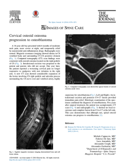
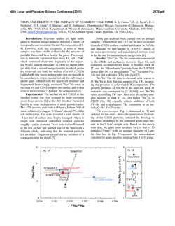
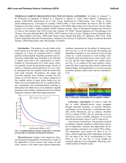
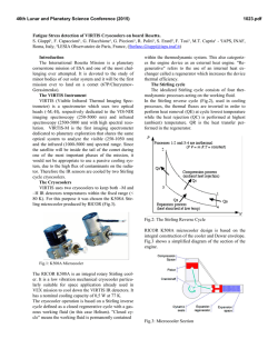
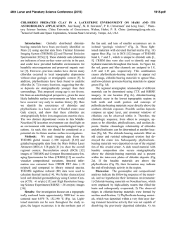
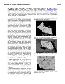
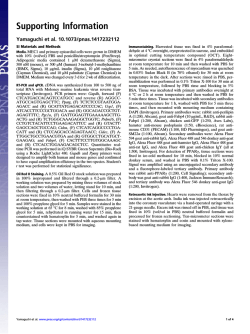
A peer-reviewed open-access journal
ZooKeys 335: 101–112 (2013)
The enigmatic Otway odd-clawed spider (Progradungula otwayensis Milledge, 1997)
doi: 10.3897/zookeys.335.6030
Research article
www.zookeys.org
101
Launched to accelerate biodiversity research
The enigmatic Otway odd-clawed spider (Progradungula
otwayensis Milledge, 1997, Gradungulidae, Araneae):
Natural history, first description of the female and
micro-computed tomography of the male palpal organ
Peter Michalik1,2, Luis Piacentini3, Elisabeth Lipke1, Martin J. Ramírez3
1 Allgemeine und Systematische Zoologie, Zoologisches Institut und Museum, Ernst-Moritz-Arndt-Universität,
J.-S.-Bach-Str. 11/12, D-17489 Greifswald, Germany 2 Research Associate, Division of Invertebrate Zoology,
American Museum of Natural History, Central Park West at 79th Street, New York, NY 10024, USA 3 Museo
Argentino de Ciencias Naturales - CONICET, Buenos Aires, Argentina
Corresponding author: Peter Michalik ([email protected])
Academic editor: Jeremy Miller | Received 2 August 2013 | Accepted 29 August 2013 | Published 25 September 2013
Citation: Michalik P, Piacentini L, Lipke E, Ramírez MJ (2013) The enigmatic Otway odd-clawed spider (Progradungula
otwayensis Milledge, 1997, Gradungulidae, Araneae): Natural history, first description of the female and micro-computed
tomography of the male palpal organ. ZooKeys 335: 101–112. doi: 10.3897/zookeys.335.6030
Abstract
The recently described cribellate gradungulid Progradungula otwayensis Milledge, 1997 is endemic to the
Great Otway National Park (Victoria, Australia) and known from only one male and a few juvenile specimens. In a recent survey we recorded 47 specimens at several localities across the western part of the Great
Otway National park. Our field data suggest that this species is dependant on the microclimate in the
hollows of old myrtle beech trees since other hollow trees were very much less inhabited. Furthermore, we
describe the female for the first time and study the male palpal organ by using X-ray microtomography.
The female genitalia are characterized by eight spermathecae which are grouped in two quartets. The
spermophor resembles the general organization of gradungulids, but is similar to Kaiya Gray, 1987 by a
convoluted appearance within the embolus. The muscle 30 is located in the cymbium and resembles the
organization of other non-entelegyne Araneomorphae.
Keywords
Taxonomy, micro-CT, palp, spermatheca, web, Nothofagus
Copyright Peter Michalik et al. This is an open access article distributed under the terms of the Creative Commons Attribution License 3.0
(CC-BY), which permits unrestricted use, distribution, and reproduction in any medium, provided the original author and source are credited.
102
Peter Michalik et al. / ZooKeys 335: 101–112 (2013)
Introduction
The family Gradungulidae consists of seven genera with 16 described species from
eastern Australia and New Zealand (Platnick 2013). The genera Macrogradungula (M.
moonya Gray, 1987) and Progradungula (P. carraiensis Forster and Gray, 1979 and P.
otwayensis Milledge, 1997) can be distinguished from the remaining gradungulids by
being cribellate. These spiders use a very conspicuous catching ladder for prey capture
(Forster et al. 1987; Gray 1983; Figs 2B–C). The enigmatic P. carraiensis is known only
from the Carrai Bat Cave in northern New South Wales (Forster and Gray 1979; Gray
1983) and thus it was remarkable when Milledge (1997) described a second species, P.
otwayensis, from the Great Otway National Park in southern Victoria (Australia). Nevertheless, only a single adult male and several immatures were reported at that time.
In the present paper, we describe the female for the first time and reconstruct the male
palpal organ by using X-ray microtomography (micro-CT). Moreover, we provide new
data on the natural history of this species.
Material and methods
We observed a total of 47 specimens (juveniles, females, one male) in the localities
depicted in Fig. 1. Several individuals were collected for further analyses. The female
genitalia were digested following the protocol of Álvarez-Padilla and Hormiga (2007).
The material was examined and documented (extended focal range images) in 80%
ethanol using a Zeiss Discovery V20 stereo microscope with a Zeiss MCr camera and
a Leica M205A with a Leica 290 camera. Editing of images to adjust brightness, contrast and color was performed using Adobe Photoshop CS4. The measurements and
description are based on Forster et al. (1987), the description of the spination follows
Ramirez (2003).
For the micro-CT analyses of the male palp, the sample was dehydrated in graded
ethanol and stained with a 1% iodine solution for 12 hours. After washing in pure
ethanol, the sample was scanned in ethanol with an Xradia MicroXCT-200 X-ray
imaging system (Carl Zeiss X-ray Microscopy Inc., Pleasanton, USA) at 20 KV and 4
W (10.0 scintillator-objective lens unit, 11 s exposure time, 2.18 µm pixel size). The
female genitalia were digested with enzymatic cleaner for contact lenses Ultrazyme,
and dried after the iodine treatment using hexamethyldisilazane (HMDS). The scan
was performed using the same system at 30 kV and 6 W (20.0 scintillator-objective
lens unit, 6 s exposure time, 1.18 µm pixel size). The obtained data were processed
using the 3D analysis software AMIRA v. 5.4.2 (Visage Imaging, Berlin, Germany).
The spermophor was reconstructed by delineation of the contours in each section and
a smooth surface was computed using the surface editor. The image stack is stored
in MorphDBase under creative commons attribution (ID: P_Michalik_20130802M-5.1, https://www.morphdbase.de/?P_Michalik_20130802-M-5.1).
The enigmatic Otway odd-clawed spider (Progradungula otwayensis Milledge, 1997)
103
Figure 1. Distribution map of P. otwayensis in the Great Otway National Park, Victoria (Australia). Red
marks indicate localities for the present study; black circles indicate localities given by Milledge (1997).
Abbreviations
ALE
AME
B
bH
Cb
E
EF
Gl
GP
m29
m30
mA
mH
PE
anterior lateral eyes
anterior median eyes
bursa
basal hematodocha
cymbium
embolus
epigastric furrow
glands
genital pockets
muscle 29
muscle 30
median apophysis
median hematodocha
process of embolus
104
PF
PLE
PME
S
Sp
St
Te
Ue
tm29
tm30
MACN
MV
ZIMG
Peter Michalik et al. / ZooKeys 335: 101–112 (2013)
postepigastric fold
posterior lateral eyes
posterior median eyes
spermophor
spermatheca
subtegulum
tegulum
uterus externus
tendon of muscle 29
tendon of muscle 30
Museo Argentino de Ciencias Naturales (Buenos Aires, Argentina)
Museum of Victoria (Melbourne, Victoria)
Zoologisches Institut und Museum Greifswald (Germany)
Taxonomy
Family Gradungulidae Forster, 1955
Genus Progradungula Forster & Gray, 1979
Progradungula otwayensis Milledge, 1997
http://species-id.net/wiki/Progradungula_otwayensis
Type material. Male holotype: AUSTRALIA: Victoria. Otway Ranges, Aire Crossing
Track, 0.5 km N of Aire River crossing, 38°42'S, 143°29'E, 31 Jan 1995, G. Milledge
(MV K3260, not examined).
Material examined. AUSTRALIA: VICTORIA: Great Otway National Park:
Little Aire Cascade Trail, E of Lavers Hill, S38.67032°, E143.49810°(GPS, ±100m),
elev. 330m, 11 Feb 2013, Nothofagus, Eucalyptus, tree ferns wet forest, hand collecting, P.Michalik & M.J.Ramírez (MJR-Loc-125), two females (MACN-Ar 30666);
Triplet Falls Trail, E of Lavers Hill, S38.67188°, E143.49673° (GPS, ±300m), elev.
300m (GPS), 10 Feb 2013, Nothofagus, Eucalyptus, tree ferns wet forest, hand collecting, P.Michalik & M.J.Ramírez (MJR-Loc-124), three females (MACN-Ar 30667,
ZIMG II/28128, ZIMG II/28129); Maits Rest Trail, W Apollo Bay, S38.75492°,
E143.55495° (GPS,±200m), elev. 240m, 13–14 Feb 2013, Nothofagus, tree ferns
wet forest, hand collecting, P.Michalik & M.J.Ramírez (MJR-Loc-126), one female
(ZIMG II/28130); Melba Gully Trail, S38°41.726', E143°22.312' (GPS, ±200m),
elev. 328m, 15 Feb 2013, Nothofagus, tree ferns wet forest, hand collecting, P.Michalik
& M.J.Ramírez (MJR-Loc-128), one male (ZIMG II/28127).
Diagnosis. This species can be distinguished from P. carraiensis by the single process on the embolus of the male palpal organ and the presence of eight spermathecae
in the female genitalia.
The enigmatic Otway odd-clawed spider (Progradungula otwayensis Milledge, 1997)
105
Figure 2. A Habitat with supporting web (blue arrow) and sturdy thread (red arrows) connecting with
the retreat in the hollow of a N. cunninghamii tree B Catching ladder in front of a hollow in a large tree
fern C Immature male of P. otwayensis holding the catching ladder.
106
Peter Michalik et al. / ZooKeys 335: 101–112 (2013)
Female (Figs 3–4).
Measurements (mm, in MV). Carapace length 6.92; carapace width 4.66;
caput width 2.66; abdomen length 8.51; abdomen width 7.18. Legs: length of segments (femur + patella/tibia + metatarsus + tarsus = total length): I 11.97 + 14.10
+ 11.57 + 2.66 = 40.30, II 9.31 + 10.77 + 8.25 + 2.53 = 30.86, III 7.98 + 9.18 +
7.85 + 2.13 = 27.14, IV 9.98 + 10.91 + 9.44 + 2.13 = 32.46, palp 2.93 + 2.66 + - +
2.79 = 8.38.
Colour pattern. Carapace yellowish brown, darker on cephalic area, particularly
in eye region. Chelicerae, maxillae and labium reddish brown. Sternum reddish brown
marginally, paler centrally. Legs yellowish brown. Abdomen fawn-coloured with dark
brown dorsal pigmentation consisting of 3 chevron markings. Carapace longer than
wide, widest between coxae II and III. Cephalic area elevated behind eyes (Fig. 3A).
Clypeus sloping, as long as the median ocular quadrangle, with a strongly curved anterior border. Fovea pit-like.
Eyes. Frontal view (Fig. 3D): anterior row slightly recurved, posterior row slightly
procurved. Dorsal view (Fig. 3B): anterior row recurved, posterior row slightly recurved. AME < PME < ALE = PLE 0.23 : 0.32 : 0.35 : 0.35. Interdistances: AMEAME 0.18, AME-ALE 0.33, ALE-PLE 0.08, PLE-PME 0.40, PME-PME 0.28. Median ocular quadrangle: length 0.58 mm, anterior width 0.55 mm, posterior width
0.87 mm. Clypeus height 0.50. AME black, remainder white.
Chelicerae. Strong, vertical, slightly divergent. Three strong prolateral teeth, evenly spaced. Five very small retromarginal teeth (or denticles) in row on basal half of
groove, with a spine on the apico-dorsal side. Stridulatory ridges absent.
Maxillae. Subparallel, external margin strongly curved and ending anteriorly in a
bluntly pointed apex (Fig. 3C). Serrula present.
Labium. Free. Length 0.83, width 0.87. Apical margin indented, lateral margin
subparallel below, sloping in toward apex above (Fig. 3C).
Sternum. Length 3.33, width 2.20. Elongate, shield-shaped with pointed apex
which extends back midway between coxae IV (Fig. 3C).
Legs. 1423. Trochanters shallowly notched. Superior claws of 1st and 2nd legs
dissimilar, with raptorial proclaws long and strongly developed (Fig. 3F), retroclaws
shorter. Inferior claws of legs I and II slender and strongly hooked. Superior claws of
legs III and IV similar (Fig. 3G). Accessory claw setae on all tarsi. Distal half of tarsus
I and II with strong ventral setae. Calamistrum short, located in second quarter of
metatarsus 4 (Fig. 3H).
Pattern of spination. (Approximate, slightly asymmetrical.) Femur I d (r2p1)r2-r1-p1-1-2-p1-2-p1-2-2-3ap v 0-p1-0-0-0; patella r 1; tibia d 2-0-2-p1-0-p1-3
v 2-0-0-2-p1-0-2-0; metatarsus d r1-p1-p1-0-0-0-0 v p1-r1-p1-p1-r1-p1-r1-2ap;
II; femur d (r2p1)-r2-2-p1-2-p1-2- 2-p1-3 v 0-p1-1-0-0; patella p 1; tibia d 2-p11-2-2-p1-p1-1-3 v 2-0-p1-r1-p1-2-0-2ap; metatarsus d 2-p1-2-r1-p1-r1-p1-p12ap v p1-r1-2-2-r1-p1-3ap; III; femur d (r3p1)-1-r1-2-p1-2-2-2-r1-3 v r1-p12-0-0; patella p 1 d 1 r 1; tibia d 2-1-p1-3-2-2 v 2-0-p1-r1-p1-2ap; metatarsus
d 2-p1-p1-r1-p1-2- 2ap v 2-p1-p1-r1-p1-p1-r1-3ap; tarsus v 0-r1-0; IV femur d
The enigmatic Otway odd-clawed spider (Progradungula otwayensis Milledge, 1997)
107
Figure 3. Somatic characters of the female of P. otwayensis. A Lateral view of prosoma and opisthosoma
(ZIMG II/28128) B Dorsal view of prosoma (MV) C Ventral view of Prosoma (MV) D Frontal view of
ocular area (ZIMG II/28128) E Ventral view of opisthosoma F Tarsus of leg I G Tarsus of leg IV H Calamistrum. I Ventral view of spinnerets. Scale bar in F–H is 500 µm.
(r4p2)-r2-1-r2-p1-2-2-2-3 v 0-p1-p1-0-0-0; patella p 1 d 1; tibia d 2-2-p1-1-3-r1
v 2-0-0-p1-p1-2ap; metatarsus d 2-p1-2-r1-2ap v p1-2-p1-p1-p1-2-r1-3ap; tarsus
v 0-1-0.
108
Peter Michalik et al. / ZooKeys 335: 101–112 (2013)
Figure 4. Female genitalia. A Lateral view of the dome-shaped genital area (compare also to Fig. 3A)
(MACN-Ar 30667) B Sagital section through genital area obtained by micro-CT (MACN-Ar 30667)
C Posterior view of genital area (MACN-Ar 30667). Spermathecae in anterior (D) and ventral view (E)
(ZIMG II/28128). Abbreviation: B bursa; EF epigastric furrow; GP genital pockets; Gl glands; PF postepigastric fold; Sp spermathecae; Ue uterus externus.
Palp. Single claw. Spines: femur d 0-0-1-1-3ap patella p 1 d 1-1, tibia d 1-1, p
0-2-0; tarsus d 2-2-1-2-3ap v 2-2-2ap.
Abdomen. Cribellum undivided, with narrow spinning field, as wide as basal span
of anterior spinnerets (Figs 3E, 3I). Spiracles of posterior lung books well separated but
joined by a transverse groove. Genital area swollen and only faintly sclerotized near the
epigastric furrow (Fig. 3E).
The enigmatic Otway odd-clawed spider (Progradungula otwayensis Milledge, 1997)
109
Figure 5. Right male palp of P. otwayensis (ZIMG II/28127). A Prolateral view; the arrow marks the
section plane of Fig. 5B B Section of the volume rendered male palp C Surface model of the spermophor
superimposed on the volume rendering of the male palp (prolateral, ventral and retrolateral views). The
cymbium, subtegulum and tegulum are partly removed in the prolateral view to show tendons, muscles and
hematodochae. The arrow points to the opening of the embolus. Abbreviation: Cb cymbium; E embolus;
m29 muscle 29; m30 muscle 30; mA median apophysis; mH median hematodocha; PE process of embolus; S spermophor; St subtegulum; Te tegulum; tm29 tendon of muscle 29; tm30 tendon of muscle 30.
110
Peter Michalik et al. / ZooKeys 335: 101–112 (2013)
Genitalia. Genital area distinct and dome-shaped (Figs 3A, 4A). Internal genitalia
simple, with eight spermathecae grouped in two quartets (Figs 4D–E). The outer spermathecae are ovoid and have a short duct. The inner spermathecae can be grouped leading into the bursa through a long, slender convoluted duct (Fig. 4). Glandular projections are mainly present on the spermathecal ducts. A fold with two distinct sclerotized
pockets is situated anterior/ventral to the spermathecae. A blind-ended invagination,
the postepigastric furrow, is situated immediately posterior to the epigastric furrow.
Male (ZIMG II/28127; Fig. 5).
Male palp. Tegulum with short median apophysis. Embolus tube-like with a solid
single hook-shaped process; tip of the embolus widened and hyaline; fundus sac-like,
spermophor with an internal band delimited by two parallel ridges (Fig. 5B), distal part of
the spermophor within the embolus partly convoluted and very thin (approximately 8µm
in diameter); m30 and tm30 in cymbium; m29 in tibia and attaching to the spermophor.
Natural history. The web structure is similar to P. carraiensis (for details see Gray
1983) and web construction is performed after sunset. The catching ladders and supporting webs (Figs 2B–C) are connected by a single sturdy thread to retreats in hollow trees Fig. 2A), often far away (up to 3 meters). As already described by Milledge
(1997), we found most of the specimens in hollows of old Nothofagus cunninghamii
trees (Fig. 2A), but also several in hollows of mountain ash (Eucalyptus regnans) trees,
the bases of large tree ferns (Fig. 2B) and under bridges on trails. On one occasion, we
had access to a large hollow mountain ash tree and found catching ladders and supporting webs of juveniles inside of it.
Distribution. Endemic to the Great Otway National Park (Victoria). In addition
to the locations reported by Milledge (1997), we found several specimens (one male,
several juveniles) at the Melba Gully Trail which is located at the western end of the
National Park (Fig. 1).
Discussion
Our data are in accordance with the previous detailed descriptions on the distribution
and natural history of Progradungula species (Forster and Gray 1979; Forster et al. 1987;
Gray 1983; Milledge 1997). The new records of P. otwayensis from the Melba Gully trail
might imply a wider distribution in the Great Otway National Park. Nevertheless, this
species seems to be dependent on hollow trees with a suitable microclimate (Gibbons and
Lindenmayer 1997; Gibbons et al. 2002), since specimens mostly occur in the oldest and
extensively hollow myrtle beech trees in the humid forests in the western part of the Great
Otway National Park, or in mountain ash trees, upon which the myrtle trees depend (Lindenmayer et al. 2000). Other habitats such as tree ferns were much less inhabited in areas
where old hollow myrtles occur (Milledge 1997). The presence of juvenile catching ladders and supporting webs inside the hollow trees suggests that early instars live exclusively
in hollows. This is also supported by the fact that we could never observe exposed juvenile
catching ladders, as we abundantly found for larger immatures and adults.
The enigmatic Otway odd-clawed spider (Progradungula otwayensis Milledge, 1997)
111
The female genitalia of P. otwayensis are very similar to P. carraiensis as the number
of spermathecae is reduced compared to other gradungulids and they are bilaterally arranged (Forster et al. 1987). Thus, we here confirm the generic placement suggested by
Milledge (1997). Additionally, we found distinct and sclerotized pockets in a fold anteriorly to the epigastric furrow which has not been reported from any other gradungulid
(Forster et al. 1987). The function of these pockets is unknown, but an interaction with
the embolic process during copulation seems to be a possibility. Moreover, in contrast
to Forster et al. (1987) we could not find a “median receptaculum”. Instead, the organization is mainly in accordance with the interpretation of Griswold et al. (2005) of the
genitalia of gradungulids.
As revealed by micro-CT analysis of the male palpal organ of P. otwayensis, the
spermophor has a thick wall and an internal band delimited by two parallel ridges,
which in the micro-CT sections appears associated with glandular tissue. As described
by Forster et al. (1987) for Kaiya Gray, 1987, the spermophor of P. otwayensis is convoluted within the embolus. The muscles (m29 and m30) and their tendons are in accordance with the description of Gradungula sorenseni Forster, 1955 by Huber (1994)
and resemble the usual organization for non-entelegyne Araneomorphae (Huber
2004). Thus, the organization might be typical for all Gradungulidae and different to
the organization found in austrochilines (Michalik and Ramírez in press).
Acknowledgments
We are very grateful to Mike Rix (WMA, Perth, Australia) for advice regarding the
localities and helpful comments on the manuscript. This study was funded by the German Science Foundation (DFG Mi 1255/5-1), CONICET (PIP 112-200801-03209)
and ANPCyT (FONCyT, PICT-2007-01393). The material was collected under the
permit number 10006549 (Department of Sustainability and Environment, Victoria).
Mark Harvey (WMA, Perth, Australia) suggested the common name for this species.
References
Álvarez-Padilla F, Hormiga G (2007) A protocol for digesting internal soft tissues and mounting spiders for scanning electron microscopy. Journal of Arachnology 35: 538–542. doi:
10.1636/Sh06-55.1
Forster RR, Gray MR (1979) Progradungula, a new cribellate genus of the spider family
Gradungulidae (Araneae). Australian Journal of Zoology 27: 1051–1071. doi: 10.1071/
Zo9791051
Forster RR, Platnick NI, Gray MR (1987) A review of the spider superfamilies Hypochiloidea and Austrochiloidea (Araneae, Araneomorphae). Bulletin of the American
Museum of Natural History 185: 1–116. http://digitallibrary.amnh.org/dspace/handle/2246/969
112
Peter Michalik et al. / ZooKeys 335: 101–112 (2013)
Gibbons P, Lindenmayer D (1997) A review of prescriptions employed for conservation of
hollow-dependent fauna in wood production forests of eastern Australia. In: Hale P, Lamb
D (Eds) Conservation outside Nature Reserves. Centre for Conservation Biology. The
University of Queensland, Brisbane, 497–505.
Gibbons P, Lindenmayer DB, Barry SC, Tanton MT (2002) Hollow selection by vertebrate
fauna in forests of southeastern Australia and implications for forest management. Biological Conservation 103: 1–12. doi: 10.1016/S0006-3207(01)00109-4
Gray MR (1983) The male of Progradungula carraiensis Forster and Gray (Araneae, Gradungulidae) with observations on the web and prey capture. Proceeding of the Linnean Society
of New South Wales 107: 51–58.
Griswold CE, Ramírez MJ, Coddington JA, Platnick NI (2005) Atlas of pylogenetic data for
entelegyne spiders (Araneae: Araneomorphae: Entelegynae) with comments on their phylogeny. Proceedings of the California Academy of Sciences 56: 1–325.
Huber BA (1994) Genital bulb muscles in entelegyne spiders. Journal of Arachnology 22: 75–76.
Huber BA (2004) Evolutionary transformation from muscular to hydraulic movements in spider (Arachnida, Araneae) genitalia: a study based on histological serial sections. Journal of
Morphology 261: 364–376. doi: 10.1002/jmor.10255
Lindenmayer DB, Mackey BG, Cunningham RB, Donnelly CF, Mullen IC, McCarthy MA,
Gill AM (2000) Factors affecting the presence of the cool temperate rain forest tree myrtle
beech (Nothofagus cunninghamii) in southern Australia: integrating climatic, terrain and
disturbance predictors of distribution patterns. Journal of Biogeography 27: 1001–1009.
doi: 10.1046/j.1365-2699.2000.00443.x
Michalik P, Ramírez M (in press) First description of the male of Thaida chepu Platnick, 1987
(Araneae, Austrochilidae) with micro-computed tomography of the palpal organ. Zookeys.
Milledge GA (1997) A new species of Progradungula Forster and Gray (Araneae: Gradungulidae) from Victoria. Memoirs of Museum Victoria 56: 65–68.
Platnick NI (2013) The world spider catalog, version 14.0, American Museum of Natural History.
http://research.amnh.org/entomology/spiders/catalog/index.html, doi: 10.5531/db.iz.0001
Ramírez MJ (2003) The spider subfamily Amaurobioidinae (Araneae, Anyphaenidae): A phylogenetic revision at the generic level. Bulletin of the American Museum of Natural History
277: 1–262. http://digitallibrary.amnh.org/dspace/handle/2246/445


© Copyright 2026