
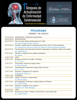
Advances in Stroke Advances in Brain Recovery and Rehabilitation
Advances in Stroke Advances in Brain Recovery and Rehabilitation 2010 Richard Zorowitz, MD; Michael Brainin, MD, FESO, FAHA Abstract—Discoveries in the past year have impacted the understanding of brain recovery and there is more of a need than ever for a foothold in recovery and rehabilitation This review reports on translational efforts, new (and old) potential drugs, various approaches to neurorehabilitation, and brain imaging that demonstrate reorganization in the human brain during stroke rehabilitation. (Stroke. 2011;42:294-297.) Key Words: brain recovery 䡲 imaging 䡲 outcomes 䡲 quality of life 䡲 rehabilitation 䡲 stem cells 䡲 stroke recovery O Downloaded from http://stroke.ahajournals.org/ by guest on October 9, 2016 BDNF after temporary occlusion of the right middle cerebral artery did not reduce infarct volume significantly, but increased the number of activated and phagocytotic microglia, suppressed tumor necrosis factor-␣ and mRNA expression, increased interleukin-10 and mRNA expression, and increased DNA-binding activity of nuclear factor- B. Overall, intranasal BDNF might protect the brain against ischemic insult by modulating local inflammation through regulation of the levels of cellular, cytokine, and transcription factor in experimental stroke. To deliver BDNF to the brain, Lee and associates developed genetically modified human neural stem cells that overexpress in a mouse stroke model.3 After inducing intracerebral hemorrhage in adult rats, a human neural stem cell line that produces 6-fold higher amounts of BDNF was transplanted into the brains. The stem cells differentiated and renewed angiogenesis of host brain and functional recovery in the animals, thereby suggesting that these cell lines could be of great value as a cellular source for experimental studies involving cellular therapy for human neurological disorders. ver the past 15 years, the focus of stroke medical advances and healthcare resources has been on acute and subacute recovery phases, which has resulted in substantial health disparities in later phases of stroke care. More recently, the field of brain recovery has seen a plethora of basic, translational, and applied experiments that deserve to be discussed, reviewed, and evaluated for further research. Unfortunately, this report can highlight only examples from the last year that appear most relevant to the authors and hold promise for clinical relevance. Translational research allows basic scientists to provide clinicians with new tools for use in patients and for assessment of their impact at the same time as clinical researchers make novel observations about the nature and progression of disease that often stimulate basic investigations.1 Pharmacotherapy allows researchers to use already available drugs and develop new medications that can protect the brain from damage and facilitate and enhance recovery. Approaches to rehabilitation allow researchers to develop new methods of facilitating recovery, enhancing compensatory strategies, and comparing techniques in the quest to determine the most effective and efficient means of rehabilitation. Finally, to better understand the mechanisms of neuroplasticity, research is using imaging and neurophysiological techniques to document the reorganization of the brain that accompanies functional improvement. The purpose of this review is to describe some of the additions to the literature that contributed to the ever growing knowledge base of neurorehabilitation of stroke. The Search for Potential New (and Not so New) Drugs Drugs also have been shown to facilitate brain recovery in animal models. Ding and associates4 used T2-, diffusionweighted, and susceptibility-weighted MRI imaging to explore whether erythropoietin (EPO) initiated at 24 hours and administered daily for 7 days after an embolic stroke assists in repairing ischemic cerebral tissue. In a randomized trial of 22 adult Wistar rats given either treatment or control after occlusion of the middle cerebral artery occlusion, they found that expansion of the ipsilateral ventricle was significantly reduced in the EPO-treated rats. The volume ratio of ipsilateral parenchymal tissue relative to the contralateral hemisphere was significantly increased after EPO treatment compared with control animals, indicating that EPO significantly Translational Research: Bench to Bedside and Back Because inflammation plays a vital role in the pathogenesis of ischemic stroke, researchers felt that brain-derived neurotrophic factor (BDNF) may protect brain tissues from ischemic injury. In 1 study, intranasal BDNF was given to rats to protect the brain from ischemic insult.2 Rats given intranasal Received December 3, 2010; accepted December 7, 2010. From the Department of Physical Medicine and Rehabilitation (R.Z.), The Johns Hopkins University School of Medicine, and the Department of Physical Medicine and Rehabilitation, Johns Hopkins Bayview Medical Center, Baltimore, MD; and the Department of Clinical Medicine and Prevention (M.B.), Danube University, Krems, Austria. Correspondence to Michael Brainin, MD, FESO, FAHA, Danube University and Danube Clinic, Department Chairman and Director, Department of Neurology, Karl Dorrekstrasse 30, Krems, Austria 3500. E-mail [email protected] © 2011 American Heart Association, Inc. Stroke is available at http://stroke.ahajournals.org DOI: 10.1161/STROKEAHA.110.605063 294 Zorowitz and Brainin Downloaded from http://stroke.ahajournals.org/ by guest on October 9, 2016 reduces atrophy of the ipsilateral hemisphere. Angiogenesis and white matter remodeling were significantly increased and occurred earlier in EPO-treated animals than in the controls as noted on T2- and diffusion-weighted images, respectively. In a Phase IIa study, Cramer and associates5 gave 15 patients 24 to 48 hours postischemic infarct with National Institutes of Health Stroke Scale scores from 6 to 24 after a 9-day course of -human chorionic gonadotropin on Days 1, 3, and 5 followed by EPO on Days 7, 8, and 9. No safety concerns were noted among clinical or laboratory measures, including screening for deep vein thrombosis and serial measures of serum hemoglobin. As we have seen over many years, other medications have not been effective. O’Collins and colleagues6 chose magnesium sulfate, melatonin, and minocycline from a library of neuroprotective agents, and these were tested in a more “realistic” model favored by the Stroke Therapy Academic Industry Roundtable. Despite the animal model, this combination of medications was not an effective neuroprotectant when infarct volume, neurological score, and 2 newly developed scales measuring general health and physiological homeostasis were measured. Brain Imaging and Neuroplasticity: Just Picture This New imaging techniques continue to be developed and applied to the detection and staging of white matter reorganization after brain injury with and without neurorestorative treatment. Jiang and colleagues7 demonstrated how variations of diffusion tensor MRI methodology could detect white matter remodeling after brain injury. In addition, Q-space diffusion tensor MRI, an emerging diffusion-weighted imaging technique that identifies the molecular diffusion probability density function without the need to assume a Gaussian distribution, can detect early-stage axonal remodeling involving randomly oriented crossing axons. Imaging also continues to be used in conjunction with rehabilitation interventions to demonstrate the efficacy of the activity. Enzinger and colleagues8 used functional MRI longitudinally to relate brain activity changes with performance gains of the lower limb after 4 weeks of treadmill training with partial body weight support. Their study in 18 chronic patients (mean age, 59.9⫾13.5 years) demonstrated not only that walking endurance improved after training, but also greater walking endurance was correlated with increased brain activity in the bilateral primary sensorimotor cortices, the cingulate motor areas, and the caudate nuclei bilaterally and in the thalamus of the affected hemisphere. New and Expanding Therapeutic Approaches In the past year, researchers continued to explore previously untested stroke rehabilitation interventions as well as better define those that have an evidence base. Wolf and associates9 compared functional improvements between stroke survivors randomized to receive constrain-induced movement therapy within 3 to 9 months (early group) with those randomized to 15 to 21 months after stroke (delayed group). Although both groups improved functionally 12 months after treatment, stroke survivors receiving constrain-induced movement ther- Advances in Brain Recovery and Rehabilitation 295 apy earlier demonstrated significant improvement than those in the delayed constrain-induced movement therapy group. However, 24 months after enrollment, there were no statistically significant differences between groups. More research was completed to determine the efficacy of treadmill training with body weight support. The MOBILISE trial10 randomized 126 stroke survivors who were unable to walk into an experimental group who received up to 30 minutes per day of treadmill walking with body weight support and a control group who received up to 30 minutes of overground walking. Six months after the training, more of the experimental group was independent in ambulation and was discharged home after rehabilitation. However, the results were not statistically significant (P⫽0.13). The role of electric stimulation also continued to be pursued. Hsu and associates11 studied 66 stroke survivors with severe motor deficit randomized to receive 0, 30, or 60 minutes of upper extremity neuromuscular electric stimulation daily for 4 weeks. In this case, both groups receiving neuromuscular electric stimulation demonstrated similar improvements as measured by the Fugl-Meyer Motor Assessment and Action Research Arm Test scales. The role of transcranial magnetic stimulation also received attention in 2010. Lindenberg and colleagues12 randomized 20 stroke survivors to receive 5 consecutive sessions of experimental or sham bihemispheric transcranial direct current stimulation with simultaneous physical/occupational therapy. Motor function was significantly greater in the experimental group and outlasted the stimulation by at least 1 week. The improvement was correlated with stronger activation of intact ipsilesional motor regions during paced movements of the affected limb. Another study of M1 theta burst stimulation (TBS) was completed by Ackerley and colleagues.13 Ten patients with chronic subcortical stroke involving the upper limb received intermittent TBS of the ipsilesional M1, continuous TBS of the contralesional M1, and sham TBS in separate sessions in conjunction with standardized training of a precision grip task. Training with real TBS improved paretic-hand grip–lift kinetics, whereas training with sham TBS resulted in deterioration of grip–lift. Ipsilesional M1 excitability increased after intermittent TBS of the ipsilesional M1 but decreased after continuous TBS of the contralesional M1, resulting in deterioration of the Action Research Arm Test. They concluded the contralesional hemisphere may play a pivotal role in recovery after stroke. Robotics continues to be an area of focus in stroke rehabilitation. In a randomized study evaluating stroke survivors with long-term upper-limb impairments, Lo and associates14 compared outcomes in 127 subjects at 12 weeks when given robot-assisted therapy, intensive comparison therapy, or usual care. Although no adverse events were reported in any subject, robot-assisted therapy did not significantly improve motor function at 12 weeks as compared with usual care or intensive therapy. However, in a secondary analysis, robot-assisted therapy improved outcomes over 36 weeks as compared with usual care but not with intensive therapy. Even virtual reality and gaming are becoming pervasive in stroke rehabilitation. Saposnik and colleagues15 devised a 296 Stroke February 2011 Downloaded from http://stroke.ahajournals.org/ by guest on October 9, 2016 randomized, single-blinded clinical trial to compare the feasibility, safety, and efficacy of virtual reality using the Nintendo Wii gaming system versus recreational therapy (playing cards, bingo, or “Jenga”). In the 17 subjects of their pilot project, the 9 subjects using virtual reality using the Nintendo Wii significantly improved in mean motor function of 7.4 seconds on the Wolf Motor Function Test after adjustment for age, baseline functional status, and stroke severity. They concluded that virtual reality using the Nintendo Wii is a potentially effective alternative to facilitate rehabilitation therapy and promote motor recovery after stroke. Finally, 2 studies in Chinese alternative medicine should be noted. First, a meta-analysis by Wu and colleagues16 concluded that “acupuncture may be effective in the treatment of poststroke rehabilitation, [but] poor study quality and the possibility of publication bias hinder the strength of this recommendation.” Second, Lee and associates17 completed a systematic review on moxibustion, a traditional Chinese method that uses the heat generated by burning herbal preparations containing Artemisia vulgaris to stimulate acupuncture points. Although moxibustion is popular in east Asian countries, their meta-analysis of 9 randomized clinical trials found only limited effectiveness of moxibustion in stroke rehabilitation. Spasticity and Disability In the year that the US Food and Drug Administration finally approved the use of onabotulinumtoxin (Botox) for upper limb spasticity, it is appropriate to include some research that showed that severe poststroke spasticity is rare but when present contributes to disability and high costs. First, Urban and colleagues18 found that spasticity developed in 42.6% of 211 subjects 6 months poststroke, but severe spasticity was relatively rare. Predictors for the development of spasticity included a severe degree of paresis and hemihypesthesia at stroke onset. Second, Lundström and associates19 estimated that the mean direct costs (ie, acute and rehabilitation hospitalization, primary health care, medication, and costs for municipality services) for a stroke survivor with spasticity was $84 195 (median, $72 816; interquartile range, $53 707) compared with $21 842 ($12 385; $17 484) for stroke survivors without spasticity. Costs for hospital care, primary care, and home or residential care also were significantly higher if the stroke survivor had spasticity. Thus, spasticity has to be viewed as 1 of several possible factors impeding motor function and treatment of spasticity-related disability should be seen as 1 of several options in chronic stroke management. Outcomes Research The Year 2010 also saw more studies that attempted to delineate good and poor outcomes after stroke. As part of the Northern Manhattan Stroke Study, Willey and associates20 reported that stroke survivors who stated that they felt depressed 7 to 10 days poststroke were at odds of severe disability compared with no disability at 1 (OR, 2.91; 95% CI, 1.07 to 7.91) and 2 years (OR, 3.72; 95% CI, 1.29 to 10.71) after stroke. However, depressed mood was not associated with overall mortality or vascular death. Rist and colleagues21 observed that among the 21 860 men enrolled in the Physicians’ Health Study, men who consumed ⬍1 drink per week had a modest beneficial association with functional outcome after stroke, but otherwise there was no strong association between increased alcohol consumption and functional outcome. Meta-Analysis and Scientific Statements: Putting It All Together Over the last year, there has been an effort to educate nursing and other members of the interdisciplinary team about the potential for recovery in the later or more chronic phases of stroke care. The American Stroke Association commissioned a scientific statement that summarizes the best available evidence and recommendations for interdisciplinary management of the needs of stroke survivors and their families during inpatient and outpatient rehabilitation and in chronic care and end-of-life settings.22 The statement makes use of the International Classification of Functioning, Disability, and Health of the World Health Organization23 as a foundation for the interdisciplinary team approach to rehabilitation and the different care settings in which stroke survivors may receive services. Batchelor and colleagues24 reviewed evidence relating to interventions that reduce falls after stroke. In the 13 studies that met their criteria, variability of falls reported was observed across the studies. Pooling of results was possible for only 2 types of interventions: exercise versus usual care (fall rate, fallers) and bisphosphonate medication versus placebo (fallers). The only intervention shown to be effective in reducing falls was vitamin D for female stroke survivors in an institutional setting, but other interventions were no more effective than usual care. Like in many other rehabilitation studies, they conclude that “further research evaluating a range of single and multifactorial interventions for fall prevention in the stroke population is required.” Conclusion: Still a Long Road Ahead Much progress has been made in stroke rehabilitation over the past months and years. The physiology of recovery is being studied intensively. Animal models of recovery are aimed at establishing the means by which pharmacological and hormonal treatments to facilitate recovery can be tested in humans. Clinical interventions that engage the stroke survivor and stimulate the brain continue to be developed at an increasing pace. Imaging techniques are assisting in confirming neuroplastic changes that intensive rehabilitation causes. However, much still needs to be done. We still do not know the types, doses, and combinations of physical and pharmacological modalities that will help specific stroke survivors. We still do not know how to initiate movement or speech when the stroke survivor has none. We still do not know how to effectively prevent cerebral damage using neuroprotective agents. We still do not know how to use cellular therapies to make the “right connections” that allow regeneration of the neural system. Over the next period, researchers must set the agenda in stroke rehabilitation so that we can further decrease the mortality and morbidity that the Zorowitz and Brainin American Stroke Association accomplished before its 2010 goal. With strong commitments from government and private funding sources alike, the world’s stroke rehabilitation can take strides to decrease impairments and improve activity and participation. That is something that will never be lost in translation. Disclosures None. References Downloaded from http://stroke.ahajournals.org/ by guest on October 9, 2016 1. Translational Research. Re-engineering the Clinical Research Enterprise. The NIH Common Fund. Available at: http://nihroadmap.nih.gov/ clinicalresearch/overview-translational.asp. Accessed November 21, 2010. 2. Jiang Y, Wei N, Lu T, Zhu J, Xu G, Liu X. Intranasal brain-derived neurotrophic factor protects brain from ischemic insult via modulating local inflammation in rats. Neuroscience. 2010 Oct 27 [Epub ahead of print]. 3. Lee HJ, Lim IJ, Lee MC, Kim SU. Human neural stem cells genetically modified to overexpress brain-derived neurotrophic factor promote functional recovery and neuroprotection in a mouse stroke model. J Neurosci. 2010;88:3282–3294. 4. Ding G, Jiang Q, Li L, Zhang L, Wang Y, Zhang ZG, Lu M, Panda S, Li Q, Ewing JR, Chopp M. Cerebral tissue repair and atrophy after embolic stroke in rat: a magnetic resonance imaging study of erythropoietin therapy. J Neurosci Res. 2010;88:3206 –3214. 5. Cramer SC, Fitzpatrick C, Warren M, Hill MD, Brown D, Whitaker L, Ryckborst KJ, Plon L. The beta-hCG⫹erythropoietin in acute stroke (BETAS) study: a 3-center, single-dose, open-label, noncontrolled, phase IIa safety trial. Stroke. 2010;41:927–931. 6. O’Collins VE, Macleod MR, Cox SF, Van Raay L, Aleksoska E, Donnan GA, Howells DW. Preclinical drug evaluation for combination therapy in acute stroke using systematic review, meta-analysis, and subsequent experimental testing. J Cereb Blood Flow Metab. 2010 Oct 27 [Epub ahead of print]. 7. Jiang Q, Zhang ZG, Chopp M. MRI evaluation of white matter recovery after brain injury. Stroke. 2010;41:S112–S113. 8. Enzinger C, MD, Dawes H, Johansen-Berg H, Wade D, Bogdanovic M, Collett J, Guy C, Kischka U, Ropele S, Fazekas F, Matthews PM. Brain activity changes associated with treadmill training after stroke. Stroke. 2009;40:2460 –2467. 9. Wolf SL, Thompson PA, Winstein CJ, Miller JP, Blanton SR, NicholsLarsen DS, Morris DM, Uswatte G, Taub E, Light KE, Lumy Sawaki L. The EXCITE Trial. Comparing early and delayed constraint-induced movement therapy. Stroke. 2010;41:2309 –2315. Advances in Brain Recovery and Rehabilitation 297 10. Ada L, Dean CM, Morris ME, Simpson JM, Katrak P. Randomized trial of treadmill training with body weight support to establish walking after stroke. The MOBILISE Trial. Stroke. 2010;41:1237–1242. 11. Hsu SS, Hu MH, Wang YH, Yip PK, Chiu JW, Hsieh CM. Dose-response relation between neuromuscular electrical stimulation and upper extremity function in patients with stroke. Stroke. 2010;41:821– 824. 12. Lindenberg R, Renga V, Zhu LL, Nair D, Schlaug G. Bihemispheric brain stimulation facilitates motor recovery in chronic stroke patients. Neurology. 2010 Nov 10 [Epub ahead of print]. 13. Ackerley SJ, Stinear CM, Barber PA, Byblow WD. Combining theta burst stimulation with training after subcortical stroke. Stroke. 2010;41: 1568 –1572. 14. Lo AC, Guarino PD, Richards LG. Robot-assisted therapy for long-term upper-limb impairment after stroke. N Engl J Med. 2010;362:1772–1783. 15. Saposnik G, Teasell R, Mamdani M, Hall J, McIlroy W, Cheung D, Thorpe KE, Cohen LG, Bayley M, for the Stroke Outcome Research Canada (SORCan) Working Group. Effectiveness of virtual reality using Wii gaming technology in stroke rehabilitation. A pilot randomized clinical trial and proof of principle. Stroke. 2010;41:1477–1484. 16. Wu P, Mills E, Moher D, Seely D. Acupuncture in poststroke rehabilitation. A systematic review and meta-analysis of randomized trials. Stroke. 2010;41:e171– e179. 17. Lee MS, Shin BC, Kim JI, Han CH, Ernst E. Moxibustion for stroke rehabilitation. Systematic review. Stroke. 2010;41:817– 820. 18. Urban PP, Wolf T, Uebele M, Marx JJ, Vogt T, Stoeter P, Bauermann T, Weibrich C, Vucurevic GD, Schneider A, Wissel J. Occurrence and clinical predictors of spasticity after acute ischemic stroke. Stroke. 2010; 41:2016 –2020. 19. Lundström E, Smits A, Borg J, Terént A. Four-fold increase in direct costs of stroke survivors with spasticity compared with stroke survivors without spasticity: the first year after the event. Stroke. 2010;41:319 –324. 20. Willey JZ, Disla N, Moon YP, Paik MC, Sacco RL, Boden-Albala B, Elkind MSV, Wright CB. Early depressed mood predicts long-term disability after stroke. The Northern Manhattan Stroke Study (NOMASS). Stroke. 2010;41:1896 –1900. 21. Rist PM, Berger K, Buring JE, Kase CS, Gaziano JM, Kurth T. Alcohol consumption and functional outcome after stroke in men. Stroke. 2010; 41:141–146. 22. Miller EL, Murray L, Richards L, Zorowitz RD, Bakas T, Clark P, Billinger SA; on behalf of the American Heart Association Council on Cardiovascular Nursing and Stroke Council. Comprehensive overview of nursing and interdisciplinary rehabilitation care of the stroke patient: a scientific statement from the American Heart Association. Stroke. 2010; 41:2402–2448. 23. World Health Organization. International Classification of Functioning, Disability and Health (ICF). Geneva, Switzerland: World Health Organization; 2008. 24. Batchelor F, Hill K, Shylie Mackintosh S, Said C. What works in falls prevention after stroke? A systematic review and meta-analysis. Stroke. 2010;41:1715–1722. Advances in Brain Recovery and Rehabilitation 2010 Richard Zorowitz and Michael Brainin Downloaded from http://stroke.ahajournals.org/ by guest on October 9, 2016 Stroke. published online January 13, 2011; Stroke is published by the American Heart Association, 7272 Greenville Avenue, Dallas, TX 75231 Copyright © 2011 American Heart Association, Inc. All rights reserved. Print ISSN: 0039-2499. Online ISSN: 1524-4628 The online version of this article, along with updated information and services, is located on the World Wide Web at: http://stroke.ahajournals.org/content/early/2011/01/13/STROKEAHA.110.605063 Data Supplement (unedited) at: http://stroke.ahajournals.org/content/suppl/2012/02/26/STROKEAHA.110.605063.DC1.html http://stroke.ahajournals.org/content/suppl/2012/03/12/STROKEAHA.110.605063.DC3.html http://stroke.ahajournals.org/content/suppl/2012/02/28/STROKEAHA.110.605063.DC2.html Permissions: Requests for permissions to reproduce figures, tables, or portions of articles originally published in Stroke can be obtained via RightsLink, a service of the Copyright Clearance Center, not the Editorial Office. Once the online version of the published article for which permission is being requested is located, click Request Permissions in the middle column of the Web page under Services. Further information about this process is available in the Permissions and Rights Question and Answer document. Reprints: Information about reprints can be found online at: http://www.lww.com/reprints Subscriptions: Information about subscribing to Stroke is online at: http://stroke.ahajournals.org//subscriptions/ Avances en recuperación cerebral y rehabilitación 2010 Richard Zorowitz, MD; Michael Brainin, MD, FESO, FAHA Resumen—Los descubrimientos realizados en el pasado año han influido de manera importante en el conocimiento de la recuperación cerebral y hay ahora mayor necesidad que nunca de un punto de apoyo para la recuperación y rehabilitación. Esta revisión presenta los esfuerzos traslacionales realizados, los nuevos (y antiguos) posibles fármacos, diversos enfoques para la neurorrehabilitación y las técnicas de imagen cerebral que muestran la reorganización del cerebro humano durante la rehabilitación del ictus. (Traducido del inglés: Advances in Brain Recovery and Rehabilitation 2010. Stroke. 2011;42:294-297.) Palabras clave: brain recovery n imaging n outcomes n quality of life n rhabilitation n stem cellsn n stroke recovery A Investigación traslacional: del laboratorio a la cabecera del paciente y de nuevo al laboratorio lo largo de los últimos 15 años, el centro de interés en cuanto a los avances médicos y los recursos de asistencia para el ictus ha estado en las fases de recuperación aguda y subaguda, y ello ha comportado disparidades sustanciales en las fases posteriores de la asistencia del ictus. Más recientemente, el campo de la recuperación cerebral ha asistido a una gran cantidad de experimentos básicos, traslacionales y aplicados que merecen un comentario, revisión y evaluación de cara a la investigación futura. Lamentablemente, en este trabajo solamente podemos resaltar algunos ejemplos del pasado año que en opinión de los autores son de la mayor relevancia y parecen prometedores en cuanto a su trascendencia clínica. La investigación traslacional permite a los especialistas en ciencias básicas proporcionar a los clínicos nuevos instrumentos que puedan usarse en los pacientes y para evaluar al mismo tiempo su impacto, cuando los investigadores clínicos hacen nuevas observaciones acerca de la naturaleza y la progresión de la enfermedad que a menudo estimulan las investigaciones básicas1. La farmacoterapia permite a los investigadores utilizar los fármacos ya disponibles y desarrollar nuevas medicaciones que puedan proteger al cerebro frente a los daños y facilitar y potenciar la recuperación. Los enfoques de rehabilitación permiten a los investigadores desarrollar nuevos métodos para facilitar la recuperación, potenciar estrategias de compensación y comparar técnicas, en su intento de determinar los medios de rehabilitación más efectivos y eficientes. Finalmente, para comprender mejor los mecanismos de neuroplasticidad, la investigación utiliza técnicas de imagen y técnicas neurofisiológicas para documentar la reorganización del cerebro que acompaña a la mejoría funcional. El objetivo de esta revisión es describir algunas de las nuevas aportaciones de la literatura que han contribuido a aumentar la base de conocimientos cada vez más amplia de la neurorrehabilitación del ictus. Dado que la inflamación desempeña un papel crucial en la patogenia del ictus isquémico, los investigadores consideraron que el factor neurotrófico de origen cerebral (BDNF) podía proteger a los tejidos cerebrales frente a la lesión isquémica. En un estudio, se administró BDNF intranasal a ratas para proteger al cerebro frente a la agresión isquémica2. En las ratas tratadas con BDNF intranasal tras una oclusión transitoria de la arteria cerebral media derecha no hubo una reducción significativa del volumen de infarto, pero sí se observó un aumento del número de células de microglía activadas y fagocitarias, una supresión del factor de necrosis tumoral-α y la expresión de su mRNA, un aumento de la interleuquina-10 y la expresión de su mRNA, y un aumento de la actividad de unión al ADN del factor nuclear-κB. Globalmente, el BDNF intranasal podría proteger al cerebro frente a la agresión isquémica al modular la inflamación local mediante la regulación a los niveles celular, de citoquinas y de factores de transcripción en el ictus experimental. Para administrar BDNF en el cerebro, Lee y colaboradores desarrollaron células madre neurales humanas modificadas genéticamente que se sobreexpresan en un modelo de ictus en el ratón3. Tras inducir una hemorragia intracerebral en ratas adultas, se trasplantó a los cerebros una línea de células madre neurales humanas que produce un cantidad 6 veces superior de BDNF. Las células madre se diferenciaron y produjeron una angiogénesis renovada en el cerebro huésped y una recuperación de los animales, lo cual sugería que estas líneas celulares podrían ser de gran valor como fuente de células para los estudios experimentales relativos al tratamiento celular de los trastornos neurológicos humanos. Recibido el 3 de diciembre de 2010; aceptado el 7 de diciembre de 2010. Department of Physical Medicine and Rehabilitation (R.Z.), The Johns Hopkins University School of Medicine, and the Department of Physical Medicine and Rehabilitation, Johns Hopkins Bayview Medical Center, Baltimore, MD; y Department of Clinical Medicine and Prevention (M.B.), Danube University, Krems, Austria. Remitir la correspondencia a Michael Brainin, MD, FESO, FAHA, Danube University and Danube Clinic, Department Chairman and Director, Department of Neurology, Karl Dorrekstrasse 30, Krems, Austria 3500. Correo electrónico [email protected] © 2011 American Heart Association, Inc. Stroke está disponible en http://www.stroke.ahajournals.org 93 DOI: 10.1161/STROKEAHA.110.605063 94 Stroke Julio 2011 La búsqueda de posibles fármacos nuevos (y no tan nuevos) Se ha demostrado también que los fármacos facilitan la recuperación cerebral en modelos animales. Ding y colaboradores4 utilizaron imágenes de RM con ponderación T2, con ponderación de difusión y con ponderación de susceptibilidad, para explorar si la eritropoyetina (EPO) iniciada a las 24 horas y administrada diariamente durante 7 días tras un ictus embólico facilitaba la reparación del tejido cerebral isquémico. En un ensayo aleatorizado realizado en 22 ratas Wistar adultas a las que se administró el tratamiento o un control tras la oclusión de la arteria cerebral media, se observó que la expansión del ventrículo (homolateral) se reducía significativamente en las ratas tratadas con EPO. El cociente de volumen del tejido del parénquima homolateral respecto al hemisferio contralateral aumentó significativamente tras el tratamiento con EPO en comparación con los animales de control, lo cual indicaba que la EPO reduce significativamente la atrofia del hemisferio homolateral. La angiogénesis y el remodelado de la sustancia blanca aumentaron significativamente y se produjeron de forma más temprana en los animales tratados con EPO, en comparación con los controles, según lo indicado por las imágenes con ponderación T2 y con ponderación de difusión, respectivamente. En un ensayo de Fase IIa, Cramer y colaboradores5 estudiaron a 15 pacientes a las 24 a 48 horas de sufrir un infarto isquémico, con unas puntuaciones de la National Institutes of Health Stroke Scale de 6 a 24 tras una tanda de 9 días de administración de β-gonadotropina coriónica humana los días 1, 3 y 5, seguido de EPO los días 7, 8 y 9. No surgió ninguna alarma de seguridad en cuanto a los parámetros clínicos y de laboratorio, incluido el examen de detección de la trombosis venosa profunda y las determinaciones seriadas de la hemoglobina sérica. A lo largo de muchos años hemos visto cómo otras medicaciones no eran eficaces. O’Collins y colaboradores6 eligieron de una biblioteca de agentes neuroprotectores el sulfato magnésico, la melatonina y la minociclina, y los estudiaron en un modelo más “realista” recomendado por la Stroke Therapy Academic Industry Roundtable. A pesar del modelo animal, esta combinación de medicaciones no fue eficaz para la neuroprotección cuando se utilizó el volumen de infarto, la puntuación neurológica y 2 escalas recientemente desarrolladas que miden la salud general y la homeostasis fisiológica. Técnicas de imagen cerebral y neuroplasticidad: simplemente visualizarlo Se continúan desarrollando técnicas de imagen que se aplican a la detección y la determinación del estadio de la reorganización de la sustancia blanca tras la lesión cerebral con o sin un tratamiento de neurorreparación. Jiang y colaboradores7 observaron que las variaciones en la metodología de RM de tensor de difusión podían detectar el remodelado de la sustancia blanca tras la lesión cerebral. Además, la RM de tensor de difusión de espacio Q, una nueva técnica de imagen con ponderación de difusión que identifica la función de densidad de probabilidad de difusión molecular sin necesidad de asumir una distribución de Gauss, puede detectar el remodelado axónico en una fase inicial, en la que hay axones con cruces de orientación aleatoria. Las técnicas de imagen se continúan utilizando también en combinación con intervenciones rehabilitadoras para demostrar la eficacia de la actividad. Enzinger y colaboradores8 utilizaron la RM funcional longitudinalmente para relacionar los cambios de la actividad cerebral con la ganancia de función de la extremidad inferior tras 4 semanas de entrenamiento en cinta sin fin con apoyo parcial del peso corporal. Su estudio llevado a cabo en 18 pacientes crónicos (media de edad, 59,9±13,5 años) demostró no sólo que el ejercicio de resistencia de caminar mejora tras el entrenamiento, sino también que una mayor capacidad de caminar estaba correlacionada con un aumento de la actividad cerebral en la corteza sensitivomotora primaria bilateral, las áreas motoras del cíngulo y los núcleos caudados bilaterales, así como en el tálamo del hemisferio afectado. Enfoques terapéuticos nuevos y en expansión Durante el pasado año, los investigadores han continuado explorando intervenciones de rehabilitación del ictus no evaluadas anteriormente, así como definiendo mejor aquellas que disponen de unas base de evidencia. Wolf y colaboradores9 compararon las mejoras funcionales de pacientes que habían sobrevivido a un ictus y a los que se asignó aleatoriamente el empleo de una terapia de movimiento inducido por constricción en los primeros 3 a 9 meses (grupo de tratamiento temprano) con las de los pacientes asignados a una terapia a los 15 a 21 meses del ictus (grupo de tratamiento tardío). Aunque ambos grupos presentaron una mejora funcional 12 meses después del tratamiento, los que recibieron la terapia de movimiento inducida por constricción de forma más temprana presentaron una mejoría significativa en comparación con los tratados de forma tardía. Sin embargo, 24 meses después de la inclusión, no hubo diferencias estadísticamente significativas entre los grupos. Se han realizado nuevas investigaciones para determinar la eficacia del entrenamiento en cinta sin fin con apoyo del peso corporal. En el ensayo MOBILISE10 se estudió a 126 pacientes que habían sobrevivido a un ictus y no podían caminar, y se les asignó aleatoriamente a un grupo experimental tratado con ejercicio en cinta sin fin con apoyo de peso corporal durante hasta 30 minutos al día o a un grupo control con ejercicio de caminar en el suelo durante hasta 30 minutos. Seis meses después del entrenamiento, hubo más pacientes del grupo experimental que mostraron una independencia en la deambulación y fueron dados de alta para regresar a su domicilio tras la rehabilitación. Sin embargo, los resultados no fueron estadísticamente significativos (p = 0,13). Se continúa investigando también el papel de la estimulación eléctrica. Hsu y colaboradores11 estudiaron a 66 pacientes que habían sobrevivido a un ictus con un déficit motor grave y les asignaron aleatoriamente la aplicación de 0, 30 ó 60 minutos de estimulación eléctrica neuromuscular en la extremidad superior diariamente durante 4 semanas. En este caso, los dos grupos tratados con estimulación eléctrica neuromuscular presentaron mejoras similares según las determinaciones realizadas con las escalas Fugl-Meyer Motor Assessment y Action Research Arm Test. En 2010 se ha prestado atención también a la estimulación magnética transcraneal. Lindenberg y colaboradores12 estudiaron a 20 pacientes que habían sobrevivido a un ictus y les Zorowitz y Brainin Avances en recuperación cerebral y rehabilitación 2010 95 asignaron aleatoriamente 5 sesiones consecutivas de estimulación con corriente continua transcraneal bihemisférica o un tratamiento simulado, junto con una terapia física/ocupacional simultánea. La función motora fue significativamente mayor en el grupo experimental, y persistió después de la simulación, al menos durante 1 semana. La mejoría estuvo correlacionada con la activación más potente de las regiones motoras homolaterales intactas durante los movimientos estimulados de la extremidad afectada. Ackerley y colaboradores13 realizaron otro estudio con la estimulación con salvas theta de M1 (TBS). Diez pacientes con ictus subcorticales crónicos que afectaban a la extremidad superior fueron tratados con TBS intermitente de la M1 homolateral a la lesión, TBS continua de la M1 contralateral respecto a la lesión o una TBS simulada en sesiones separadas, junto con un entrenamiento estandarizado de la tarea de aprensión de precisión. El entrenamiento con TBS real mejoró la cinética de prensión-elevación de la mano parética, mientras que la TBS simulada produjo un deterioro de la prensión-elevación. La excitabilidad de la M1 homolateral a la lesión aumentó tras la TBS intermitente de la M1 homolateral pero se redujo tras la TBS continua de la M1 contralateral, causando un deterioro en la Action Research Arm Test. Los autores llegaron a la conclusión de que el hemisferio contralateral a la lesión puede desempeñar un papel clave en la recuperación tras el ictus. La robótica continúa siendo un campo de interés en la rehabilitación del ictus. En un estudio aleatorizado en el que se evaluó a pacientes que habían sobrevivido a un ictus y presentaban un deterioro de la función de la extremidad superior a largo plazo, Lo y colaboradores14 compararon los resultados obtenidos en 127 pacientes a las 12 semanas al utilizar un tratamiento robotizado, un tratamiento de comparación intensivo o la asistencia habitual. Aunque no se registraron acontecimientos adversos en ningún paciente, el tratamiento robotizado no mejoró de forma significativa la función motora a las 12 semanas en comparación con la asistencia habitual o la terapia intensiva. Sin embargo, en un análisis secundario, el tratamiento robotizado mejoró los resultados a lo largo de 36 semanas, en comparación con la asistencia habitual pero no en comparación con el tratamiento intensivo. Incluso la realidad virtual y los juegos se están introduciendo de manera generalizada en la rehabilitación del ictus. Saposnik y colaboradores15 diseñaron un ensayo clínico aleatorizado y ciego simple para comparar la viabilidad, seguridad y eficacia de la realidad virtual utilizando el sistema de juego de Nintendo Wii en comparación con el tratamiento recreativo (jugar a cartas, bingo o “jenga”). En los 17 pacientes de su proyecto piloto, los 9 que utilizaron la realidad virtual con la Nintendo Wii mejoraron significativamente en la función motora media de 7,4 segundos en la Wolf Motor Function Test tras introducir un ajuste para la edad, el estado funcional basal y la gravedad del ictus. Los autores llegaron a la conclusión de que la realidad virtual con el empleo de la Nintendo Wii puede ser una alternativa efectiva para facilitar el tratamiento de rehabilitación y fomentar la recuperación motora tras el ictus. Por último, deben señalarse 2 estudios de medicina alternativa china. El primero de ellos, un metanálisis de Wu y colaboradores16 llegó a la conclusión de que “la acupuntura puede ser eficaz en el tratamiento de la rehabilitación tras el ictus, [pero] la mala calidad de los estudios y la posibilidad de un sesgo de publicación limitan la fuerza de esta recomendación.” En segundo lugar, Lee y colaboradores17 llevaron a cabo una revisión sistemática de la moxibustión, un método tradicional chino que utiliza el calor generado por la quema de preparados de plantas medicinales que contienen Artemisia vulgaris para estimular los puntos de acupuntura. Aunque la moxibustión es popular en los países del oriente asiático, el metanálisis de 9 ensayos clínicos aleatorizados solamente encontró una efectividad limitada de la moxibustión en la rehabilitación del ictus. Espasticidad y discapacidad En el año en el que la Food and Drug Administration de EEUU ha autorizado finalmente el uso de la onabotulinumtoxina (Botox) para la espasticidad de la extremidad superior, es apropiado mencionar algunas investigaciones que han mostrado que la espasticidad grave tras el ictus es muy poco frecuente pero, cuando se da, contribuye a causar la discapacidad y unos costes elevados. En primer lugar, Urban y colaboradores18 observaron que se produjo una espasticidad en el 42,6% de 211 pacientes 6 meses después del ictus, pero la espasticidad grave fue relativamente infrecuente. Los factores que predecían la aparición de espasticidad eran un grado elevado de paresia y hemihipoestesia al inicio del ictus. En segundo lugar, Lundström y colaboradores19 calcularon que los costes directos medios (es decir, hospitalización aguda y de rehabilitación, asistencia sanitaria primaria, medicación y costes de los servicios sociales) para un paciente que sobrevivía a un ictus con espasticidad eran de $84.195 (mediana, $72.816; rango intercuartiles, $53.707) en comparación con $21.842 ($12.385; $17.484) para los supervivientes a un ictus sin espasticidad. Los costes de asistencia hospitalaria, atención primaria y cuidados domiciliarios o en residencia fueron también significativamente mayores si el superviviente del ictus presentaba espasticidad. Así pues, la espasticidad debe considerarse uno de los varios factores posibles que impiden la función motora y el tratamiento de la discapacidad asociada a la espasticidad debe considerarse una de las varias opciones existentes para el tratamiento crónico del ictus. Investigación sobre parámetros de valoración En el año 2010 hemos asistido también a nuevos estudios que han intentado delimitar el buen o mal resultado después de un ictus. Como parte del estudio Northern Manhattan Stroke Study, Willey y colaboradores20 indicaron que los pacientes que sobrevivían a un ictus e indicaban que estaban deprimidos 7 a 10 días después del episodio tenían una mayor probabilidad de presentar una discapacidad grave que de no presentar discapacidad al cabo de 1 año (OR, 2,91; IC del 95%, 1,07 a 7,91) y de 2 años (OR, 3,72; IC del 95%, 1,29 a 10,71) después del ictus. Sin embargo, el estado de ánimo deprimido no se asoció a la mortalidad global ni a la muerte de causa vascular. Rist y colaboradores21 observaron que, en los 21.860 varones incluidos en el estudio Physicians’ Health Study, que consumían < 1 bebida por semana hubo una asociación favorable modesta con los resultados funcionales tras 96 Stroke Julio 2011 el ictus, pero por lo demás no se observó ninguna asociación intensa entre el aumento del consumo de alcohol y el resultado funcional. Metanálisis y declaraciones científicas: combinarlo todo En el pasado año se ha realizado un gran esfuerzo por formar al personal de enfermería y otros miembros del equipo interdisciplinario acerca del potencial de recuperación en las fases posteriores o más crónicas de la asistencia del ictus. La American Stroke Association encargó la elaboración de una declaración científica que resumiera la mejor evidencia disponible y las recomendaciones para el manejo interdisciplinario de las necesidades de los pacientes que han sobrevivido a un ictus y sus familias durante la rehabilitación del paciente hospitalizado o ambulatorio y en los contextos de asistencia crónica y terminal22. Esa clasificación utiliza la clasificación International Classification of Functioning, Disability, and Health de la Organización Mundial de la Salud23 como base para el abordaje con un equipo interdisciplinario de la rehabilitación y los diferentes contextos en los que se presta asistencia a los pacientes que han sobrevivido a un ictus. Batchelor y colaboradores24 revisaron la evidencia relativa a las intervenciones que reducen las caídas tras el ictus. En los 13 estudios que cumplían sus criterios, se observó la variabilidad de las caídas descritas en los estudios. Solamente fue posible combinar los resultados para 2 tipos de intervenciones: ejercicio frente a asistencia habitual (frecuencia de caídas, pacientes que sufrieron alguna caída) y medicación de bisfosfonato en comparación con placebo (pacientes que sufrieron alguna caída). La única intervención para la que se demostró una eficacia en la reducción de las caídas fue la vitamina D en las mujeres que habían sobrevivido a un ictus y estaban internadas, pero las demás intervenciones no fueron más eficaces que la asistencia habitual. Como en muchos otros estudios de rehabilitación, los autores concluyen que “serán necesarias nuevas investigaciones para evaluar toda una gama de intervenciones individuales o multifactoriales para la prevención de las caídas en la población con ictus”. Conclusión: queda un largo camino por recorrer En los últimos meses y años se han realizado grandes avances en la rehabilitación del ictus. Se está estudiando intensamente la fisiología de la recuperación. Los modelos animales de la recuperación tienen como objetivo establecer los medios con los que poner a prueba los tratamientos farmacológicos y hormonales para facilitar la recuperación en el ser humano. Se continúan desarrollando a un ritmo creciente intervenciones clínicas que involucran a los pacientes que han sobrevivido a un ictus y estimulan el cerebro. Las técnicas de imagen están facilitando la confirmación de los cambios neuroplásticos que causa la rehabilitación intensiva. Sin embargo, queda mucho por hacer. Todavía no conocemos los tipos, dosis y combinaciones de modalidades de terapia física y de tratamiento farmacológico que serán útiles para pacientes específicos que han sobrevivido a un ictus. Todavía no conocemos cómo iniciar el movimiento o el habla cuando el paciente que ha sobrevivido a un ic- tus no los tiene. Todavía no conocemos cómo prevenir de manera efectiva el daño cerebral con el empleo de agentes neuroprotectores. Todavía no conocemos cómo utilizar las terapias celulares para establecer las “conexiones correctas” que permitan la regeneración del sistema neural. En el próximo periodo, los investigadores deberán establecer los puntos a considerar en la rehabilitación del ictus, de manera que podamos reducir en mayor medida la mortalidad y la morbilidad que la American Stroke Association ha establecido antes de su objetivo de 2010. Con un compromiso claro por parte de la administración y de las fuentes de financiación privadas, la rehabilitación del ictus en el mundo puede tener resultados en la reducción del deterioro y la mejora de la actividad y la participación. Esto es algo que no se puede olvidar. Declaraciones de conflictos de intereses Ninguna. Bibliografía 1. Translational Research. Re-engineering the Clinical Research Enterprise. The NIH Common Fund. Available at: http://nihroadmap.nih.gov/ clinicalresearch/overview-translational.asp. Accessed November 21, 2010. 2. Jiang Y, Wei N, Lu T, Zhu J, Xu G, Liu X. Intranasal brain-derived neurotrophic factor protects brain from ischemic insult via modulating local inflammation in rats. Neuroscience. 2010 Oct 27 [Epub ahead of print]. 3. Lee HJ, Lim IJ, Lee MC, Kim SU. Human neural stem cells genetically modified to overexpress brain-derived neurotrophic factor promote functional recovery and neuroprotection in a mouse stroke model. J Neurosci. 2010;88:3282–3294. 4. Ding G, Jiang Q, Li L, Zhang L, Wang Y, Zhang ZG, Lu M, Panda S, Li Q, Ewing JR, Chopp M. Cerebral tissue repair and atrophy after embolic stroke in rat: a magnetic resonance imaging study of erythropoietin therapy. J Neurosci Res. 2010;88:3206 –3214. 5. Cramer SC, Fitzpatrick C, Warren M, Hill MD, Brown D, Whitaker L, Ryckborst KJ, Plon L. The beta-hCGerythropoietin in acute stroke (BETAS) study: a 3-center, single-dose, open-label, noncontrolled, phase IIa safety trial. Stroke. 2010;41:927–931. 6. O’Collins VE, Macleod MR, Cox SF, Van Raay L, Aleksoska E, Donnan GA, Howells DW. Preclinical drug evaluation for combination therapy in acute stroke using systematic review, meta-analysis, and subsequent experimental testing. J Cereb Blood Flow Metab. 2010 Oct 27 [Epub ahead of print]. 7. Jiang Q, Zhang ZG, Chopp M. MRI evaluation of white matter recovery after brain injury. Stroke. 2010;41:S112–S113. 8. Enzinger C, MD, Dawes H, Johansen-Berg H, Wade D, Bogdanovic M, Collett J, Guy C, Kischka U, Ropele S, Fazekas F, Matthews PM. Brain activity changes associated with treadmill training after stroke. Stroke. 2009;40:2460 –2467. 9. Wolf SL, Thompson PA, Winstein CJ, Miller JP, Blanton SR, NicholsLarsen DS, Morris DM, Uswatte G, Taub E, Light KE, Lumy Sawaki L. The EXCITE Trial. Comparing early and delayed constraint-induced movement therapy. Stroke. 2010;41:2309 –2315. 10. Ada L, Dean CM, Morris ME, Simpson JM, Katrak P. Randomized trial of treadmill training with body weight support to establish walking after stroke. The MOBILISE Trial. Stroke. 2010;41:1237–1242. 11. Hsu SS, Hu MH, Wang YH, Yip PK, Chiu JW, Hsieh CM. Dose-response relation between neuromuscular electrical stimulation and upper extremity function in patients with stroke. Stroke. 2010;41:821– 824. 12. Lindenberg R, Renga V, Zhu LL, Nair D, Schlaug G. Bihemispheric brain stimulation facilitates motor recovery in chronic stroke patients. Neurology. 2010 Nov 10 [Epub ahead of print]. 13. Ackerley SJ, Stinear CM, Barber PA, Byblow WD. Combining theta burst stimulation with training after subcortical stroke. Stroke. 2010;41: 1568 –1572. 14. Lo AC, Guarino PD, Richards LG. Robot-assisted therapy for long-term upper-limb impairment after stroke. N Engl J Med. 2010;362:1772–1783. 15. Saposnik G, Teasell R, Mamdani M, Hall J, McIlroy W, Cheung D, Thorpe KE, Cohen LG, Bayley M, for the Stroke Outcome Research Canada (SORCan) Working Group. Effectiveness of virtual reality using Wii gaming technology in stroke rehabilitation. A pilot randomized clinical trial and proof of principle. Stroke. 2010;41:1477–1484. 16. Wu P, Mills E, Moher D, Seely D. Acupuncture in poststroke rehabilitation. A systematic review and meta-analysis of randomized trials. 13. 14. 15. 16. 17. 18. 19. 20. 21. 22. 23. 24. brain stimulation facilitates motor recovery in chronic stroke patients. Neurology. 2010 Nov 10 [Epub ahead of print]. Ackerley SJ, Stinear CM, Barber PA, Byblow WD. Combining theta burst stimulation with training after subcorticalZorowitz stroke. Stroke. 2010;41: y Brainin 1568 –1572. Lo AC, Guarino PD, Richards LG. Robot-assisted therapy for long-term upper-limb impairment after stroke. N Engl J Med. 2010;362:1772–1783. Saposnik G, Teasell R, Mamdani M, Hall J, McIlroy W, Cheung D, Thorpe KE, Cohen LG, Bayley M, for the Stroke Outcome Research Canada (SORCan) Working Group. Effectiveness of virtual reality using Wii gaming technology in stroke rehabilitation. A pilot randomized clinical trial and proof of principle. Stroke. 2010;41:1477–1484. Wu P, Mills E, Moher D, Seely D. Acupuncture in poststroke rehabilitation. A systematic review and meta-analysis of randomized trials. Stroke. 2010;41:e171– e179. Lee MS, Shin BC, Kim JI, Han CH, Ernst E. Moxibustion for stroke rehabilitation. Systematic review. Stroke. 2010;41:817– 820. Urban PP, Wolf T, Uebele M, Marx JJ, Vogt T, Stoeter P, Bauermann T, Weibrich C, Vucurevic GD, Schneider A, Wissel J. Occurrence and clinical predictors of spasticity after acute ischemic stroke. Stroke. 2010; 41:2016 –2020. Lundström E, Smits A, Borg J, Terént A. Four-fold increase in direct costs of stroke survivors with spasticity compared with stroke survivors without spasticity: the first year after the event. Stroke. 2010;41:319 –324. Willey JZ, Disla N, Moon YP, Paik MC, Sacco RL, Boden-Albala B, Elkind MSV, Wright CB. Early depressed mood predicts long-term disability after stroke. The Northern Manhattan Stroke Study (NOMASS). Stroke. 2010;41:1896 –1900. Rist PM, Berger K, Buring JE, Kase CS, Gaziano JM, Kurth T. Alcohol consumption and functional outcome after stroke in men. Stroke. 2010; 41:141–146. Miller EL, Murray L, Richards L, Zorowitz RD, Bakas T, Clark P, Billinger SA; on behalf of the American Heart Association Council on Cardiovascular Nursing and Stroke Council. Comprehensive overview of nursing and interdisciplinary rehabilitation care of the stroke patient: a scientific statement from the American Heart Association. Stroke. 2010; 41:2402–2448. World Health Organization. International Classification of Functioning, Disability and Health (ICF). Geneva, Switzerland: World Health Organization; 2008. Batchelor F, Hill K, Shylie Mackintosh S, Said C. What works in falls prevention after stroke? A systematic review and meta-analysis. Stroke. 2010;41:1715–1722. clinical predictors of spasticity after acute ischemic stroke. Stroke. 2010; 41:2016 –2020. 19. Lundström E, Smits A, Borg J, Terént A. Four-fold increase in direct costs stroke survivors with spasticity compared with stroke survivors Avances enofrecuperación cerebral y rehabilitación 2010 97 without spasticity: the first year after the event. Stroke. 2010;41:319 –324. 20. Willey JZ, Disla N, Moon YP, Paik MC, Sacco RL, Boden-Albala B, Elkind MSV, Wright CB. Early depressed mood predicts long-term disability after stroke. The Northern Manhattan Stroke Study (NOMASS). Stroke. 2010;41:1896 –1900. 21. Rist PM, Berger K, Buring JE, Kase CS, Gaziano JM, Kurth T. Alcohol consumption and functional outcome after stroke in men. Stroke. 2010; 41:141–146. 22. Miller EL, Murray L, Richards L, Zorowitz RD, Bakas T, Clark P, Billinger SA; on behalf of the American Heart Association Council on Cardiovascular Nursing and Stroke Council. Comprehensive overview of nursing and interdisciplinary rehabilitation care of the stroke patient: a scientific statement from the American Heart Association. Stroke. 2010; 41:2402–2448. 23. World Health Organization. International Classification of Functioning, Disability and Health (ICF). Geneva, Switzerland: World Health Organization; 2008. 24. Batchelor F, Hill K, Shylie Mackintosh S, Said C. What works in falls prevention after stroke? A systematic review and meta-analysis. Stroke. 2010;41:1715–1722. 4 Stroke 日本語版 Vol. 6, No. 2 Stroke Vol. 42; 294-297 Advances in Stroke 2010 脳機能回復とリハビリテーションの進歩 2010 Advances in Brain Recovery and Rehabilitation 2010 Richard Zorowitz, MD; Michael Brainin, MD, FESO, FAHA 本報では,臨床的に重要な昨年の報告に焦点をあ て,脳卒中神経リハビリテーション(リハ) の知識の 基盤を広げる文献をいくつか提示する。 橋渡し研究:BDNF が細胞・サイトカイン・転写因 子等を調節することで,虚血性脳卒中の病因に重要 な局所炎症を調節し虚血から脳を保護する可能性が, BDNF を 経 鼻 投 与 し た 動 物 モ デ ル で 示 さ れ た 1。 BDNF を過剰発現したヒト神経幹細胞をラット脳へ 移植し BDNF が脳へ良好に輸送された。 将来性ある薬剤の探索:エリスロポエチンで脳の回 復効果が検討され,MCA 閉塞ラットでは脳萎縮を減 らし血管再生や白質再構築に効果を認めた。ヒトの 第 II 相試験が行われ安全性に問題はなかった 2。 脳イメージングと神経可塑性:治療の有無による脳 障害後の白質再構築や病期分類の検出に新しいイ メージング法が用いられている。MRI 拡散テンソル により,脳障害後の白質再構築が見出され,Q-space 拡散テンソルでは交差する早期の軸索再構築が検出 された 3。fMRI では訓練後の改善と脳活動増加の相 関が示された。 新しい治療法:3 ~ 9 カ月と 15 ~ 21 カ月の患者群で 非麻痺手抑制リハによる上肢の機能改善効果を比較 し,治療後 12 カ月で早期リハ群に機能改善を認めた が,24 カ月では両群に差はなかった。体重免荷型ト レッドミルで統計的有意差はないものの治療 6 カ月 の独歩自宅退院数が多かった。電気刺激療法では, 上肢の神経筋電気刺激で運動障害の改善を認めた。 経頭蓋磁気刺激では運動機能の有意な改善を認め, その改善は障害肢運動中での対側運動野の活動と相 関した 4。M1 のθバースト刺激で,上肢麻痺手把握 運動に改善効果がみられた。ロボット工学は他のリ ハ法と比べ優位性は十分に証明されていない。任天 堂 Wii ゲームによる仮想現実は,脳卒中後のリハビ リ療法を容易にし,運動回復を促進させた 5。鍼灸の 有効性は十分に証明されていない。 痙縮と身体障害:米国食品医薬品局が上肢の痙縮へ Botox 使用を承認した。しかし,脳卒中後高度な痙 縮の発症率は低く Botox は費用が高いので,痙縮の みに囚われるのではなく,慢性脳卒中患者の治療法 の 1 つと考えるべきである。 転帰の分析:脳卒中後 7 ~ 10 日間うつ症状がある患 者は重度障害のオッズ比が高いが,うつ症状は死亡 率と関係なかった。週 1 回未満の飲酒は脳卒中後の 機能回復に多少有益であったが,飲酒量と機能回復 に強い関連はなかった。 メタ解析と科学的声明:リハ中や慢性・終末期に患 者や家族が必要とする学際的管理につき有用なエビ デンスや提言を集約した科学的声明を米国心臓協会 が発表した。 結論:まだ道半ば。昨年は脳卒中リハに関する数多 くの進展がみられたが,取り組まなければならない 問題は数多く存在する。さらに死亡率や罹患率をさ らに減らすために研究者は脳卒中リハの検討すべき 課題を決めねばならない。強力な政府の関与と民間 出資により,世界の脳卒中リハが脳卒中後の障害を 減らすことが可能である。 (文責:福山 秀直) 代表的な引用文献 1.Jiang Y, et al. Intranasal brain-derived neurotrophic factor protects brain from ischemic insult via modulating local inflammation in rats. Neuroscience. 2010 Oct 27 [Epub ahead of print]. 4.Lindenberg R, et al. Bihemispheric brain stimulation facilitates motor recovery in chronic stroke patients. Neurology. 2010 Nov 10 [Epub ahead of print]. 2.Cramer SC, et al. The beta-hCG+erythropoietin in acute stroke (BETAS) study: a 3-center, single-dose, open-label, noncontrolled, phase IIa safety trial. Stroke. 2010; 41: 927-931. 5.Saposnik G, et al., for the Stroke Outcome Research Canada (SORCan) Working Group. Effectiveness of virtual reality using Wii gaming technology in stroke rehabilitation. A pilot randomized clinical trial and proof of principle. Stroke. 2010; 41: 1477-1484. 3.Jiang Q, et al. MRI evaluation of white matter recovery after brain injury. Stroke. 2010; 41: S112-S113. stroke6-2.indb 4 11.9.27 10:53:37 AM
© Copyright 2026