
In vitro antibacterial activity of peptides isolated from Areca catechu
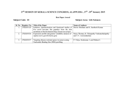
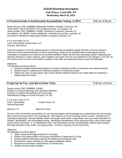
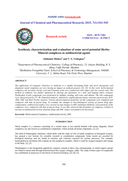
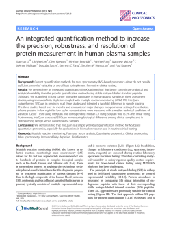
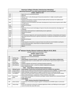
Available online at www.scholarsresearchlibrary.com
Scholars Research Library
Der Pharmacia Lettre, 2015, 7 (1):1-7
(http://scholarsresearchlibrary.com/archive.html)
ISSN 0975-5071
USA CODEN: DPLEB4
In vitro antibacterial activity of peptides isolated from Areca catechu Linn.
Dibya Jyoti Hazarika and Kaushal Sood*
Centre for Studies in Biotechnology, Dibrugarh University, Dibrugarh, Assam
_____________________________________________________________________________________________
ABSTRACT
Antimicrobial Peptides (AMPs) from plantsrepresent a group of diverse biologically active protein molecules. In
recent years, a wide variety of AMPs have been isolated and characterized for their biological properties from
variousplants. The current study was designed to screen Areca catechu Linn.for the presence of antibacterial
peptides. The present works is most probably the first to report two antimicrobial peptides (68 kDa and 65 kDa)
from A. catechu Linn. with activity against S. epidermidis, E. coliand P. mirabilis.
Keywords: Antimicrobial peptides, Areca catechu, SDS-PAGE, Antibacterial activity
_____________________________________________________________________________________________
INTRODUCTION
Nature may be regarded as the best combinatorial chemist with its numerous therapeutic agents to help man in his
struggle for survival against microbial infections. The history of use of natural products as therapeuticsis as ancient
as human civilization and, for a long time, minerals, plants and animal products have been the main sources of drugs
[1]. Medicinal plants have played a prime role in maintaining human health through their inclusion in human diet as
vegetables, spices, tonics and masticatory products. Several preclinical and clinical studies have examined various
medicinal properties such as anti-inflammatory, antioxidant, anti-microbial, anthelmintic, anti-cancer,
cytoprotective, hepatoprotective, etc. in a huge number of medicinal plants. The medicinal properties of plants have
often been attributed to their secondary metabolites, also often referred to as the phytochemicals [2]. Apart from the
biologically active phytochemicals, Antimicrobial Peptides (AMPs) from plantsrepresent a group of diverse
biologically active protein molecules. Antimicrobial peptides have been recognized as potent alternatives to the
contemporary antibiotics [3, 4].AMPs are small molecular weight proteins which have broad spectrum antimicrobial
activity against bacteria, viruses, and fungi. These peptides are reported to be evolutionarily conserved and have
both hydrophobic and hydrophilic sidechains that enable the molecule to be soluble in aqueous environments and at
the same time facilitate their entry into the lipid-rich membranes [5].AMPs target a previously under-appreciated
‘microbial Achilles heel’, a design feature of the microbial cellular membrane. This feature distinguishes broad
species of microbes from multicellular plants and animals [4]. The present study was aimed at screening the crude
protein extracts of Areca catechu Linn.forantimicrobial peptides as the same have not yet been reported for the
antimicrobial peptides.
Areca catechu Linn.is a tree with an annulate stem. The stem is surrounded by a crown of pinnate leaves. The
leaflets are numerous, the petioles expanded into a broad, tough, sheath-like growth at the lower end; the
inflorescence is a spathe which is compressed and glabrous; the spadices are much-branched, bearing ebracteate
male and female flowers. The male flowers are small and numerous; the female flowers are solitary or in groups of
two or three and much larger than the male; bisexual flowers have also been recorded; the fruits are ovoid or oblong,
smooth and orange or scarlet when fully ripe. They are single-seeded and the endosperm or seed-kernel, popularly
called the "arecanut", is greyish brown and ruminate, with reddish brown lines. It isa widely cultivated plant in
eastern countries like India, Bangladesh, Ceylon, Malaya, the Philippines and Japan. In India, the plant is widely
distributed in coastal regions, from Maharashtra to Kerala and Tamil Nadu. It also grows in the Deccan Plateau,
1
Scholar Research Library
Dibya Jyoti Hazarika and Kaushal Sood
Der Pharmacia Lettre, 2015, 7 (1):1-7
______________________________________________________________________________
Assam, Meghalaya, West Bengal, and the Andaman and Nicobar Islands. The traditional uses of the plant are
summarized in Table 1.
Table 1: Medicinal uses of the plant
Plant part used
Used as
Raw
Nut
Kernel of green
fruit
Root
Ointment(in combination with other ingredients)
Along with opium
Astringent and stimulant (chewed with betel peeper and
lime)
Juice
Used in
Anaemia, fits, leucoderma, leprosy, obesity and
worms
Nasal ulcers
Intestinal trouble
Regular purpose
Liver diseases
Figure 1: Areca Catechu Linn. Tree
(Insert: Nuts)
Vernacular Names:
English
: Beetel nut, Areca nut
Assamese
: Tamol, Guwa
Sanskrit
: Akoth
Bengali
:Supari
Hindi
: Chamarpushpa, Supari
Manipuri
:Kwapambi, Kwamaru
Marathi
: Pophal, Pugaphal, Supari
Botanical Classification:
Kingdom
: Plantae
Division
: Magnoliophyta
Class
: Liliopsida
Sub-class
: Arecidae
Order
: Arecales
Family
: Arecaceae
Genus
: Areca
Species : catechu Linn.
2
Scholar Research Library
Dibya Jyoti Hazarika and Kaushal Sood
Der Pharmacia Lettre, 2015, 7 (1):1-7
______________________________________________________________________________
MATERIALS AND METHODS
Plant Material
Nuts of Areca catechu were selected for the present study. The nuts were collected and washed immediately with
sterile distilled water. The outer cover was removed and the kernel was weighed and preserved aseptically for
further use.
Extraction of Total Proteins
The total proteins were extracted from the kernel by the method of Aliahmadiet al.(2011) [6]. Protein isolation
buffer containing 50 mM phosphate buffer (pH 7), 2 mM EDTA, 5% glycerol and 50 mMNaClwas used for
extracting proteins. Extraction buffer (cold) was added to the material (10:1 v/w) and homogenized in a mortar
pestle. The mixture was incubated 2 hours at 4ºC in a shaking incubator (Certomat, Sartorius Stedim, Germany). It
was then centrifuged at 12,000 rpm for 20 min at 4ºC (Sigma 3-30K Refrigerated Centrifuge, Germany). The
supernatant was sterilized using 0.22 µm membrane filter. The crude protein solution thus obtained was stored at
4ºC until further use.
Estimation of Proteinsby Bradford Assay
The proteins in the crude solutionwere quantified by the Bradford assay[7]. A standard curve was prepared using
bovine serum albumin (BSA) with concentrations in the range of 100 to 2000 µg/ml. 2.0 ml of the assay reagent was
mixed with 40 µl of the standard solution and incubated for 15 minutes.The absorbance was measured at 595 nm
using a spectrophotometer (Shimadzu UV-1800 spectrophotometer, Japan). The regression equation for absorbance
versus concentration was determined and the protein content in the sample was calculated using the equation.
In vitro Antibacterial Activity Assay
Test Organisms
Standard bacterial strains were obtained from IMTECH, Chandigarh. Two Gram Positive bacteria- Bacillus subtilis
MTCC 441; Staphylococcus epidermidis MTCC 435 and two Gram Negative bacteria-Escherichia coliMTCC 739;
Proteus mirabilis MTCC 1429 were used for the study. The cultures of test organisms were maintained in Nutrient
Broth (Hi-Media, Mumbai).
Antimicrobial Susceptibility Test
The samples were screened for antibacterial activity in vitro by agar-well diffusion method [8].Muller Hinton Agar
(Hi-Media, Mumbai) was prepared according to manufacturer’s instructions. Glass petri plates (Diameter 100 mm)
were sterilized prior to use. 20 ml ofagar medium was poured into the petri plates under laminar air flow in a
biosafety hood and was allowed to solidify. After solidification,100 µl of inoculum was spread on the agar plate
using a sterile L- spreader. Wells (diameter 6 mm) were boredin the agar plates using a sterile glass well-borer. Each
well was loaded with 50 µl of the test sample. Protein extraction buffer was used as negative control and Ofloxacin
(5 µg/ml) was used as the standard drug. The plates were incubated at 25°C for 24 hours after which the diameter of
the zone of inhibition formed around the well was measured. The experiment was repeated thrice and the mean
diameter of the zone of inhibition was determined.
Purification of Antibacterial Peptides
The proteins present in the crude solution were precipitated using acetone. Acetone solution was cooled to -20°C
and was added to the protein solution in 1:1, 1:2 and 1:4 (v/v) ratios. The mixture was then centrifuged at 20,000
rpm for 5 minutes at 4 oC. The protein palette was allowed to air dry and re-dissolved in phosphate buffer.The
protein fractions thus obtained were screened for antibacterial activity as mentioned in the previous section.
Determination of Minimum Inhibitory Concentration (MIC) and Minimum Bactericidal Concentration
(MBC)
MIC and MBC were determined by the procedure described by Ericsson and Sherris (1971) [9] with some
modifications. Two-fold dilutions of the protein fraction with antibacterial activity were prepared in nutrient broth.
50 µl of the inoculums were added to each test tube. Positive control tubes were prepared with 1 ml of broth and 50
µl of the inoculums and no sample. Negative control tubes for each dilution were also prepared and were maintained
without the inoculums. All tubes were incubated at 37°C for 18 hours and then examined for growth by observing
for increase in turbidity at 620 nm.The minimum concentration of protein fraction which exhibited the inhibition of
bacteria was considered as the minimum inhibitory concentration (MIC). A loopful from each of the test and control
tubes was then streaked onto petri plates containing nutrient agar medium. The plates were incubated at 37°C for 18
hours. After incubation, the minimum concentration that did not show the growthof bacteria was considered as
minimum bactericidal concentration (MBC).
3
Scholar Research Library
Dibya Jyoti Hazarika and Kaushal Sood
Der Pharmacia Lettre, 2015, 7 (1):1-7
______________________________________________________________________________
Determination of temperature stability of the proteins:
The protein fraction with antimicrobial activity was subjected to thermal treatment by incubation at -20°C, 0°C, 8°C,
25°C, 40°C and 70°C for 4 hours. The concentration of proteins in the sample was determined before and after
temperature treatment by the Bradford assay [7] as described in the previous sections. The sample was also assayed
for antimicrobial activity by methods outlined in the previous sections.
Characterization of Antibacterial Peptides by SDS- PAGE
The antimicrobial peptides present in the protein fraction were characterized by SDS- PAGE [10]. Electrophoresis
was performed at 100 volts for 55 minutes. At the end of the run, the protein bands were visualized by silver staining
[11].
RESULTS AND DISCUSSION
A standard graph for Bradford assay was prepared using 2000 µg/ml stock of Bovine Serum Albumin (BSA) and the
amount of proteins in the solution was calculated from the following regression equation (Figure 2)1.40
1.20
A 595
1.00
0.80
0.60
y = 0.0006x - 0.0142
R² = 0.99
0.40
0.20
0.00
0
500
1000
1500
2000
2500
Concentration of BSA (in µg/ml)
Figure 2: Bradford Assay: Standard Graph
Y= 0.0006 x – 0.0142; R2 = 0.99
where, y = absorbance at 595 nm, x= concentration in µg/ml
The total protein content ofA. catechu kernel was determined to be 1205.89 ± 29.17µg/ml.
The total proteins of A. catechu were observed to inhibit the test strains except B. subtilis. Among the Gram Positive
and Gram Negative bacteria, Gram Negative bacteria were observed to be more susceptible to the proteins of A.
catechu (Figure 3). The negative control did not inhibit the test strains. The proteins present in the crude solution
were fractionated by precipitation with acetone at different ratios and the individual fractions were then screened for
protein content and antimicrobial activity (Table 2). The results showed that the proteins were absent in fractions 1
and 3 which were obtained with 1:1 and 1:4 ratio (v/v) of acetone respectively, while the fraction 2 obtained using
acetone in 1:2 ratio (v/v) contained 500.33 ± 21.67µg/ml of proteins.Moreover, only fraction 2 exhibited the
inhibition of S. epidermidis, E. coli and P. mirabilis.Similar to the case of the crude sample, B. subtilis was not
inhibited by any of the fractions.
4
Scholar Research Library
Dibya Jyoti Hazarika and Kaushal Sood
Der Pharmacia Lettre, 2015, 7 (1):1-7
______________________________________________________________________________
Diameter of Zone of Inhibition (in mm)
16
14
12
10
8
6
4
2
0
B. subtilis
S. epidermidis
E. coli
P. mirabilis
Figure 3: Antimicrobial activity of Total Proteins of A. catechu
Table 2: Protein Content and Antimicrobial activity of Proteins Fractions of A. catechu
Diameter of zone of inhibition
(in mm)
B. subtilis S. epidermidis E. coli P. mirabilis
na
na
na
na
500.33 ± 21.67
na
10 ± 1
12 ± 1
11 ± 1
na
na
na
na
na= No Activity
Results expressed as mean ± sd of three replicates
Fraction
Sample: Acetone
(v/v)
1
2
3
1:1
1:2
1:4
Protein Content
(in µg/ml)
Figure 4: Effect of Temperature on Antimicrobial Activity of A. catechu Fraction 2
The minimum concentration of fraction 2 required inhibiting the growth of the bacterial strains and the
concentration required to exert bactericidal effect was determined and is presented in Table 3. The MIC values were
observed to be in the range of 250 µg/ml to 500 µg/ml while the MBC values were observed to range from 1000
µg/ml and above. Among the three strains, P. mirabilis was observed to be more susceptible to the AMPs obtained
from A. catechu (MIC= 250µg/ml; MBC= 1000 µg/ml). The results suggest that the AMPs from A. catechu may
have potential applications against E. coli and P. mirabilis which are commonly responsible for various infections of
gut and urinary tract in humans, especially in immune-compromised individuals.The effect of temperature on the
5
Scholar Research Library
Dibya Jyoti Hazarika and Kaushal Sood
Der Pharmacia Lettre, 2015, 7 (1):1-7
______________________________________________________________________________
activity of the antimicrobial peptides of fraction 2 was studied and the results are presented in Figure 4. It was
observed that fraction 2 retained antimicrobial activity against the test strains even after exposures to extremely low
(- 20 oC) and extremely high (70 oC) temperatures for 4 hours. Optimum activity was observed in the temperature
range of 25 oC to 40 oC. The AMPs obtained in fraction 2 were further characterized by SDS-PAGE (Figure 4). The
electrophoretogram revealed the presence of two polypeptides of 68 kDa and 65 kDa.
Table 3: MIC and MBC of the Antimicrobial Peptides from A. catechu
Strain
S. epidermidis
E. coli
P. mirabilis
MIC (in µg/ml)
500
500
250
MBC (in µg/ml)
> 1000
1000
1000
68
65
Figure 5: SDS-PAGE Electrophoretogram of A. catechu Protein Fraction 2 (Lane 4 & 5)
Lane 2 (Marker)
The main groups of antimicrobial peptides found in plants are grouped into three types–thionins, defensins and lipid
transfer proteins[12].The thionins constitute a family of basic peptides with low molecular weight (~5 kDa) and are
rich in basic and sulfur-containing residues. The members of this group share high sequential and structural
similarities and exert toxic effects against bacteria, fungi, yeast, animal and plant cells. Several thionins have been
isolatedfrom barley, wheat, oats, rye and sugar beet [13- 17]. Other reported thionins from plants includeviscotoxins and phoratoxins from mistletoe species, and crambin from the cruciferous plant Crambeabyssinica.
Thionins from cereals and Pyrulariapuberaand some other dicotyledonous plants have been shown to contain
disulfide bonds.Thioninhave been reported to exert their effects on the membrane through binding with
phospholipids[18]. Another group of plant antimicrobial peptides known as thedefensinsare small 45–54 amino
acids long cationic peptidesthat are widely distributed among dicots and monocots.These were originally grouped
with the thionins and were defined as -thionins. The lipid transfer proteins have the ability to facilitate the transfer
of phospholipids among natural or artificial membranes and by linking to fatty acids invitro[12]. Plant lipid transfer
proteins are quite abundant in plants and include two groups, LTP1 and LTP2. Members of the plant LTP1 family
are about 10 kDa in size and consist of 90-95 amino acids.They are basicin nature with isoelectric points between 9
and 10.The LTP2 family members showsimilarity with the LTP1 family but are only about 7 kDa in size. They
contain about 70 amino acids on average and a signal peptide. Antifungal and antibacterial activities have been
reported for LTPs of various plants including barley, maize, spinach, and A. thaliana[19- 21]. The peptides reported
in the present work are distinct from the major classes of AMPs. Further works are required to identify the sequence
of the peptides for understanding their structure and biological functions.
Although A. catechu has been reported for its various medicinal properties, specially antibacterial and antiviral
activities[22- 24] to the best of our knowledge, there have been no reports onthe antibacterial activity of the peptides
isolated from the kernels of the A. catechu nuts. Therefore, the present works is most probably the first to report two
antimicrobial peptides (68 kDa and 65 kDa) from A. catechu. Further works towards the purification and sequencing
of the antimicrobial peptides reported in the present study are being pursued.
6
Scholar Research Library
Dibya Jyoti Hazarika and Kaushal Sood
Der Pharmacia Lettre, 2015, 7 (1):1-7
______________________________________________________________________________
REFERENCES
[1] S. M. K. Rates, Toxicon, 2001, 39 (5): 603-613.
[2] M. M. Cowan, Clin. Microbiol.Rev.,1999, 12 (4), 564–582.
[3] H.G. Boman,Annu Rev Immunol, 1995, 13, 61–92
[4] M. Zasloff, Nature, 2002, 415, 389–395
[5] A. Izadpanah, R. L. Gallo,. Journal of the American Academy of Dermatology, 2005, 52(3), 381-390.
[6]A. Aliahmadi, R. Roghanian, G. Emtiazi, A. Ghassempour, Iranian journal of microbiology,2011,3(2),104.
[7] M. M. Bradford, Anal Biochem, 1976,72, 248–254
[8] T. I. Mbata, L. Debiao, A. Saikia, African Journal of Biotechnology, 2008, 7 (10),1571-1573.
[9] J. M. Ericsson,J. C. Sherris, Acta PatholMicrobiol Scand, 1971,217 (supp l), 1-90.
[10] U. K. Laemmli, Nature, 1970, 227, 680-685.
[11] S. F. D. S.Groth, R. G. Webster, A. Datyner, BiochemBiophys Acta,1963, 71, 377–391.
[12] M. S. Castro, W. Fontes, Protein and Peptide Letters,2005, 12(1), 11-16.
[13] F. J. Colilla, A. Rocher, E. Mendez,
FEBS Lett.1990,270, 191–194.
[14] E. Mendez, A. Moreno, F. Colilla, F. Pelaez, G. G. Limas, R. Mendez, F. Soriano, M. Salinas, C. De Haro, Eur.
J. Biochem, 1990, 194, 533– 539.
[15] R. Fernandes de Caleya, B. Gonzalez-Pascual, F. Garcia-Olmedo, P. Carbonero.Appl. Microbiol.1972, 23, 998–
1000.
[16] K. M. Kragh, J. E. Nielsen, K. K. Nielsen, S. Dreboldt, J. D. Mikkelsen,Mol. Plant- Microbe Interact, 1995, 8,
424– 434.
[17] Y. Zhang, K. Lewis. FEMS Microbiol. Lett.,1997,149, 59–64.
[18]B. Stec, O. Markman, U. Rao, G. Heffron, S. Henderson, L.P.Vernon,V.Brumfeld, M. M. Teeter, J Pept
Res,2004,64, 210–224.
[19] F. R. Terras, I. J. Goderis, F. Van Leuven, J. Vanderleyden, B. P. Cammue, W.F.Broekaert,Plant Physiol 1992,
100, 1055–1058.
[20] A. Molina, A. Segura, F. Garcia-Olmedo,FEBS Lett,1993,316, 119–122.
[21] A. Segura, M. Moreno, F. Garcia-Olmedo, FEBS Lett.,1993, 332, 243–246.
[22] H. Lalithkumari, M. Sirsi, Indian Journal of Experimental Biology,1965. 7, 66.
[23] J.L.N. Sastry;DravyagunaVijnana: Fundamental Principles of Pharmaco-therapeutics in Ayurveda, Varanasi,
Chaukham bhaOrientalia, India,2005,2.
[24]R. Reena, N. Authikat, A. Michael, Pharmacognosy, 2009, 1(1), 42-45.
7
Scholar Research Library
© Copyright 2026