
PDF version - Andersen Lab - California Institute of Technology
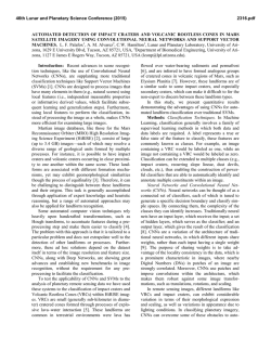
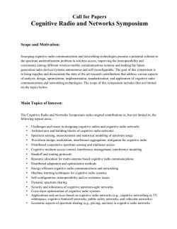
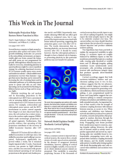
Cognitive Based Neural Prosthetics*
R. A. Andersen and S. Musallam
J. W. Burdick and J. G. Cham
Division of Biology
California Institute of Technology
Pasadera, CA 91125
Division of Engineering and Applied Science
California Institute of Technology
Pasadera, CA 91125
[email protected]
Abstract – Intense activity in neural prosthetic research
has recently demonstrated the possibility of robotic interfaces
that respond directly to the nervous system. The question
remains of how the flow of information between the patient and
the prosthetic device should be designed to provide a safe,
effective system that maximizes the patient’s access to the
outside world. Much recent work by other investigators has
focused on using decoded neural signals as low-level commands
to directly control the trajectory of screen cursors or robotic
end-effectors. Here we review results that show that high-level,
or cognitive, signals can be decoded during reaching arm
movements. These results, coupled with fundamental
limitations in signal recording technology, motivate an
approach in which cognitive neural signals play a larger role in
the neural interface. This proposed paradigm predicates that
neural signals should be used to instruct external devices,
rather than control their detailed movement. This approach
will reduce the effort required of the patient and will take
advantage of established and on-going robotics research in
intelligent systems and human-robot interfaces.
Index Terms – Neural prosthetics, brain-machine interfaces.
I. INTRODUCTION
Recent advances in neural prosthetics research have
demonstrated the possibility of computer interfaces and
robot arms that interact directly with the nervous system [1][6]. The development of neural prosthetic systems can one
day allow persons with lost motor function due to spinal
cord injury, stroke or neurodegenerative disorders to regain
the ability to communicate and interact with their
surroundings. Such systems will arguably represent the
ultimate human-robot interface, with the robotic device
literally becoming an extension of the patient’s conscious
sense of self.
Despite recent breakthroughs, many challenges still
remain [7]. The development of neural prosthetics requires
advances across many disciplines, including neuroscience,
engineering, neurosurgery and neural informatics. An
underlying challenge is to improve technology for acquiring
neural signals and maintaining signal quality for long
periods of time. Long-term tracking of neural signals is
difficult due to the invasive nature of prevalent recording
techniques. Another challenge is to understand how the brain
encodes movements such as reaches, and how to use these
signals for neural prosthetic applications. The question
remains of how the flow of information between the patient
and the prosthetic device should be designed to maximize
the safety and effectiveness of neural prosthetic systems.
Given these challenges, in this paper we review and
elaborate upon a framework for neural prosthetics that
*
emphasizes the use of cognitive signals in interacting with
external devices [8][15]. Most recent successes in neural
prosthetics have used signals primarily from the motor and
pre-motor cortices (see Figure 1) to directly control the
trajectory (position and velocity) of a screen cursor or robot
arm. This approach, though proven successful, has its
limitations. Using neural signals to directly control a robot
arm for a wide variety of movements, manipulations and
postures will likely place higher demands on the number and
quality of signals that need to be recorded. We believe that
neural prosthetics should acquire signals to instruct external
devices, rather than to control every detail of their
movement. Recent work by some of the authors, reviewed
here, shows that it is possible to capture the intention, or
goal, of reaching movements, and also the expected reward,
or motivation, of the subject before the reach is made.
Utilizing such cognitive variables may decrease the effort
required of the user and reduce the informational burden
placed on the neural recording interface.
Future neural prosthetic devices will likely require an
integration of both higher-level (cognitive) and lower-level
(motor) brain activity. This approach can take advantage of
established and on-going robotics research in intelligent and
supervisory systems and human-robot interfaces in order to
develop new paradigms that can adapt to the needs and
cognitive states of the user while maintaining safety.
Brain
Parietal
Reach
Region
Electrode
Primary Motor
Cortex
Premotor
Cortex
Spikes
Field Potential
Micro-electrode
Spinal
cord
Figure 1. Signals used for neural prosthetic applications. In patients with
lost motor function, microelectrodes can be used to record the neural
activity (spiking or field potentials) from brain areas that encode
movements such as reaching and grasping to drive external devices such as
robot arms.
This work is partially supported by National Eye Institute, DARPA, ONR, the Boswell Foundation, NSF, the Christopher Reeve Paralysis Foundation, the
Sloan-Swartz Center for Theoretical Neurobiology at Caltech, and the Human Frontier Science Program.
decode
velocity
Visual
feedback
Endeffector
Target
joystick
Figure 2. In current studies, non-human primates learned to control an endeffector using brain signals alone. Signals recorded during normal arm
movement tasks were used to build a decode model that could predict the
desired trajectory (primarily the instantaneous velocity of the end-effector).
II. RECENT ADVANCES IN NEURAL PROSTHETICS
Several investigators have demonstrated systems in which
neural signals from non-human primates were used to
control the motion of an end effector (a screen cursor or a
robotic arm) [2][3][4]. In these studies, the activity of
populations of neurons was recorded using arrays of
microelectrodes implanted primarily in motor and pre-motor
cortex (see Figure 1). The electrode arrays recorded the
electrical or “spike” activity of individual or groups of
neurons while the subjects performed arm movement tasks.
This activity was then used to calibrate models, usually
linear regression models, that relate the firing rate of the
neurons to the observed movements. The subjects were then
shown capable of moving the end-effector in “brain control”
tasks in which current neural activity and the calibrated
models were used to predict the end-effector trajectory
without actual movement of the subject’s arm.
In addition, similar progress has been made in using
EEG-based signals to derive neuroprosthetic commands
from motor related areas [6]. Though EEG methods are less
invasive, their poor spatial resolution may limit the amount
of information that can be obtained with them.
These groups showed performance well above the
chance level, and, more interestingly, reported that the
performance of the subjects in the brain control task
improved over time, giving evidence that the neural system
learned to use the prosthetic device by adapting its output. In
fact, [2] showed that the subjects learned to compensate for
the dynamics of the robotic arm used. This is an encouraging
result that allows a certain margin of error in the acquisition
and decoding of neural signals, as the system can be relied
upon to adapt to these errors.
While these studies have shown the possibility of
neural-controlled prosthetic devices, and have sparked
renewed interest in the field, the question arises of what
other types of signals could be used in a neural interface.
These studies used neuronal populations located primarily in
motor and pre-motor cortex, which tend to encode the
specific commands sent to our muscles for the control of
limb movements. More important than the particular brain
region the signals are derive from, however, is the type of
information that is decoded from them.
In the current approaches, the neural activity is used as a
control signal to directly specify where the location of the
end-effector needs to be at any instant in time. In many
cases, the primary signal decoded from the neural activity
was the velocity of the end-effector. Thus, the trajectories of
the brain-controlled end-effector were the result of velocity
control on the part of the subject, who had to continually
command online corrections based only on visual feedback
of the task (see Figure 2). Here the neural activity is used for
low-level control commands, and the effectiveness of the
neural prosthetic is limited to the subject’s ability to perform
the closed-loop task.
It could be argued that motor cortex neurons should be
used exclusively for interfacing to external devices, given
their demonstrated ability to adapt to different tasks. In this
approach, the motor cortex is used as a generic source of
neural signals that can be adapted to communicate cognitive
states and/or control movements of robot arms. However,
there are at least two reasons not to depend solely on motor
cortex areas. The first is that this dependence creates an
informational bottleneck that will reduce the number of
cognitive variables that can be read out at any one time. For
example, a patient’s mood could be determined by asking
him or her to move a cursor on a computer interface to
answer sets of questions about their emotional status.
However, this would preclude the patient from performing
other tasks at the same time. It would be more efficient to
decode this signal directly from an area that processes the
mood of the subject.
The second reason is that the normal functional
architecture of motor cortex is for generating commands for
movement trajectories. While it may be possible for motor
cortex to be treated like an undifferentiated neural network
and trained to perform any task, it has been shown that
neural networks trained to do a large number of different
tasks tend to do each one poorly compared to being trained
to perform a small number of tasks [9].
Can higher-level, or cognitive, signals be used to
interface to an assistive neural prosthetic device? In the next
section, we review current work that shows that cognitive
signals such as reach goals and reach motivation can be used
in a neural prosthetic system.
III. COGNITIVE NEURAL SIGNALS
In theory, cognitive control signals appropriate for
reaching tasks could be derived from many higher cortical
areas related to sensory-motor integration in the parietal and
frontal lobes. Here we focus on the posterior parietal reach
region (PRR) and the dorsal premotor cortex (PMd). Again,
the primary distinction is not the specific brain area where
the signals are obtained, but rather the type of information
that is being decoded, and how that information is used.
Similar approaches to the one presented can be used for
interpreting cognitive signals from other brain areas. It is
likely that some areas will yield better results than others
depending on the cognitive signals to be decoded and the
parts of the brain that are damaged.
PRR in non-human primates lies within a broader area
of cortex called the posterior parietal cortex (PPC) [10][11]
(see Figure 1). The PPC is located functionally at a
transition between sensory and motor areas and is involved
in sensory-motor integration, that is, it helps transform
sensory inputs into plans for action. PRR is known to be
primarily active when a subject is preparing and executing a
movement [11][13]. However, the region receives direct
visual projections and vision is perhaps its primary sensory
input.
Neural activity in PRR has been found to encode the
targets for a reach in visual coordinates relative to the
current direction of gaze (also called retinal or eye-centered
coordinates) [13]. In other words, this area contains
information about where in the subject’s field of view the
subject is planning on reaching towards. This coding of
planning information in visual coordinates underscores the
cognitive nature of the signal within PRR. It is coding the
desired goal of a movement, rather than the intrinsic limb
variables required to reach to the target. The human
homologue of PRR has recently been identified in fMRI
experiments [14].
Recent experiments with monkeys have demonstrated
that reach goals decoded from PRR can be used to drive a
neural prosthetic computer interface to position a cursor on a
screen [15]. These experiments are described in more detail
in the following section.
the cued location. If the animals moved their arm, the trial
was cancelled and no reward was given. This approach was
necessary because the monkeys cannot simply be instructed
to “think about reaching to the target without actually
reaching to it.”
Thus, the reach goals were decoded from activity
present when the monkeys were planning the reach
movements, but otherwise were sitting motionless in the
dark and were not making eye movements. Figure 4a shows
a typical result, which shows the cumulative accuracy of the
brain control system for four target locations, as the
experimental session progresses. As shown in Figure 4b,
only a small number of cells were required for successful
performance of the task, with performance increasing with
the number of neurons. Figure 4c shows neural activity in
both the reaching and brain control tasks from a recording
site in PRR, demonstrating that the cognitive signals in the
brain control task were free of any sensory or motor related
activity.
In addition, the animals showed considerable learning in
the brain control task, as evidenced by a significant increase
in their performance over the course of one to two months
[15]. This behaviour is consistent with a number of studies
of cortical plasticity [16], and the time scale for learning is
similar to that seen in motor cortex for trajectory decoding in
previous studies [2][4]. In this case, the improvement in
performance was found to be related to an increase in the
A. Decoding the goal of a reach
In the experiments, arrays of electrodes were placed in
the medial intraparietal area (MIP), a portion of PRR, area 5
(also in the posterior parietal cortex), and the PMd. These
electrodes record the electrical activity of neurons in the
vicinity of the electrode tips. Each experimental session
began with the monkeys performing a series of reaches to
touch different locations on a computer screen (Figure 3a).
As shown in the figure, the reaching task consisted of four
phases. First, the monkey is instructed to fixate on the center
cue. Next, a target location is presented for a brief period of
time. The target then disappears followed by a delay period.
Finally, the monkey is given a “go” signal instructing him to
reach to the location where the target was.
The neural activity recorded during the delay period, in
which the monkey presumably plans the reach movement,
was used to build a database that relates the firing rate of the
neurons to the target location. For example, as illustrated in
Figure 3a, the database would store which location in the eye
field a particular neuron exhibited higher firing rates when
movements were planned to that location.
After enough trials were performed to build an
acceptable database, the monkey was switched to the “brain
control” task. Here, the monkeys were instructed with a
briefly flashed cue to plan to reach to different locations but
without making a reach movement (Figure 3b). The activity
during the delay period was then compared to that in the
database and, using a Bayesian decode algorithm, the
location where the monkeys were planning the reach was
predicted. If the predicted reach direction corresponded to
the cued location, then the animals received a drop of fluid
reward and visual feedback was provided by re-illuminating
a
Reach Task
“Target cue”
“Delay period”
Trial time
“Reach”
Record
Build
database
Neuron 1
Neuron
2
Neuron
6
Neuron 3
Receptive fields
of neurons
Neuron 4
“Fixate”
Neuron 5
Eye field
b
Brain Control Task
“Fixate”
“Target cue”
“Delay period”
“Reward cue”
Trial time
Decode
reach goal
Record
Figure 3. Normal reaching and brain control tasks used in decoding reach
goal and expected reward cognitive signals. Adapted from [19].
amount of information encoded by the neurons in the brain
control task [15], which was calculated using a mutual
information measure. This measure quantifies the degree to
which a neuron’s firing rate encodes a particular direction.
In essence, the neurons tuned themselves to increase
performance in the task. Plastic behaviour such as this will
be important in enabling patients to optimize neural
prosthetic systems with training.
B. Decoding expectation
It has also been shown possible to decode neural activity
that foretells a subject’s expectation of a reward when
performing a task. Signals related to reward prediction are
cumulative percent correct
a
100
80
60
40
chance level
20
4 targets
0 0
50 100 150 200 250 300
trial number
b
c
Reach
Delay period
Brain control
Figure 4. Results of brain control tasks in which cognitive signals (reach
goals) were used to drive a cursor to target positions on a screen. a) Task
performance as a function of time for an eight-target brain control task. b)
Performance as a function of the number of neurons used in the decode
model. c) Comparison of neural activity during normal reaching and brain
control tasks (b and c adapted from [15]).
found in a number of brain areas [17]. In area LIP of the
PPC, which is involved in planning and executing eye
movements, it was found that cells code the expected value
of rewards [18]. Using an eye motion task, it was found that
the neurons increased their activity when the animal
expected a larger reward or the instructed eye movement
was more likely to be in the cells’ preferred locations.
Similar effects have recently been found for PRR neurons
for amount of reward in both the reaching and brain control
task previously described [15]. PRR cells are also more
active and better encode reach goals when the animal is cued
to expect a higher probability of reward at the end of a
successful trial. Remarkably, PRR cells also encode reward
preference, with higher activity seen when the monkey
expects delivery of a preferred citrus juice reward rather
than water.
This expectation signal tells us something about the
cognitive state of the subject, possibly indicating the
subject’s motivation or interest in the task’s outcome.
Moreover, this expectation of reward could be read out
simultaneously with the intended reach goal using offline
analysis of the brain control trials [15], thus showing that
multiple cognitive variables can be read from the brain at the
same time.
IV. NEUROPROSTHETIC CONTROL SYSTEMS BASED ON
INTELLIGENT DEVICES AND SUPERVISORY CONTROL
A neural prosthetic system must be flexible and capable
of adapting to the needs and cognitive states of the patient. It
is not clear how the sole use of motor-based signals can
provide enough information for such subtle and complex
interaction. Given that it is possible to obtain cognitive
signals, we have proposed a framework for the control of
neural prosthetics that takes advantage of the high-level
nature of these signals. In this approach, cognitive signals
like the intended goal of a reach or the motivation or
expected reward of a reach are used to instruct the neural
prosthetic system, rather than control it directly, as
illustrated in Figure 5.
In this case, interaction between the patient and the
mechanical device is monitored by an intelligent supervisory
system [20]. This system monitors the cognitive state of the
patient, and combines it with knowledge of the workspace
and patient information such as gaze direction to assess the
situation and calculate the most appropriate course of action.
The cognitive neural signals can operate much like “body
language” by providing, on-line and in parallel with readouts
of other cognitive variables, the preferences, mood, and
motivational level of the patient. Implants in emotional
centers could also provide real time readouts of the patient’s
emotional states, such as alarm, urgency or displeasure. This
augmentation of the information channels derived from the
patient is particularly important for locked-in patients that
cannot move or speak.
Given this information, combined with readings of the
patient’s intended reaching goals from regions such as
PRR, the intelligent system can then compute the
appropriate trajectory and posture of the robotic arm. For
example, given the Cartesian coordinates of an intended
object for grasping and knowledge of the environment, a
Eye movement tracking
Knowledge of workspace
Supervisory
controller
Cognitive
states (motivation,
attention, emotion)
Trajectory
generator
PRR Reach Goals
Motor / Pre-motor cortex
cues or adjustments
Joint Controller
Joint
commands
and
feedback
Visual
feedback
Robot arm
Figure 5. The proposed framework emphasizes parallel decoding of high-level cognitive variables (reach
goals, motivation) and low-level motor variables under the supervision of an intelligent system, which
manages the interaction between the patient and the robot arm.
robotic motion planner [21] can determine the detailed joint
trajectories that will transport a prosthetic hand to the
desired location in the safest, most efficient way.
This trajectory can then be sent to the robot arm’s lowlevel joint controller for execution. Sensors embedded in the
mechanical arm ensure that it follows the commanded
trajectories, thereby replacing the function of proprioceptive
feedback that is often lost in paralysis. Other sensors can
allow the artificial arm and gripper to avoid sudden
obstacles and regulate the interaction forces with its
surroundings, including grasping forces, thereby replacing
somatosensory feedback.
If available, motor signals can augment low level plans
to help “guide” the end-effector to the appropriate action by
providing cues or corrections to the trajectory planned by the
supervisory controller. Thus, future applications are likely to
involve recordings from many areas to read out a substantial
number of cognitive and motor variables. Results from
cognitive-based and motor-based approaches will likely be
combined in single prosthetic systems to capitalize on the
benefits of both.
IV. CONCLUSIONS AND FUTURE WORK
We have reviewed a cognitive based framework for the
control of neural prosthetic systems, based on evidence that
such cognitive signals can be directly read from the nervous
system. In this approach, neural signals are used to instruct
an intelligent supervisory system, rather than directly control
an external device such as a robot arm. The proposed
supervisory system in turn manages the interaction between
the user and the external device. This approach has many
potential benefits for both the neuroprosthetic user and for
the implementation of the prosthetic system.
The proposed framework would reduce the effort
required of the user in executing tasks such as reaching,
since they are not continually involved in the task of
controlling the position and trajectory of the robot arm.
What we are proposing is to use the intrinsic organization of
the nervous system to provide multiple hierarchical channels
of communication and control. Task execution will more
closely resemble normal function, in which low-level control
of limb movements often occurs without conscious attention.
Similarly, the hierarchical nature of supervisory control
should allow patients to learn much more quickly how to
command a new device.
For the systems engineer, this approach has the benefit
that to adapt the neural interface to different
electromechanical devices (e.g. different types of robotic
arms or communication devices), only the lowest level of the
control hierarchy need be re-engineered for the specific
mechanical device. In addition, the approach may possibly
reduce the number of signals needed to be extracted from the
nervous system, since lower numbers of variables may be
needed to specify cognitive states such as goals and
intentions in comparison to those needed to control a wide
set of movements of an articulated robot arm. This in turn
can alleviate the informational burden placed on the neural
recording device, making them more practical.
Finally, this approach can take advantage of the long
history and on-going research in robotics that focuses on
intelligent systems, autonomous navigation and path
planning, and human-robot interaction. In turn, this
application will present exciting new challenges for these
areas of robotics research.
Future neural prosthetic devices will likely require an
integration of both higher-level (cognitive) and lower-level
(motor) brain activity. By using activity from several
different parts of the brain and decoding a number of
cognitive variables, a neural prosthetic can provide a patient
with maximum access to the outside world.
ACKNOWLEDGMENT
We would like to thank the members of the Andersen
lab at Caltech, especially C. Buneo, B. Pesaran, B. Corneil
and B. Greger. We also thank E. Branchaud and Z. Nenadic.
REFERENCES
[1] Wessberg, J., et al., Real-time prediction of hand trajectory by
ensembles of cortical neurons in primates. Nature, 2000. 408(6810): p.
361-365.
[2] Carmena, J.M., M.A. Lebedev, R.E. Crist, J.E. O'Doherty, D.M.
Santucci, D.F. Dimitrov, P.G. Patil, C.S. Henriquez, M.A.L. Nicolelis,
Learning to control a brain-machine interface for reaching and
grasping by primates. PLoS, 2003. 1: p. 193-208.
[3] Serruya, M.D., Hatsopoulos, N.G., Paninski, L., Fellows, M.R.,
Donoghue, J.P., Instant neural control of a movement signal. Nature,
2002. 416: p. 141-142.
[4] Taylor, D.M., Tillery, S.I.H., Schwartz, A.B., Direct cortical control of
3D neuroprosthetic devices. Science, 2002. 296: p. 1829-1832.
[5] Kennedy, P.R. and R.A. Bakay, Restoration of neural output from a
paralyzed patient by a direct brain connection. Neuroreport, 1998.
9(8): p. 1707-11.
[6] Wolpaw JR, B.N., McFarland DJ, Pfurtscheller G, Vaughan TM,
Brain-computer interfaces for communication and control. Clin
Neurophysiol, 2002. 113: p. 767-791.
[7] Donoghue, J. P., Connecting cortex to machines: recent advances in
brain interfaces. Nature Neurosci, 2002 vol. 5, p. 1085-1088.
[8] Andersen, R.A., Burdick, J.W., Musallam, S., Pesaran, B., and Cham,
J.G. Cognitive Neural Prosthetics. Trends in Cognitive Science (In
press).
[9] Kosslyn, S.M., C.F. Chabris, C.J. Marsolek, and O. Koenig,
Categorical versus coordinate spatial relations: computational analyses
and computer simulations. Journal of Experimental Psychology, 1992.
18: p. 562-577.
[10] Andersen, R.A. and C.A. Buneo, Intentional maps in posterior
parietal cortex. Ann. Review Neuroscience, 2002. 25: p. 189-220.
[11] Snyder, L.H., Batista, A.P., Andersen, R.A., Coding of intention in
the posterior parietal cortex. Nature, 1997. 386(6621): p. 167-170.
[12] Sakata, H., et al., Neural mechanisms of visual guidance of hand
action in the parietal cortex of the monkey. Cereb Cortex, 1995. 5(5):
p. 429-38.
[13] Batista, A.P., et al., Reach plans in eye-centered coordinates. Science,
1999. 285: p. 257-260.
[14] Connolly, J.D., R.A. Andersen and M.A. Goodale, FMRI evidence for
a 'parietal reach region' in the human brain. Exp Brain Res, 2003. 153:
p. 140-145.
[15] Musallam, S., B.D. Corneil, , B. Greger, H. Scherberger, and R.A.
Andersen, Cognitive control signals for neural prosthetics. Science,
2004. 305: p. 258-262.
[16] Buonomano, D.V., M.M. Merzenich, Cortical plasticity: from
Synapses to Maps. Ann. Review Neuroscience, 1998. 21: p. 149-186.
[17] Schultz, W., Neural coding of basic reward terms of animal learning
theory, game theory, microeconomics and behavioural ecology.
Current Opinion in Neurobiology, 2004. 14: p. 139-147.
[18] Platt, M.L. and P.W. Glimcher, Neural correlates of decision variables
in parietal cortex. Nature, 1999. 400(6741): p. 233-8.
[19] Buneo, C. A., Jarvis, M. R., Batista, A. P. and Andersen, R. A.
Properties of spike train spectra in two parietal reach areas. Exp. Brain
Res (2003) 153:134-139.
[20] Sheridan, T.B., Telerobotics, Automation, and Human Supervisory
Control. 1992, Cambridge, MA: MIT Press.
[21] Murray, R.M., S. Li, and S.S. Sastry, A Mathematical Introduction to
Robotic Manipulation. 1994, Boca Raton, FL: CRC Press.

© Copyright 2026