
Role of Homeobox Genes in Tooth Morphogenesis: A Review
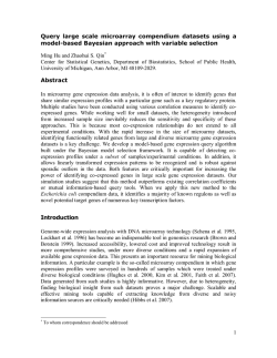
Dentistry Section DOI: 10.7860/JCDR/2015/11067.5606 Review Article Role of Homeobox Genes in Tooth Morphogenesis: A Review Sreevalli Suryadeva1, Mohammadi Begum2 ABSTRACT In oral cavity, disturbances due to genetic alterations may range from lack of tooth development to morphological defects. Due to technical advances in genetic engineering and molecular biology, valuable information regarding dentofacial growth could be studied in detailed manner. This helped us to explain the aetiology and pathogenesis of many dentofacial disorders. The success in treatment lies first in determining the aetiology of tooth anomalies and finally differentiating the effect of genes and environment on the orofacial diseases of that particular individual. Several genes belonging to class II homeobox families are expressed during odontogenesis however homeobox genes are not directly imvolved in tooth formation as they are not directly expressed in the first branchial arch derivatives. Keywords: Homeobox genes, Tooth development, Transcription growth factors INTRODUCTION Body organisation requires cell differentiation and morphogenesis which are controlled by gene expression. Gene expression is defined as an activation of a gene that results in production of polypeptide/ protein that can activate/deactivate other genes with the influence of transcription factors (growth factors). Every organism has a unique body pattern because of the influence of Homeobox genes. These seem to be the master genes that help in development of individual structures from different areas of the body. They are likely to be an important fundamental in evolution of the specialized body parts of many animal species and the differences between different organisms can be due to different modes of action of homeobox genes [1,2]. A homeobox is a DNA sequence found within genes that are involved in the regulation of patterns of anatomical development (morphogenesis) in human beings. The homeobox is about 180 base pairs long. It encodes a protein domain (homeodomain) which when expressed (as protein) help in binding with the DNA. The homeodomain is capable of recognizing and binding to specific DNA sequences. During embryogenesis, through the early recognition property of the homeodomain, the homeoproteins are believed to regulate the entire expression of genes and also direct the formation of many body structures. Homeobox genes encode transcription factors that can regulate expression of other genes. This domain is first identified in Drosophila (fruit fly). These play an important role in specifying cell, identity and position ing during embryonic development and mutations in these genes can cause developmental disturbances in tooth genesis like of specific structures as well as changes in the dentistry of a body part causing phenotypic changes in the patterning of an organism due to mutations [3]. WHAT ARE HOX GENES? Genes containing Homeobox sites are first identified in Hox cluster that is group of genes located on the same chromosome, each coding for a particular protein which is regulated by the same cellular mechanisms. These cluster sequences are highly conserved during evolution without any change in the pattern for hundreds of years. During tooth morphogenesis, expression of these homeobox genes is directly under the control of signalling cascades initiated by the Journal of Clinical and Diagnostic Research. 2015 Feb, Vol-9(2): ZE09-ZE12 interaction of certain growth factors and receptors on the surface of the target cells [4,5]. These genes are essential metazoan genes which determine the identity of embryonic regions along the anterioposterior axis. They encode homeodomain containing transcriptional regulators that can operate differential genetic programs along the anteroposterior axis. Humans contain Hox genes in four clusters, called HOXA, HOXB, HOXC, or HOXD, on chromosomes 2,7,12, and 17. HOX gene influence appears to be in human tooth buds between 18 and 24 week of embryonic development [6]. HOMEOBOX GENES INVOLVED IN DENTINOGENESIS Information regarding tooth organogenesis was found by doing number of trails on mouse embryo’s as experimental material. All these studies show that there is a direct genetic control on odontogenesis, which determines the position, number, size and shape of the teeth [7]. Tooth formation undergoes different stages like bud, cap and bell stage during its developmental process. During the bell stage cyto differentiation occurs which lead to the formation of enamel, dentin, Periodontal ligament which is a supporting structure of the tooth [8,9]. Like other development processes during the embryonic phase morpho differentiation of teeth occurs under the influence of first branchial arch, where complex interactions between the stomodaeal epithelium which is ectodermal derivative and the underlying mesenchyme which cranial neural crest derivative take place. More than 300 genes are involved in this processes and prominent role is played by the transcription factors that have a homeodomain. The homeodomain consists of 60 aminoacids with a helix-turn-helix DNA binding protein and is encoded by a homeobox sequence. Not only homeodomain facilitates it binding with DNA but transcription factors also contain a transactivation domain that interacts with a RNA polymerase and these transcription factors are in turn involved in the regulation of homeobox gene expression sites thus having a role in activation of genes in embryogenesis [10,11]. ROLE OF GROWTH FACTORS IN ODONTOGENESIS During embryonic development, the neural crest cells differentiate into most of the skeletal and connective tissue structures of the 9 Sreevalli Suryadeva and Mohammadi Begum, Role of Homeobox Genes in Tooth Morphogenesis craniofacial region. Establishment of pattern in the craniofacial region is determined partly by the axial origin of the neural crest cells present within each arch and partly by regional epithelial mesenchymal interactions mediated by several growth factors signaling pathways. Important factors belong to Fibroblast growth factors (FGF) and Transforming growth factors (TGF, containing BMP4-bone morphogenetic protein 4), the family of Wnt (Wingless) and morphogenesis molecule Shh (Sonic hedgehog) [12]. The general pattern of dentition is developed much before the teeth erupt into the oral cavity. Generally tooth formation is genetically a complicated processes controlled in two different ways: on one end by specifying the type, position, size of each tooth bud; and on the other end by the processes of enamel and dentin formation. All the above mentioned transcription factors define spatially the domains of expression of the homeobox genes in the developing jaws. Every combination of homeobox genes expressed is code that specifies the type of the tooth that has to be formed [13]. Proximal area of the molars to be developed is patterned by the expression of growth factors like FGF8 and FGF9, while BMP4 is expressed in the distal region of the incisors [14,15]. There are many genes that are involved in the formation of tooth buds belong to signalling pathways with functions in regulating other organ morphogenesis. This explains the reason why the mutation of these genes have pleiotropic effects in addition to nonsyndromic /syndromic related dental abnormalities [16,17]. MUTATIONS-TOOTH AGENESIS Congenital absence of one or more teeth is the frequent anomaly in humans. In hypodontia where the patients missing up to five permanent teeth, excluding the 3rd molars; Oligodontia where there is absence of more than six teeth (excluding 3rd molar). Anodontia that is the congenital absence of teeth in both primary and permanent dentition. The above conditions may be or may not be associated with genetic disorders. Frequency of cases of non syndromic hypodontia/Oligodontia is 80% when missing a tooth. But most of the agenesis is due to genetic origin. So far, only eight genes have been diagnosed to be associated with syndromic ad non-syndromic hypodontia, oligodontia and agenesis. Those are MSX1, PAX9, AXIN2, EDA1, DLX. MSX-1 TRANSCRIPTION FACTORS (Muscle segment homeobox containing transcription factors): MSX-1 gene is located on the shorth arm of chromosome 4 (4p16.1.) This gene has a homeobox sequence and two exons that encode a homeodomain-a 297 aminoacid protein. MSX-1 plays an important role in craniofacial and tooth development. This gene inhibits cell differentiation by maintaining high levels of cyclin DI expression and Cdk-4 activity, thus preventing the cells to respond to proliferative factors. Mutations occurring in exon 1 and 4 and exon- 2, have been associated with hypodontia in relation to 2nd premolars and 3rd molars [18]. MSX-1 phenotypes due to protein deficiency depend on the location of the mutations and their effect on the structure and function of protein. Mostowska et al., [19,20] identified an Argnine to Proline substitution in position 31 of the MSX-1 gene homeodomain that caused hypodontia and was transmitted as autosomal dominant trait. Pax Genes (Paired Box) This is a highly conserved gene in humans (14q12-q13), encoding a transcription factor that is involved in organogenesis that can trigger cellular differentiation. Mutations in exon 1, 2 and 4 mostly in exon 2 were associated with non syndrome oligodontia. According to Stockton mutation in the PAX9 gene modified the open reading frame that is frame shift mutation causing premature termination of translation. This disease is transmitted as autosomal dominant trait [21]. Three different mutations leading to substitution of arginine with proline in the homeodomain (Arg26Pro), glutamic acid with lysine (Glu9Lys) and leucine-to- proline (Leu21Pro) affecting all permanent first molars [22]. 10 www.jcdr.net Axin2 Gene (Axin Inhibition Protein 2) This gene plays an important role in the regulation of the stability of beta catenin, which is involved in the Wnt (Wingless) signalling pathway. AXIS inhibition protein 2 gene polymorphic variants may be associated with both colorectal carcinomas and tooth agenesis (oligodontia/hypodontia).These mutations activate the Wnt pathways which prove the importance of this signalling pathway in the normal development of teeth [23]. Eda1 Gene Ectodysplasin-A (EDA) is a protein that in humans is encoded by the EDA gene. Mutations in this gene cause X-linked hypohydrotic ectodermal dysplasia which is a rare disease. Non-sense and point mutations occurring in EDA gene cause only hypo/oligodontia which is non syndromic tooth agenesis [24]. Dlx Gene Distal less is a family of homeodomain transcription factors which are related to the craniofacial morphogenesis. Mutation in these genes results in abnormalities of first brachial arch derivatives like mandible and calvarias. Failure of development of molars occurs due loss of function mutation of this gene [25]. MOLECULAR CASCADE OF RECIPROCAL SIGNALING EVENTS IN TOOTH DEVELOPMENT [Table/Fig-1] [26] Tooth formation is considered to be a more complex process, which also is genetically controlled in two different ways: on one side, each tooth organ is specified by its type, size and position and on the other, by the processes of formation of enamel and dentin. Different genes involved in the formation of teeth belong to signaling pathways with functions in regulating the morphogenesis of other organs [27]. This explains the fact that mutations in these genes have pleiotropic effects in addition to causing non-syndromic dental abnormalities and dental anomalies associated with different genetic syndromes. There are different molecular signalling that regulate tooth development and it is possible to observe that the molecular signals are expressed in different stages of odontogenesis. Regarding initiation stage, a genetic model explaining the regulation of the expressed genes has been proposed by Beiand Maas [28] and by Zhang et al., [29]. The expression of Msx 1 in the dental mesenchyme is initially by epithelially derived Bmps and Fgfs. Interestingly, Bmp4 cannot induce Fgfs, neither the contrary, suggesting that Bmp4 and Fgf8 act by independant pathways in inducing dental mesenchyme. The arrest of tooth development in Msx 1 mutant mice was associated with a down-regulation of Bmp4, Fgfe, Lef1, Ptc, Dlx 2 and Syndecan 1 in the molar mesenchyme. This suggests that Msx 1 is placed upstream of those genes. In addition mesenchymal Bmp4 provides a positive feedback signal for maintenance of Msx 1 expression [30]. Recent studies have clarified some aspects of the molecular signalling that occur during the bud stage of odontogenesis. The down regulation of Lef 1 and Dlx 2 in the epithelial bud is caused by the down regulation of Bmp4 in the molar mesenchyme. This was deduced from the observations that addition of exogenous BMP4 could partly rescue the tooth phenotype and induces Lef 1 and Dlx 2 expression in the Msx1 mutant molar tooth germ. Moreover, mesenchymal BMP4 is also required for the maintenance of Shh and Bmp2 expression in dental epithelium [31] and may be responsible for inducing the formation of enamel knot in tooth epithelium [32]. On the other hand, over-expression of Bmp4 in the wild type molar mesenchyme represents Shh and Bmp2 expression in the enamel knot, suggesting that Shh and Bmp2 may not be critical signals in regulating the formation of tooth cusps [33]. Similar to the many other embryonic organs, the mammalian tooth development also relies largely on epithelial-mesenchymal interactions. It is also Journal of Clinical and Diagnostic Research. 2015 Feb, Vol-9(2): ZE09-ZE12 www.jcdr.net Sreevalli Suryadeva and Mohammadi Begum, Role of Homeobox Genes in Tooth Morphogenesis [Table/Fig-1]: Schematic representation of the molecular signaling in odontogenesis of molar mouse tooth reported that approximately 8% of the newborn double-mutants generated exhibited clefts in the mandible and tongue, whereas the mandibular processes of the double-mutant mice generated lacked the midline symphysis and were fused. In these double-mutants, either a single incisor arrested in the bud stage or no incisors were present. The arrested incisor tooth buds showed decreases in the expression of Pax-9 and patched. Furthermore, in these double mutants, most of Meckel’s cartilage was absent. The phenotypic abnormalities in Prx-l and Prx-l/Prx-2 mutants indicate redundant but essential roles for Prx-l and Prx-2 in the signaling network regulating epithelial-mesenchymal interactions that promote outgrowth and skeletogenesis in the mandible [34]. CONCLUSION A dentist himself must have thorough knowledge of genetic research in order to observe the various abnormalities and to intervene early to remedy the situation, and in more complex cases, recommend Journal of Clinical and Diagnostic Research. 2015 Feb, Vol-9(2): ZE09-ZE12 patients to specialists in medical genetics and/or genetic counseling. According to the National Institute of Dental and Craniofacial Research Genetics (2008) in the U.S. more than 700 are craniofacial disorders from the approximately 5500 known genetic disorders in humans. Only in 20% of all known diseases could have been genetically determined. Teeth are serially homologous structures, which allow the localization and quantification of the effects of specific gene mutations. Tooth development may be divided in multiple stages, where the number, size and type of teeth are sequentially determined. Furthermore, it is also possible to determine the phase of odontogenesis affected by these conditions. These features make tooth development an important system to understand the intricate molecular mechanisms that regulate development and provide a link between development and evolutionary genetics. The genetic causes of dental pathologies are multiple causing phenotypic changes and the severity of which is dependant on the affected gene, the type and location of mutations.All the causes of 11 Sreevalli Suryadeva and Mohammadi Begum, Role of Homeobox Genes in Tooth Morphogenesis dental diseases is still not known, but their genetic basis is never a neglected factor. Hence, it can be stated that tooth morphogenesis occurs by numerous genetic and epigenetic factors not just by a single gene and also most of the developmental defects in teeth usually occur as a result of mutations in genes encoding signalling molecules and transcriptional factors. REFERENCES [1] PA Mossey. The heritability of malocclusion Part-1 Genetics, Principles and terminology Br J Orthod.1999;26(2):103-13. [2] ThesleffI. The genetic basis of tooth development and dental defects. Am J Med Genet A. 2006;140:2530-35. [3] Mc Collum MA, Sharpe PT. Developmental genetics and early hominid craniodental evolution. Bioessays. 2001;23:481-93. [4] Campbell NA, Reece JB, Mitchell LG. Biology. Addison-Wesley Longman, Inc, 1999. [5] Zarnea G, Popescu O.Dictionar de Microbiologie generala si Biologie Moleculara. Edit. Acad.Romane, Bucuresti,2011. [6] DG Grier, A Thompson, A Kwasniewska, GJ McGonigle, HL Halliday, TR Lappin. Review Article on The pathophysiology of HOX genes and their role in cancer. J Pathol. 2005;205:154–71. [7] Theseleff I. Epithelial-Mesenchymal signalling regulating tooth morphogenesis. Cell Sci. 2003;116:1647-48. [8] Theseleff I Pirinen S. Dental anamolies: Genetics. Encyclopedia of Life Sciences, John Wiley & sons, Ltd 2005. [9] Tucker AS. Tooth morphogenesis and patterning molecular genetics. Encyclopedia of life sciences, JohnWiley & sons LTD 2009. [10] Thesleff I. Homeobox genes and growth factors in regulation of craniofacial and tooth morphogenesis. Acta Odontol scand. 1995;53:129-34. [11] Sharpe TP. Homeobox genes in initiation and shape of teeth during development in mammalian embryos In: Evolution of teeth Ed Teaford M.F Smith, M.M. si Ferguson,M.W.J. Cambridge Univ. Pres, New York,2000. [12] Haworth KE, Healy C, Morgan P, Sharpe PT. Regionalisation of early head ectoderm is regulated by endoderm and prepatterns the orofacial epithelium. Development. 2004;131:4797-806. [13] Chen Y, Bei M, Woo I, Maas R. Msx1 controls inductive signalling in mammalian tooth morphogenesis. Development.1996;122:3035-44. [14] Mandler M, Neubuser A. FGF signalling is necessary for the specification of the odontogenic mesenchyme. Dev Biol. 2001;240:548-59. [15] Tucker AS, Matthews KL, Sharpe PT. Transformation of tooth type induced by inhibition of BMP signalling. Science. 1998;282:1136-38. [16] Kurisu K, Tabata MJ. Human genes for dental anomalies. Oral Dis. 1997;3: 22328. www.jcdr.net [17] Kurisu K, Tabata MJ. Hereditary diseases with tooth anomalies and their casual genes. Kaibogaku Zasshi. 1998;73:201-08. [18] Matalova E, Fleischmannova J, Sharpe PT, Tucker AS. Tooth agenesis: from molecular genetics to molecular dentistry. J. Dental Res. 2008;87:617-23. [19] Mostowska A, Kobielak A, Trzeciak WH. Molecular basis of non-syndromic tooth agenesis: mutations of MSX1 and PAX9 reflect their role in patterning human dentition. Europ. J Oral Sci. 2003;11:365-70. [20] Vastardis H, Karimbux N, Gutua SW, Seidman JG, Seidman CE. A human MSX 1 homeodomain missensemutation causes selective tooth agenesis. Nat Genet. 1996;13:417-21. [21] Stockton DW, Das P, Goldenberg M, D’Souza RN, Patel P. Mutation of PAX9 is associated with oligodontia. Nat. Genet. 2000;24:18-19. [22] Zhao J,Hu Q,Chen Y, Luo S, Bao L, Xu Y. A novel missense mutation in the paired domain of human PAX9 causes oligodontia. Amer J Med Genet. 2007;143A:259297. [23] Lammi L, Arte S, Somer M, et al. Mutations in AXIN2 cause familial tooth agenesis and predispose to colorectal cancer. Amer J Hum Genet. 2004;74:1043-50. [24] Song S, Han D, Qu H, et al. Gene mutations underlie non syndromic oligodontia. J Dental Res. 2009;88:126-31. [25] Thomas BL, Liu JK, Rubenstein JL, Sharpe PT. Independent regulation of DLx2 expression in the epithelium and mesenchyme of the first branchial arch. Development. 2000;127:217-24. [26] RM Scarel-Caminaga, S Pasetto, E Ribeiro da Silva, RCR Peres. Genes and tooth development: reviewing the structure and function of some key players. Braz J Oral Sci. 2(7):339-47. [27] Ghergie M, Cocîrla E, et al. Genes and dental disorders. Clujul Medical. 2013;86(4): 305-08. [28] Bei M, Maas R. Fgfs and Bmp4 induce both Msx 1-independent and Msx 1dependent signalling pathways in early tooth development. Development. 1998;125:4325-33. [29] Zhang Y, Zhao X, Hu Y, St Amand T, Zhang M, Ramamurthy R, etal. Msx1 is required for the induction of patched by sonic hedgehog in the mammalian tooth germ. Dev Dyn. 1999;215:45-53. [30] Chen YP, Bei M, Woo I, Satokata I, Maas R, Msx1 controls inductive signalling in mammalian tooth morphogenesis. Development. 1996;122:3035-44. [31] Zhang Y, Zhang Z, Zhao X, Yu X, Hu Y, Geronimo B, et al. A new function of BMP 4 in regulation of sonic hedgehog expression in the mouse tooth germ development. 2000; 127:1431-43. [32] Jernvall J, Aberg T, Kettunen P, Keranen S, Thesleff I. The life history of an embryonic signaling center: BMP-4 induces p21 and is associated with apoptosis in the mouse tooth enamel knot. Development. 1998;125:161-69. [33] Zhao Z, Stock D, Buchanan A, Weiss K. Expression of Dlx genes during the development of murine dentition. Dev Genes Evol. 2000;210:270-75. [34] Doshi RR and Patil AS. A role of genes in craniofacial growth. IIOABJ. 2012;3(2):19–36. PARTICULARS OF CONTRIBUTORS: 1. Associate Professor, Department of Orthodontics, Drs. Sudha and Nageswara Rao Pinnamineni Siddhartha Institute of Dental Sciences, Gunnavaram, Vijayawada, Andhara Pradesh, India. 2. Assistant Professor, Department of Orthodontics, Drs. Sudha and Nageswara Rao Pinnamineni Siddhartha Institute of Dental Sciences, Gunnavaram, Vijayawada, Andhara Pradesh, India. NAME, ADDRESS, E-MAIL ID OF THE CORRESPONDING AUTHOR: Dr. Mohammadi Begum Khan, Room No.109, Post Graduate Students Girls Hostel, Drs. Sudha and Nageswara Rao Pinnamineni Siddhartha Institute of Dental Sciences Campus, Gunnavaram, Vijayawada, Andhara Pradesh-521 286, India. E-mail: [email protected] Financial OR OTHER COMPETING INTERESTS: None. 12 Date of Submission: Sep 05, 2014 Date of Peer Review: Nov 16, 2014 Date of Acceptance: Dec 03, 2014 Date of Publishing: Feb 01, 2015 Journal of Clinical and Diagnostic Research. 2015 Feb, Vol-9(2): ZE09-ZE12


© Copyright 2026