
Download as a PDF
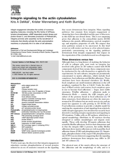
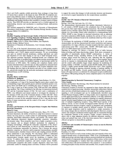
JOURNAL OF
GENETICS AND
GENOMICS
J. Genet. Genomics 37 (2010) 159−172
www.jgenetgenomics.org
Cytokinesis and cancer: Polo loves ROCK’n’ Rho(A)
Jing Li *, Jue Wang, Hong Jiao, Ji Liao, Xingzhi Xu *
Laboratory of Cancer Biology, College of Life Science, Capital Normal University, Beijing 100048, China
Received for publication 24 December 2009; revised 8 February 2010; accepted 9 February 2010
Abstract
Cytokinesis is the last step of the M (mitosis) phase, yet it is crucial for the faithful division of one cell into two. Cytokinesis failure is
often associated with cancer. Cytokinesis can be morphologically divided into four steps: cleavage furrow initiation, cleavage furrow
ingression, midbody formation and abscission. Molecular studies have revealed that RhoA as well as its regulators and effectors are important players to ensure a successful cytokinesis. At the same time, Polo-like kinase 1 (Plk1) is an important kinase that can target many
substrates and carry out different functions during mitosis, including cytokinesis. Recent studies are beginning to unveil a closer tie between Plk1 and RhoA networks. More specifically, Plk1 phosphorylates the centralspindlin complex Cyk4 and MKLP1/CHO1, thus recruiting RhoA guanine nucleotide-exchange factor (GEF) Ect2 through its phosphopeptide-binding BRCT domains. Ect2 itself can be
phosphorylated by Plk1 in vitro. Plk1 can also phosphorylate another GEF MyoGEF to regulate RhoA activity. Once activated,
RhoA-GTP will activate downstream effectors, including ROCK1 and ROCK2. ROCK2 is among the proteins that associate with Plk1
Polo-binding domain (PBD) in a large proteomic screen, and Plk1 can phosphorylate ROCK2 in vitro. We review current understandings
of the interplay between Plk1, RhoA proteins and other proteins (e.g., NudC, MKLP2, PRC1, CEP55) involved in cytokinesis, with particular emphasis of its clinical implications in cancer.
Keywords: Polo-like kinase 1; RhoA GTPase; Rho kinase; cytokinesis
Introduction
Cytokinesis, the last step of the M (mitosis) phase, involves physically dividing the cytoplasm of a single cell to
form two daughter cells. This is a crucial step in cell cycle
and has been widely studied in many model organisms:
budding yeast, fission yeast, Drosophila, Caenorhabditis
elegans, Xenopus, Dictyostelium, plants, and vertebrate
Abbreviation: Plk1, Polo-like kinase 1; GEF, guanine nucleotide-exchange
factor; GAP, GTPase-activating protein; ROCK, Rho-associated coiledcoil-forming kinase; MLC, myosin light chain.
* Corresponding authors. Tel: +86-10-6890 9575; Fax: +86-10-6890 6307.
E-mail address: [email protected] (J. Li);
[email protected] (X. Xu)
DOI: 10.1016/S1673-8527(09)60034-5
cells (Normand and King, 2010). In animal cells the contractile ring carries out the cytokinesis step and is composed of the actin cytoskeleton and its motor molecule,
myosin II (referred to as myosin in this review). But what
are the regulatory proteins for the spatial and temporal
events of cytokinesis? The small GTPase of Rho (Ras homologous) families are among the first proteins to be identified. Mammalian Rho GTPases comprise 20 intracellular
signaling molecules, and can be subdivided into three major subsets: Rho, Rac and Cdc42 (Narumiya and Yasuda,
2006). They cycle between the inactive GDP-bound form
and the active GTP-bound form. The cycling of Rho
GTPases between these two states is regulated by three
sets of proteins, guanine nucleotide-exchange factors
(GEFs), GTPase-activating proteins (GAPs) and guanine
160
Jing Li et al. / Journal of Genetics and Genomics 37 (2010) 159−172
nucleotide-dissociation inhibitors (GDIs). All three subsets
of Rho GTPases are implicated in cytokinesis in different
organisms, but RhoA is the most critical in mammalian
cells. During cytokinesis, both induction and progression
of the contractile ring depend on RhoA activation (Piekny
et al., 2005).
Besides the cytoskeleton system and its interacting Rho
GTPase, a successful cytokinesis also requires key protein
kinases and signaling networks to coordinate the position
of chromosomes in relative of the cell cortex. Cyclindependent kinases (Cdks), Aurora B and Polo-like kinases
(Plks) are important kinases that not only regulate cytokinesis, but also are crucial regulators of other mitotic events
(Glotzer, 2005; Barr and Gruneberg, 2007). There are several conserved Plks in humans, and we will only focus on
Plk1 in this review, since it is believed that the major function is attributed to Plk1. Recently, some substrates of Plk1
have been identified to be involved in cytokinesis, including PRC1 (Protein Regulator of Cytokinesis 1) (Neef et al.,
2007), CEP55 (CEntrosome Protein 55) (Fabbro et al.,
2005), NudC (Nuclear-distribution gene C) (Zhou et al.,
2003) and MKLP2 (mitotic-kinesin-like protein 2) (Neef et
al., 2003). Also among these substrates are the Rho proteins: Rho GEF Ect2 (Epithelial cell transforming gene 2)
(Niiya et al., 2006), Rho GAP HsCyk-4 (Burkard et al.,
2009; Wolfe et al., 2009) and MKLP1/CHO1 (Liu et al.,
2004). Moreover some of the RhoA downstream effectors
are found to bind to the Plk1 Polo-box domain (PBD),
including the Rho-associated coiled-coil-forming kinase
(ROCK) (Lowery et al., 2007). ROCK is also phosphorylated by Plk1 in vitro (Lowery et al., 2007). Thus Plk1 and
Rho GTPases are intricately linked with each other during
the cytokinesis process.
It has been widely known that cytokinesis failure results
in polyploidy and increased genome instability, which are
frequently observed in cancer cells. In fact, Plk1, RhoA
and their interacting proteins are all reported to be deregulated in some cancers. As more and more proteins involved
in tumorigenesis are found to play a role in cytokinesis,
such as Chk1 (Peddibhotla et al., 2009) and BRCA2
(Daniels et al., 2004), it has become apparent that cytokinesis and cancer are interconnected. This review will focus
on these recent new findings in vertebrate cells and will
explore its potential implication in cancer therapy, but observations from yeast and other organisms are discussed
where appropriate.
The structure of Plk1 and its function in cytokinesis
Plk1 is a serine/threonine kinase that orchestrates the
mitotic process. It was first discovered in Drosophila, as
polo mutants fail to undergo a normal mitosis (Sunkel and
Glover, 1988). And Plk1 homologues have been identified
in many eukaryotes (Table 1). Plk1 has been shown to play
key roles during different stages of mitosis, including mitotic entry, bipolar spindle formation, chromosome segregation and cytokinesis (Barr et al., 2004; van de Weerdt
and Medema, 2006).
The structure of Plk1 is conserved across different species, with a serine/threonine kinase domain at its
N-terminus and a regulatory domain, the PBD, at its
C-terminus (Fig. 1A). Plk1 is activated by phosphorylation
at Thr210 within the kinase domain. All Plks have a conserved PBD, and PBD has been identified as a phosphopeptide-binding motif (Elia et al., 2003). Indeed, studies
Table 1
Homologues of relevant proteins in eukaryotes
Saccharomyces
cerevisiae
Schizosaccharomyces
pombe
Drosophila
melanogaster
Caenorhabditis
elegans
Mammals
Polo-like kinase 1
Cdc5
Plo1
Polo
Plc1
Plk1
Rho A
Rho1
Rho1
Rho
Rho A
Rho A
ROCK
NA
NA
Rok/Drok
LET-502
ROCK
RhoGEF/Ect2
Tom2, Tus1
NA
Pebble(Pbl)
Let-21
Ect2
GAP
NA
NA
Tumbleweed/MgcGAP50C
Cyk-4
MgcRacGAP/HsCyk-4
MKLP1
NA
NA
Pavarotti
ZEN-4
MKLP1/CHO1/Kif23
MYPT1
NA
NA
MYPT/Mbs
MEL-11
MYPT1
NA: no homologs available.
Jing Li et al. / Journal of Genetics and Genomics 37 (2010) 159−172
161
Fig. 1. Diagrams of protein structures. A: Plk1 consists of a serine/threonine kinase domain at its N-terminus and a regulatory domain and the polo box
domain (PBD) at its C-terminus. B: Ect2 consists of two phosphopeptide-binding BRCT domains at its N-terminus and a tandem array of Dbl-homology
(DH) domain and pleckstrin-homology (PH) domain at its C-terminus C: Cyk4 consists of an N-terminal coiled-coil domain and a C-terminal RhoGAP
domain. D: MKLP1 consists of an N-terminal motor domain and a short coiled-coil region. E: PRC1 contains a central-spindle targeting at its N-terminus,
and a central microtubule binding domain.
of Plk1 and its substrates have established a common
theme that Plk1 can dock to specific phosphorylated targets through its PBD domain.
Early evidence showing Plk1’s function in cytokinesis
comes from S. pombe Plo1 (Ohkura et al., 1995). Plo1
activity correlates with division septum formation, as upor down-regulation of Plo1 both affects the division septum. The study of the role of Plk1 in mammalian cell cytokinesis is hampered by the fact that Plk1 depletion
causes early mitotic defects. But overproduction of Plk1
results in multinucleation in mammalian cells, indicative
of cytokinesis failure. Plk1’s substrates during cytokinesis
include MKLP2 and NudC. Both MKLP2 and NudC have
motor protein activity (MKLP2 is a kinesin and NudC is a
component of the dynein), and both localize to the central
spindle. Plk1 phosphorylates MKLP2 at Ser528 and phosphorylated MKLP2 binds with Plk1 PBD (Neef et al.,
2003). When this phosphorylation is blocked, cells show
cytokinesis defects. NudC RNA interference (RNAi) results in multinucleation and midbody arrest (Zhou et al.,
2003). NudC is phosphorylated by Plk1 at Ser274 and
Ser326 in vitro, and phosphorylation-deficient mutants will
not rescue the cytokinesis defects of NudC RNAi.
Direct evidence of Plk1’s involvement in RhoA mediated cytokinesis pathway comes from chemical studies
(Brennan et al., 2007; Burkard et al., 2007; Petronczki et
al., 2007; Santamaria et al., 2007). Burkard et al. (2007)
disrupted Plk1 and substituted it with a mutant Plk1as.
Plk1as has an enlarged catalytic pocket that can accommodate bulky purine analogs (e.g., 1-NM-PP1, or 3-MB-PP1).
Since these analogs will not fit into the wild-type Plk1,
they can specifically block the activity of Plk1as. When the
purine analogs are applied during anaphase, Plk1’s recruitment to the central spindle is blocked, which prevents
162
Jing Li et al. / Journal of Genetics and Genomics 37 (2010) 159−172
cleavage furrow formation and cell division. Closer examination reveals that RhoA localization to the cleavage
cortex is disrupted, and so are the RhoA downstream targets including citron kinase and anillin. Ect2’s recruitment
to the equatorial cortex and central spindle is also affected.
Another method directly uses Plk1’s small-molecule inhibitor BI 2536 (Petronczki et al., 2007). BI2536 treatment
abolishes Ect2 localization to the central spindle, but Cyk4
and Mklp1 are not affected. Other inhibitors such as
BTO-1 and ZK-Thiazolidinone (TAL) have the same effect
(Brennan et al., 2007; Santamaria et al., 2007). These
studies reveal that Plk1 plays important roles during the
last stages of cell cycle.
Plk1 and RhoA: caught in the act of cytokinesis
There are four morphological stages of cytokinesis: division site positioning and cleavage furrow initiation;
cleavage furrow ingression; midbody formation; and abscission (Fig. 2). We are going to review the roles of Plk1,
RhoA and their relationship at each distinct stage. Table 2
summarizes the currently known Plk1 substrates and interacting proteins involved in cytokinesis.
Fig. 2. The mitotic cell cycle. A: HeLa cells in different phases of mitosis were stained with anti-tubulin (green) antibodies and DAPI. B: Schematic
diagrams of the spindle assembly in relative to the chromosomes during mitosis. Red circle demarcates the contractile ring.
Table 2
Some of the known Plk1 substrates and interacting proteins in cytokinesis and their phosphorylation sites
Protein
Phosphorylation site
Biological function of phosphorylation
Reference
Rock2
Multiple sites
Binds with Plk1 PBD, activates ROCK2 activity
Lowery et al., 2007
MgcRacGAP/HsCyk-4
Ser157
Binds to the BRCT domain of Ect2, thus recruiting Ect2 to the midzone
Burkard et al., 2009;
Wolfe et al., 2009
CHO1/MKLP1
Ser904, Ser905
Binds with Plk1 PBD, essential for cytokinesis
Liu et al., 2004
MyoGEF
Thr574
Regulates RhoA activity
Asiedu et al., 2008
MKLP2
Ser528
Binds with Plk1 PBD, essential for cytokinesis
Neef et al., 2003
NudC
Ser274,Ser26
Essential for cytokinesis
Zhou et al., 2003
Cep55
Ser436
Essential for cytokinesis
Fabbro et al., 2005
Ect2
Thr412 (by Cdk1)
Binds with Plk1 PBD, regulates RhoA recruitment and activation
Niiya et al., 2006
PRC1
Thr578, Thr602
Binds with Plk1 PBD in an anaphase-specific manner, essential for cytokinesis
Neef et al., 2007
Jing Li et al. / Journal of Genetics and Genomics 37 (2010) 159−172
Division site positioning and cleavage furrow initiation
In different organisms, division site positioning is established at different points in the cell cycle. Cytokinesis
initiation in various systems was reviewed by Oliferenko
et al. (2009). In animal cells the site of cell division is
chosen during mitosis. More specifically, it is determined
in late anaphase and/or telophase. During this stage, the
mitotic spindle pulls the sister chromatids apart, and in the
mean time emits signals to initiate cytokinesis. Earlier
micromanipulation experiments show that when mitotic
spindle orientation in star fish eggs is changed, the position
of the cortical contractile ring is also altered (Rappaport
and Ebstein, 1965), indicating that the spindle position
specifies the cleavage furrow formation. The mitotic spindle consists of the interdigital central spindle and polar
astral microtubules. Central spindle may send positive
signals to the cell cortex nearest to the spindle midzone to
specify cleavage sites, while the polar astral spindle may
inhibit the cortical contractility. These distinct groups of
spindles cooperate together to signal the furrowing site
(Bringmann and Hyman, 2005; Glotzer, 2009).
How can the spindle microtubule control the cortical
contractility? The answer lies in the small GTPase RhoA.
RhoA is crucial for furrowing, as biochemical inactivation
or depletion of RhoA will lead to cleavage furrow formation failure (Piekny et al., 2005). Activated RhoA localizes
to a narrow cortical zone within the cleavage furrow, and
spindle displacement can perturb this localization pattern
(Bement et al., 2005). Activated GTP-bound RhoA in turn
induces F-actin assembly and activates myosin function,
thus promoting the contractility of cell cortex. The specificity of RhoA activation is achieved by localizing specific
RhoA regulators on the microtubules. The regulators that
have been identified so far include Ect2, Cyk4, MyoGEF
(myosin-interacting GEF), p0071, and phospholipids (details will be discussed below).
Ect2 was originally isolated as a proto-oncogene from
epithelial cells that are capable to transform (Miki et al.,
1993). The C-terminus of Ect2 contains a tandem array of
Dbl-homology (DH) domain and pleckstrin-homology (PH)
domain (Fig. 1B). It is the C-terminus that confers Ect2 the
ability to catalyze guanine nucleotide exchange on RhoA
(Saito et al., 2004). Besides the DH/PH cassette, Ect2 contains N-terminal tandem BRCT (BRCA1-C Terminal) domains, and the central S domain that contains the nuclear
localization signals. The BRCT domains associate with the
163
C-terminal DH/PH domain and blocks its ability of guanine nucleotide exchange (Saito et al., 2004). Ect2 localizes in the nucleus during interphase in an inactive state,
becomes activated and localizes to the mitotic spindles
during metaphase, and finally appears at the midbody
structure during cytokinesis. Both Ect2 antibody injection
and Ect2 RNAi inhibits cytokinesis and leads to multinucleated cells (Saito et al., 2004). These results demonstrate
that Ect2 plays a crucial role to activate RhoA.
How is Ect2 recruited to the central spindle? It turns out
that the centralspindlin complex recruits Ect2. The
tetrameric centralspindlin complex is composed of a dimer
of the kinesin 6 protein MKLP1 (also known as Kif23) and
a dimer of the GAP Cyk4 (also known as RacGAP1 or
MgcRacGAP) (Mishima et al., 2002). The interaction between MKLP1 and Cyk4 is evolutionarily conserved, and
they localize to the central spindle in an inter-dependent
manner, where they promote the microtubule bundling
(Mishima et al., 2002). The N-terminus of Cyk4 binds to the
neck linker of MKLP1 (Fig. 1, C and D), and assembles into
a stable centralspindlin complex (Pavicic-Kaltenbrunner et
al., 2007). Cyk4 binds to and stabilizes activated Ect2, allowing Ect2 to interact with RhoA (Yuce et al., 2005). As a
result, Ect2 activates RhoA and signals to the overlying
equatorial cortex, leading to contractile ring formation and
cleavage furrow ingression. The centralspindlin complex
travels along central spindles as higher-order clusters and
accumulates at the midbody. Centralspindlin clustering is
critical for microtubule bundling and motility, as well as
midbody formation (Hutterer et al., 2009). When the centralspindlin complex localizes to the central spindle, it restricts Ect2 within the narrow zone, leading to RhoA’s
narrow activation. When this localization pattern is disrupted, RhoA will be activated in a much broader range,
and cells will fail to form a furrow. In Xenopus,
MgcRacGAP not only anchors active RhoA to the activity
zone, but also promotes local RhoA inactivation that provides constant Rho to the GTPase cycle (Miller and Bement, 2009), consistent with earlier findings that the GAP
activity is essential for cytokinesis in mammals (Hirose et
al., 2001).
It may seem paradoxical that both Rho GEF and GAP
are required for RhoA activation. When MgcRacGAP antisense morpholino oligonucleotides are applied, the
Xenopus embryos display cytokinesis defects as well as a
broader zone of RhoA activity (Miller and Bement, 2009).
Only wild-type MgcRacGAP, but not a GAP activity mu-
164
Jing Li et al. / Journal of Genetics and Genomics 37 (2010) 159−172
tant allele of MgcRacGAP can rescue the defects, suggesting that the GAP activity is required for RhoA inhibition,
which in turn is required for cytokinesis. Thus the “Rho
flux model” speculates that a weaker GAP activity than the
GEF activity at the cleavage furrow will lead to RhoA activation, but both the GAP and GEF are required. The
other “Rac inactivation” model states that RacGAP is required to inhibit Rac GTPase and activate RhoA at the
same time (Canman et al., 2008). Further experiments may
reconcile these two different models, but there is no doubt
that GAP is essential for RhoA activity.
Recent investigations show that both GEF Ect2 and
Cyk4 are Plk1 substrates. Ect2 is phosphorylated during
the G2/M transition (Niiya et al., 2006). In vitro Ect2 can
be phosphorylated by Cdk1 and Plk1. Cdk1 phosphorylation at Thr412 creates a phosphospecific binding pocket
(SpTP) for Plk1 PBD (Niiya et al., 2006). When the
Thr412 residue is mutated to Ala, RhoA activation is diminished. In contrast, the phosphomimic Ect2 T412D mutant still exhibits significant action of RhoA. Ect2 overproduction induces cortical hyperactivity that leads to cell
death, but Ect2-T412A overproduction has no such effect
(Niiya et al., 2006). Furthermore, this interaction is not
limited to mammalian cells. In budding yeasts two RhoA
GEFs (Tus1 and Rom2) are both substrates of Cdc5 in
vitro and in vivo (Yoshida et al., 2006). Thus it seems a
conserved pathway for Plk1 to regulate the recruitment and
activation of RhoA by regulating Rho GEF’s localization
to the division site.
Since the interaction between Ect2’s BRCT domains and
Cyk4 is dependent on phosphorylation (Yuce et al., 2005),
there has been speculation that Cyk4 might be phosphorylated prior to binding to Ect2’s BRCT domains, which bind
phosphopeptides. Indeed, two independent investigations
show that Plk1 specifically binds and phosphorylates Cyk4
at Ser157, thus creating a docking site for the Ect2 BRCT
domains (Burkard et al., 2009; Wolfe et al., 2009). When
Ser157 is mutated to a nonphosphorylated form, Ect2 fails
to localize at the midzone, leading to cleavage furrowing
errors (Burkard et al., 2009; Wolfe et al., 2009).
Besides Ect2, RhoA also has other regulators, including
GEF MyoGEF (Wu et al., 2006), the armadillo protein
p0071 (Wolf et al., 2006) and phospholipids (Yoshida et al.,
2009). MyoGEF also contains the DH and PH domains.
MyoGEF disruption by RNAi results in binucleated and
multinucleated cells and decreased RhoA activation (Wu et
al., 2006). MyoGEF localizes to the central spindle, and
interacts with Ect2, and MyoGEF RNAi leads to Ect2 and
RhoA mislocalization during cytokinesis (Asiedu et al.,
2009). MyoGEF and Plk1 colocalize at the spindle pole
and central spindle, and MyoGEF localization is dependent
on Plk1 (Asiedu et al., 2008). Plk1 phosphorylates MyoGEF on Thr574 in vitro (Asiedu et al., 2008). In vivo experiments show that the MyoGEF T574A mutant dramatically decreases MyoGEF phosphorylation. MyoGEF
T574A displays decreased GEF activity towards RhoA,
suggesting that Plk1 can regulate RhoA activity through
phosphorylating MyoGEF.
The armadillo protein p0071 localizes at the midbody
(Wolf et al., 2006). Its upregulation or downregulation
leads to cytokinesis defects and induces apoptosis. It turns
out that p0071 interacts with Ect2 and RhoA, and alteration of p0071 expression deregulates RhoA activity (Wolf
et al., 2006). p0071 localization is dependent on kinesin-II
member Kif3b (Keil et al., 2009).
Evidence that phospholipids promote RhoA localization
comes from the budding yeast. During septation and abscission in the yeast cells, Rho1 bud neck targeting requires that
the Rho1 polybasic sequence binds to acidic phospholipids,
including phosphatidylinositol 4,5-bisphosphate (PIP2)
(Yoshida et al., 2009). In animal cells, this mechanism
might be further facilitated by phosphatidylinositol 3,4,5trisphosphate, which is not present in the budding yeast.
Cleavage furrow ingression
After RhoA is temporally and locally activated, it will
recruit and activate downstream effectors to induce cleavage furrow ingression (Fig. 3). Downstream targets include
proteins to stimulate actin polymerization, as well as
ROCK and citron kinase that stimulate myosin activity
(Matsumura, 2005).
Myosin comprises two heavy chains, two essential light
chains and two regulatory myosin light chains (MLC).
MLC Ser19 phosphorylation stimulates myosin ATPase
activity, and Thr18 phosphorylation promotes myosin assembly (Matsumura, 2005). During mitosis, MLC is
phosphorylated at Ser1, 2 and 9 by CDK1 to inhibit myosin ATPase activity. CDK1 deactivation at anaphase allows MLC dephosphorylation at these sites. Thus specific
MLC phosphorylation site controls contractile ring assembly. Expression of a nonphosphorylation mutant of MLC
in fly and culture cells both results in cytokinesis failure
(Jordan and Karess, 1997; Komatsu et al., 2000).
Jing Li et al. / Journal of Genetics and Genomics 37 (2010) 159−172
Fig. 3. Regulation of the contractile ring formation by the RhoA
GTPase. RhoA is activated by GEF (Ect2, MyoGEF) and inactivated by
GAP (Cyk4). RhoA-GTP is concentrated at the cleavage site and induces
actin filament assembly. In the meantime, RhoA activates downstream
effectors (ROCK and citron kinase), which induce MCL phosphorylation
and thereby myosin II filament formation. Thus the contractile ring, which
is composed of the actin filaments and myosin II, is assembled. In this
diagram, proteins marked in bold are currently known Plk1 substrates.
MLC phosphorylation is controlled by three kinases:
ROCK, citron kinase, MLCK (Myosin Light Chain
Kinase), and also reversibly controlled by myosin phosphatase. ROCK localizes to the cleavage furrow, and inhibition of ROCK with Y-27632 causes cleavage delay, but
not affecting cytokinesis initiation and completion
(Kosako et al., 2000). Mammalian cells have two ROCK
isoforms: ROCK1 (Rokβ) and ROCK2 (Rokα). Knockdown of either ROCK1 or ROCK2 in mice does not display
cytokinesis defects (Thumkeo et al., 2003; Shimizu et al.,
2005), which might be caused by the redundancy between
these two kinases. Double-knockout mice will help to elucidate the function of ROCK in cytokinesis.
Both cytology and biochemistry studies show that Plk1
and ROCK2 interact. Plk1 and ROCK2 colocalize at the
midbody during cytokinesis (Lowery et al., 2007). Plk1
coimmunoprecipitates with ROCK2 in a phosphorylation-dependent and mitosis-specific manner. Plk1 can
phosphorylate ROCK2 in vitro, and phosphorylated
ROCK2 interacts with Plk1 PBD (Lowery et al., 2007).
Plk1 and RhoA act in a synergistic fashion to activate
ROCK2 in vitro and in vivo.
The second kinase, citron kinase, also localizes in the
cleavage furrow. Its overproduction upregulates cortex
165
contractility, suggesting that it positively regulates myosin
activity (Madaule et al., 1998). It phosphorylates MLC at
Ser19/Thr18 both in vitro and in vivo (Yamashiro et al.,
2003). Citron kinase knockout mice complete embryonic
development, but some neuronal precursor cells display
abnormal cytokinesis and massive apoptosis (Di Cunto et
al., 2000). These cells express ROCK proteins at the normal level, suggesting that although ROCK and citron
kinase might be redundant in activating myosin, they may
have other distinct regulatory function.
MLCK is a third kinase that can phosphorylate MLC in
the cleavage furrow. It is activated by Ca2+/calmodulin and
also by phosphorylation. MLCK inhibition in cultured
mammalian cells leads to cytokinesis failure (Normand
and King, 2010). MLCK might also be regulated by phospholipids, because PIP2 hydrolysis is important for inositol-1,4,5-trisphosphate (IP3)-induced calcium release
(Wong et al., 2007).
The only known phosphatase of MLC is myosin phosphatase, which consists of a targeting subunit MYPT1
(Myosin Phosphatase Targeting Subunit 1), a catalytic
subunit PP1Cβ and an additional small subunit (Baumann
et al., 2007; Matsumura and Hartshorne, 2008). During
anaphase, both ROCK and Aurora B kinase phosphorylate
MYPT1 in the furrow to inhibit its phosphatase activity
(Kawano et al., 1999; Yokoyama et al., 2005), which may
lead to increase of MLC phosphorylation at the cleavage
furrow and signal for cytokinesis. MYPT1 is recently reported to be phosphorylated by Cdk1 in a mitosis-specific
fashion, which generates a binding motif for Plk1 PBD
(Yamashiro et al., 2008). MYPT1 antagonizes Plk1 activity,
as MYPT1 depletion increases Plk1-T210 phosphorylation
(Yamashiro et al., 2008). But whether this interaction
functions in cytokinesis remains to be elucidated. MYPT1
is recently found to be a regulatory subunit of serine/threonine-protein phosphatase 6 catalytic subunit
(PP6C) and function in the homologous recombination
pathway (unpublished data). It is a possible scenario that
PP6C is targeted by MYPT1 to dephosphorylate MLC.
Formation of the midbody
Cleavage furrow ingression continues until the actomyosin contractile ring comes into close proximity to the central spindle. Although proteomic approaches have been
used to identify the midbody components (Skop et al.,
2004; Chen et al., 2009a), its precise function is not yet
166
Jing Li et al. / Journal of Genetics and Genomics 37 (2010) 159−172
understood in detail. It is hypothesized that the midbody
maintains a state of constriction until abscission is completed.
The centralspindlin complex also plays an essential role
in this step. MKLP1 has a splice variant called CHO1.
CHO1 is shown to be required for midbody formation in
mammalian cells in RNAi studies (Matuliene and Kuriyama, 2002, 2004). CHO1 contains an extra F-actin interacting domain, indicating that it may function to link the
actin with the centralspindlin complex. Plk1 interacts with
CHO1 during anaphase and telophase, and the PBD domain of Plk1 is responsible for this association (Liu et al.,
2004). Also needed is the stalk domain of CHO1. When
CHO1 is depleted, Plk1 fails to localize at the midbody, and
cells turn multinucleated with more centrosomes. Plk1 can
phosphorylate CHO1 in vitro, and Ser904 and Ser905 are the
major phosphorylation sites. When a non-phosphorylated
form of CHO1 is expressed, cells display cytokinesis defects (Liu et al., 2004).
Some proteins that localize to the midbody have been
studied in detail, and one of them is PRC1. It is critical for
midbody formation in mammalian cells. In anaphase,
PRC1 localizes to the central spindle where its main function is to bundle microtubules (Mollinari et al., 2002). Depletion of PRC1 leads to abscission failure without affecting the cleavage furrow ingression. Domain analysis reveals that PRC1 has two separate domains that target
PRC1 to distinct subcellular structures (Mollinari et al.,
2002) (Fig. 1E). Its N-terminus interacts with kinesin Kif4
and is targeted to the midzone, where PRC1 recruits the
centralspindlin complex and other kinesin proteins (Zhu
and Jiang, 2005). On the contrary, the central region of
PRC1 is required for microtubule binding and bundling
activity (Mollinari et al., 2002). In metaphase CDK1
phosphorylates PRC1 at T470 and T481 (Neef et al., 2007).
These phosphorylation events prevent Plk1 binding. As
CDK1 activity decreases in anaphase, Plk1 phosphorylates
PRC1 at T578 and T602, creating its own docking site.
Thus PRC1 binds to Plk1 in an anaphase-specific manner,
which is essential for cytokinesis. Consistent with this,
CDK1 phosphorylation-deficient mutations result in premature binding with Plk1 and mitotic block (Neef et al.,
2007).
Plk1 also regulates the katanin protein that severs
microtubules at the midbody and functions both in mitosis
and meiosis (McNally et al., 2002). In Xenopus, Plx1
colocalizes with katanin at spindle poles in vivo and puri-
fied Plx1 increases the microtubule-severing activity of
katanin in vitro (McNally et al., 2002). Katanin is also essential for post-mitotic differentiation events in vertebrate
neurons and in Arabidopsis.
Abscission
Abscission is the final step of cytokinesis. During this
stage, the microtubule bundles start to compact and disappear (Fig. 2). The centrosome protein Cep55, which localizes to the midbody in a centralspindlin-dependent manner,
is essential for this step (Zhao et al., 2006). The absence of
Cep55 will lead to midbody formation defects (Zhao et al.,
2006). Cdk1/Erk2 phosphorylates Cep55 at Ser425/Ser428,
which is required for Cep55’s interaction with Plk1. Plk1
further phosphorylates Cep55 at Ser436. When Ser436 is
mutated, cells display cytokinesis failure (Fabbro et al.,
2005). Cep55 also has microtubule-bundling function that
is essential for the midbody formation (Zhao et al., 2006).
Recently, it was shown that the peptidyl-prolyl isomerase
Pin1 enhances the Plk1-dependent phosphorylation of
Cep55 (van der Horst and Khanna, 2009). Moreover,
Cep55 is stabilized post-translationally during mitosis in a
Pin1-dependent manner, and Cep55’s stable protein level
is essential for proper execution of cytokinesis (van der
Horst et al., 2009).
Because abscission involves the membrane organization
changes, the membrane trafficking system is crucial for
this step. These include the secretory pathway, the endocytic pathway and the ESCRT machinery (Endosomal
Sorting Complex Required for Transport) (Normand and
King, 2010). Depletion of components of these pathways
has been shown to lead to cytokinesis defects. Alix and
Tsg101 (Tumor-Susceptibility Gene 101) are components
of the ESCRT network, and they are recruited to the midbody by interacting with Cep55 (Carlton and Martin-Serrano, 2007).
Implication of cytokinesis in cancer
It has been widely known that cytokinesis failure will
lead to polyploidy. But some body tissues are polyploid by
nature. For instance, megakaryocyte (MK) cells are unique
among mammalian cells in that they are the naturally
polyploid hematopoietic cells that can give rise to platelets.
Polyploidization is intrinsic to this differentiation event.
Jing Li et al. / Journal of Genetics and Genomics 37 (2010) 159−172
MK cell polyploidization occurs by endomitosis, which
was recently shown to be caused by late cytokinesis failure
related to defects in Rho/Rock signaling (Lordier et al.,
2008). Thus in certain tissues Rho and ROCK can be the
targets of physiological regulation.
Apart from the naturally polyploid cells, failure of cytokinesis in normal cells can cause cell death or lead to
genome amplification, which is characteristic of many
cancers. When diploid and tetraploid cultures are isolated
from p53-null cells, only the tetraploid cells are transformed in vitro after carcinogen exposure (Fujiwara et al.,
2005). In addition, only the tetraploid cells will give rise to
malignant mammary epithelial cancers after transplanted
into nude mice. These results indicate that tetraploidy,
which is frequently the product of cytokinesis failure, can
promote tumor development in p53-null cells. Further
studies show that the p53-deficient tetraploid cells display
aneuploidy, genomic rearrangements and amplification
(Fujiwara et al., 2005). Thus, cytokinesis failure, polyploidization and erroneous chromosome segregation may
form a positive feedback loop to promote tumorigenesis.
Indeed, a lot of DNA damage checkpoint proteins are
recently shown to be involved in cytokinesis. These proteins have been shown to be implicated in cancer. For instance, Chk1 is a critical component of the DNA damage
checkpoint network that functions in DNA replication,
intra-S phase and the G2/M phase transition. Chk1+/− mice
display Aurora B mislocalization. Moreover, Chk1 abrogation leads to cytokinesis regression and binucleation
(Peddibhotla et al., 2009). Another example is BRCA2,
which is a breast cancer susceptibility protein. People carrying germ-line mutations inactivating BRCA2 are predisposed to breast cancer. RNAi experiments in HeLa cells
to knock down BRCA2 lead to cytokinesis defects (Daniels
et al., 2004). Furthermore, cytokinesis failure is also observed when BCCIP1, a BRCA2-interacting protein, is
downregulated (Meng et al., 2007).
More proteins are found to be at the intersection of cytokinesis and tumorigenesis. The peptidyl-prolyl isomerase
Pin1 that was mentioned above localizes to the midbody
ring and regulates the final stages of cytokinesis by binding to Cep55 (van der Horst and Khanna, 2009). In Pin1
knockout mice, embryonic fibroblasts show a cytokinesis
delay, and depletion of Pin1 from HeLa cells also causes
cytokinesis defects. Pin1 is also deregulated in many tumors,
including breast, prostate and lung cancer (Bao et al.,
2004). Overexpression of Pin1 promotes tumor growth,
167
while inhibition of Pin1 causes tumor cell apoptosis.
Therefore, many Pin1 inhibitors have been developed and
could be used as a novel type of anticancer drug by blocking cell cycle progression (Xu and Etzkorn, 2009). In fact,
Cep55 itself is found to be upregulated in breast, colorectal
and lung cancers, and Cep55 could act as a novel breast
cancer-associated antigen (Inoda et al., 2009).
Not surprisingly, both Plk1 and Rho proteins are long
known to be implicated in cancer. An example is the relationship between Plk1 and p53. Plk1 depleted cells undergo apoptosis, activate caspase 3 and form fragmented
nuclei (Liu and Erikson, 2003). Plk1 is later found to
physically interact with p53 (through both coimmunoprecipitation and colocalization studies) and inhibit p53’s
pro-apoptotic function (Ando et al., 2004). Plk1 might
negatively regulate p53 through Topors, a ubiquitin and
SUMO E3 ligase (Yang et al., 2009). Plk1 phosphorylates
Topors on Ser718 in vivo, and phosphorylated Topors inhibits p53’s sumoylation, while enhancing its ubiquitination (Yang et al., 2009). Another target of Plk1 to regulate
p53 might be Mdm2, as Plk1 also phosphorylates Mdm2 at
Ser260, thus stimulating Mdm2-mediated p53 turnover
(Dias et al., 2009). Moreover, Plk1 knockdown decreased
Mdm2 protein level (Kreis et al., 2009). On the other hand,
using both p53 binding-defective human papillomavirus
type-16 E6, and p53 RNAi, Incassati et al. (2006) show
that p53 represses Plk1 expression, suggesting that Plk1 is
a target of p53. Thus Plk1 and p53 may form a feedback
loop to function in tumorigenesis. Other members of the
p53 family include p63 and p73, both of which regulate
cell survival and apoptosis in tumors. Plk1 phosphorylates
p63 at Ser52, which decreases p63’s protein stability and
suppresses apoptosis (Komatsu et al., 2009). Plk1 interacts
and colocalizes with p73, and phosphorylates p73 at Thr27
(Koida et al., 2008; Soond et al., 2008). The CDK inhibitor
p21WAF1/CIP (referred to as p21 afterwards) is induced by
p73 in HeLa cells (p53-deficient), and the long-term suppression of Plk1 increases p21 protein level (Kreis et al.,
2009). These data strongly suggest a relationship between
Plk1 and p53 protein families.
As Plk1 plays a pivotal role in regulating mitosis, Plk1
is upregulated in many tumors, such as melanomas and
lymphomas, and could be used as a prognostic marker for
some cancers (Strebhardt and Ullrich, 2006). Plk1 has two
functional distinct domains, thereby providing investigators two targets within the same protein to develop
anti-proliferative drugs. The small molecule inhibitors that
168
Jing Li et al. / Journal of Genetics and Genomics 37 (2010) 159−172
have been developed against Plk1 have been useful in dissecting its cellular functions and might be useful in the
clinic. For instance, BI 2536 is developed as an anti-cancer
drug that blocks Plk1 activity with high potency in vitro
and in vivo (Mross et al., 2008).
Rho GTPase functions in a broad spectrum of cell metabolism, including cytoskeleton dynamics, cell cycle progression, transcriptional regulation and cell survival, which
are all important for tumorigenesis (Vega and Ridley,
2008). RhoA is shown to be involved in all stages of cancer progression. In breast cancers, in particular, RhoA is
not only upregulated, but also involved in the tumor metastasis (Lin and van Golen, 2004). The RhoA/ROCK signaling pathway has been proposed to regulate actomyosin-based cortical contractility that leads to cell invasion
(Vega and Ridley, 2008). Thus it is not surprising to find
that RhoA protein levels are significantly increased in
breast, lung and colon cancers (Gomez del Pulgar et al.,
2005).
RhoA GEF Ect2 is a proto-oncogene. Ect2 is found to
be overexpressed in glioma patients (Sano et al., 2006). In
these patients, RhoA activation is broadened, and cytokinesis initiation is affected, both of which correlate with
Ect2’s function in RhoA regulation. The other activator,
MyoGEF, is highly expressed in invasive breast cancer cell
lines and infiltrating ductal carcinomas. It regulates the
invasion activity of MDA-MB-231 breast cancer cells
through activation of RhoA and RhoC (Wu et al., 2009).
DLC1 (Deleted in Liver Cancer 1) encodes a RhoGAP, and
has been identified as a tumor suppressor gene that is deleted in cancers of the breast, colon and lung (Xue et al.,
2008). Other GAPs, such as DLC2, DLC3, p190RhoGAP
and GRAF are also implicated in cancer progression.
As far as RhoA effectors are concerned, so far there is
no evidence showing that Citron is involved in tumor, but
Citron interacting protein Kif14 is overproduced in breast
and lung cancers (Normand and King, 2010). ROCK is
upregulated in cancers. Moreover, ROCK not only interacts with p21 in vivo in Ras-transformed cells (Lee and
Helfman, 2004), but also upregulates p21 in prostate cancer cells (Xiao et al., 2009). Upon stimulating a conditional active ROCK-estrogen receptor fusion protein, cyclin D1, p21, cyclin A levels are elevated, while p27Kip1
levels are reduced (Croft and Olson, 2006). Current therapeutic strategies targeting Rho signaling in cancer have
been twofold: one directly targets Rho, the other focuses
on inhibiting Rho downstream effectors, such as ROCK.
The ROCK inhibitor fasudil not only is used to treat cardiovascular diseases, but also inhibits tumor progression in
human and rat tumor models (Ying et al., 2006). Another
ROCK inhibitor, Y-27632, can block Ras-mediated transformation of NIH3T3 cells (Sahai et al., 1999).
Outlooks and future directions
Are there more RhoA activators? Ect2, MyoGEF,
p0071 are among the RhoA activators that have been
identified so far. But to date over 70 RhoGEF, 60
RhoGAPs have been identified (Vega and Ridley, 2008),
suggesting that there might be more RhoA activators. In
light of the intricate interplay between Plk1 and RhoA,
we speculate that more RhoA activators would turn out to
be regulated by Plk1.
Upon cytokinesis completion, RhoA may need to be inactivated for cleavage furrow disassembly (Chalamalasetty
et al., 2006). But how is RhoA inactivated? Recent evidence suggests that it may be regulated at the protein level.
RhoA is shown to be ubiquitinated by Cul3-BACURD
ubiquitin ligase complexes (Chen et al., 2009b). When
Cul3 is lacking, RhoA degradation is affected, and actin
stress fibers are aberrant. Dysfunction of the Cul3-BACURD
complex attenuates the migration potential of HeLa cells
and mouse embryonic fibroblasts, and affects convergent
extension during gastrulation in Xenopus embryos (Chen
et al., 2009b). However, the authors did not examine
whether cells display any cytokinesis defects. Alternatively,
RhoA might be inactivated by the RhoGDI. It has been
known that GDP-Rho is prenylated and bound at the
translation site by specific RhoGDIs until RhoGDF
(RhoGDI-displacement factor) liberation, transported to its
specific membrane location and activated by RhoGEF. It is
conceivable that premature release of RhoA by RhoGDI
will lead to RhoA deregulation. But so far only RhoGDIs
in Dictyostelium are shown to be involved in cytokinesis
(Imai et al., 2002; Rivero et al., 2002). Mammals have
three isoforms of RhoGDI. Are they also involved in cytokinesis? Could they also be regulated by Plk1 phosphorylation? These questions await further studies.
It is now clear that cytokinesis and cancer are intricately
linked. And targeting proteins involved in cytokinesis
could become a common theme for tumor therapy. Further
studies will reveal more Plk1 substrates and more RhoA
regulating proteins that are involved in the cytokinesis
Jing Li et al. / Journal of Genetics and Genomics 37 (2010) 159−172
pathway, thus providing more targets for novel drug development that can be taken to the clinic.
Acknowledgements
This work was supported by the National Natural Science Foundation of China (NSFC) (No. 30700420), Beijing Nova Program (No. 2007B062), Scientific Research
Program of Beijing Municipal Commission of Education
(No. KM200810028013), Scientific Research Foundation
for the Returned Overseas Chinese Scholars from Beijing
Municipal Commission of Human Resources (No.
085402600) and also from State Education Ministry (SRF
for ROCS, SEM) to J.L. X.X. was supported by the startup
fund from CNU, NSFC funds (No. 30570371, 90608014,
and 30711120570), the Program for New Century Excellent Talents in University (No. NCET-06-0187), Beijing
Natural Science Foundation Program and Scientific Research Key Program of Beijing Municipal Commission of
Education (No. KZ200810028014), and Funding Project
for Academic Human Resources Development in Institutions of Higher Learning Under the Jurisdiction of Beijing
Municipality (PHR(IHLB)). We thank members of the
Xu’s lab for helpful discussion and comments on the
manuscript.
References
Ando, K., Ozaki, T., Yamamoto, H., Furuya, K., Hosoda, M., Hayashi,
S., Fukuzawa, M., and Nakagawara, A. (2004). Polo-like kinase 1
(Plk1) inhibits p53 function by physical interaction and phosphorylation. J. Biol. Chem. 279: 25549−25561.
Asiedu, M., Wu, D., Matsumura, F., and Wei, Q. (2008). Phosphorylation of MyoGEF on Thr-574 by Plk1 promotes MyoGEF localization
to the central spindle. J. Biol. Chem. 283: 28392−28400.
Asiedu, M., Wu, D., Matsumura, F., and Wei, Q. (2009). Centrosome/spindle pole-associated protein regulates cytokinesis via promoting the recruitment of MyoGEF to the central spindle. Mol. Biol.
Cell 20: 1428−1440.
Bao, L., Kimzey, A., Sauter, G., Sowadski, J.M., Lu, K.P., and Wang,
D.G. (2004). Prevalent overexpression of prolyl isomerase Pin1 in
human cancers. Am. J. Pathol. 164: 1727−1737.
Barr, F.A., and Gruneberg, U. (2007). Cytokinesis: placing and making
the final cut. Cell 131: 847−860.
Barr, F.A., Sillje, H.H., and Nigg, E.A. (2004). Polo-like kinases and the
orchestration of cell division. Nat. Rev. Mol. Cell Biol. 5: 429−440.
Baumann, C., Korner, R., Hofmann, K., and Nigg, E.A. (2007). PICH,
a centromere-associated SNF2 family ATPase, is regulated by Plk1
169
and required for the spindle checkpoint. Cell 128: 101−114.
Bement, W.M., Benink, H.A., and von Dassow, G. (2005). A microtubule-dependent zone of active RhoA during cleavage plane specification. J. Cell Biol. 170: 91−101.
Brennan, I.M., Peters, U., Kapoor, T.M., and Straight, A.F. (2007).
Polo-like kinase controls vertebrate spindle elongation and cytokinesis. PLoS One 2: e409.
Bringmann, H., and Hyman, A.A. (2005). A cytokinesis furrow is positioned by two consecutive signals. Nature 436: 731−734.
Burkard, M.E., Randall, C.L., Larochelle, S., Zhang, C., Shokat,
K.M., Fisher, R.P., and Jallepalli, P.V. (2007). Chemical genetics
reveals the requirement for Polo-like kinase 1 activity in positioning
RhoA and triggering cytokinesis in human cells. Proc. Natl. Acad. Sci.
USA 104: 4383−4388.
Burkard, M.E., Maciejowski, J., Rodriguez-Bravo, V., Repka, M.,
Lowery, D.M., Clauser, K.R., Zhang, C., Shokat, K.M., Carr, S.A.,
Yaffe, M.B., and Jallepalli, P.V. (2009). Plk1 self-organization and
priming phosphorylation of HsCYK-4 at the spindle midzone regulate
the onset of division in human cells. PLoS Biol. 7: e1000111.
Canman, J.C., Lewellyn, L., Laband, K., Smerdon, S.J., Desai, A.,
Bowerman, B., and Oegema, K. (2008). Inhibition of Rac by the
GAP activity of centralspindlin is essential for cytokinesis. Science
322: 1543−1546.
Carlton, J.G., and Martin-Serrano, J. (2007). Parallels between cytokinesis and retroviral budding: a role for the ESCRT machinery. Science 316: 1908−1912.
Chalamalasetty, R.B., Hummer, S., Nigg, E.A., and Sillje, H.H. (2006).
Influence of human Ect2 depletion and overexpression on cleavage
furrow formation and abscission. J. Cell Sci. 119: 3008−3019.
Chen, T.C., Lee, S.A., Hong, T.M., Shih, J.Y., Lai, J.M., Chiou, H.Y.,
Yang, S.C., Chan, C.H., Kao, C.Y., Yang, P.C., and Huang, C.Y.
(2009a). From midbody protein-protein interaction network construction to novel regulators in cytokinesis. J. Proteome Res. 8:
4943−4953.
Chen, Y., Yang, Z., Meng, M., Zhao, Y., Dong, N., Yan, H., Liu, L.,
Ding, M., Peng, H.B., and Shao, F. (2009b). Cullin mediates degradation of RhoA through evolutionarily conserved BTB adaptors to
control actin cytoskeleton structure and cell movement. Mol. Cell 35:
841−855.
Croft, D.R., and Olson, M.F. (2006). The Rho GTPase effector ROCK
regulates cyclin A, cyclin D1, and p27Kip1 levels by distinct mechanisms. Mol. Cell Biol. 26: 4612−4627.
Daniels, M.J., Wang, Y., Lee, M., and Venkitaraman, A.R. (2004).
Abnormal cytokinesis in cells deficient in the breast cancer susceptibility protein BRCA2. Science 306: 876−879.
Di Cunto, F., Imarisio, S., Hirsch, E., Broccoli, V., Bulfone, A.,
Migheli, A., Atzori, C., Turco, E., Triolo, R., Dotto, G.P., Silengo,
L., and Altruda, F. (2000). Defective neurogenesis in citron kinase
knockout mice by altered cytokinesis and massive apoptosis. Neuron
28: 115−127.
Dias, S.S., Hogan, C., Ochocka, A.M., and Meek, D.W. (2009).
Polo-like kinase-1 phosphorylates MDM2 at Ser260 and stimulates
MDM2-mediated p53 turnover. FEBS Lett. 583: 3543−3548.
Elia, A.E., Rellos, P., Haire, L.F., Chao, J.W., Ivins, F.J., Hoepker, K.,
Mohammad, D., Cantley, L.C., Smerdon, S.J., and Yaffe, M.B.
(2003). The molecular basis for phosphodependent substrate targeting
170
Jing Li et al. / Journal of Genetics and Genomics 37 (2010) 159−172
and regulation of Plks by the Polo-box domain. Cell 115: 83−95.
Fabbro, M., Zhou, B.B., Takahashi, M., Sarcevic, B., Lal, P., Graham,
M.E., Gabrielli, B.G., Robinson, P.J., Nigg, E.A., Ono, Y., and
Khanna, K.K. (2005). Cdk1/Erk2- and Plk1-dependent phosphorylation of a centrosome protein, Cep55, is required for its recruitment to
midbody and cytokinesis. Dev. Cell 9: 477−488.
Fujiwara, T., Bandi, M., Nitta, M., Ivanova, E.V., Bronson, R.T., and
Pellman, D. (2005). Cytokinesis failure generating tetraploids promotes tumorigenesis in p53-null cells. Nature 437: 1043−1047.
Glotzer, M. (2005). The molecular requirements for cytokinesis. Science
307: 1735−1739.
Glotzer, M. (2009). The 3Ms of central spindle assembly: microtubules,
motors and MAPs. Nat. Rev. Mol. Cell. Biol. 10: 9−20.
Gomez del Pulgar, T., Benitah, S.A., Valeron, P.F., Espina, C., and
Lacal, J.C. (2005). Rho GTPase expression in tumourigenesis: evidence for a significant link. Bioessays 27: 602−613.
Hirose, K., Kawashima, T., Iwamoto, I., Nosaka, T., and Kitamura, T.
(2001). MgcRacGAP is involved in cytokinesis through associating
with mitotic spindle and midbody. J. Biol. Chem. 276: 5821−5828.
Hutterer, A., Glotzer, M., and Mishima, M. (2009). Clustering of centralspindlin is essential for its accumulation to the central spindle and
the midbody. Curr. Biol. 19: 2043−2049.
Imai, K., Kijima, T., Noda, Y., Sutoh, K., Yoda, K., and Adachi, H.
(2002). A Rho GDP-dissociation inhibitor is involved in cytokinesis
of Dictyostelium. Biochem. Biophys. Res. Commun. 296: 305−312.
Incassati, A., Patel, D., and McCance, D.J. (2006). Induction of
tetraploidy through loss of p53 and upregulation of Plk1 by human
papillomavirus type-16 E6. Oncogene 25: 2444−2451.
Inoda, S., Hirohashi, Y., Torigoe, T., Nakatsugawa, M., Kiriyama, K.,
Nakazawa, E., Harada, K., Takasu, H., Tamura, Y., Kamiguchi, K.,
Asanuma, H., Tsuruma, T., Terui, T., Ishitani, K., Ohmura, T.,
Wang, Q., Greene, M.I., Hasegawa, T., Hirata, K., and Sato, N.
(2009). Cep55/c10orf3, a tumor antigen derived from a centrosome
residing protein in breast carcinoma. J. Immunother. 32: 474−485.
Jordan, P., and Karess, R. (1997). Myosin light chain-activating phosphorylation sites are required for oogenesis in Drosophila. J. Cell Biol.
139: 1805−1819.
Kawano, Y., Fukata, Y., Oshiro, N., Amano, M., Nakamura, T., Ito,
M., Matsumura, F., Inagaki, M., and Kaibuchi, K. (1999). Phosphorylation of myosin-binding subunit (MBS) of myosin phosphatase
by Rho-kinase in vivo. J. Cell Biol. 147: 1023−1038.
Keil, R., Kiessling, C., and Hatzfeld, M. (2009). Targeting of p0071 to
the midbody depends on KIF3. J. Cell Sci. 122: 1174−1183.
Koida, N., Ozaki, T., Yamamoto, H., Ono, S., Koda, T., Ando, K.,
Okoshi, R., Kamijo, T., Omura, K., and Nakagawara, A. (2008).
Inhibitory role of Plk1 in the regulation of p73-dependent apoptosis
through physical interaction and phosphorylation. J. Biol. Chem. 283:
8555−8563.
Komatsu, S., Yano, T., Shibata, M., Tuft, R.A., and Ikebe, M. (2000).
Effects of the regulatory light chain phosphorylation of myosin II on
mitosis and cytokinesis of mammalian cells. J. Biol. Chem. 275:
34512−34520.
Komatsu, S., Takenobu, H., Ozaki, T., Ando, K., Koida, N., Suenaga,
Y., Ichikawa, T., Hishiki, T., Chiba, T., Iwama, A., Yoshida, H.,
Ohnuma, N., Nakagawara, A., and Kamijo, T. (2009). Plk1 regulates liver tumor cell death by phosphorylation of TAp63. Oncogene
28: 3631−3641.
Kosako, H., Yoshida, T., Matsumura, F., Ishizaki, T., Narumiya, S.,
and Inagaki, M. (2000). Rho-kinase/ROCK is involved in cytokinesis through the phosphorylation of myosin light chain and not ezrin/radixin/moesin proteins at the cleavage furrow. Oncogene 19:
6059−6064.
Kreis, N.N., Sommer, K., Sanhaji, M., Kramer, A., Matthess, Y.,
Kaufmann, M., Strebhardt, K., and Yuan, J. (2009). Long-term
downregulation of Polo-like kinase 1 increases the cyclin-dependent
kinase inhibitor p21WAF1/CIP1. Cell Cycle 8: 460−472.
Lee, S., and Helfman, D.M. (2004). Cytoplasmic p21Cip1 is involved in
Ras-induced inhibition of the ROCK/LIMK/cofilin pathway. J. Biol.
Chem. 279: 1885−1891.
Lin, M., and van Golen, K.L. (2004). Rho-regulatory proteins in breast
cancer cell motility and invasion. Breast Cancer Res. Treat. 84: 49−60.
Liu, X., and Erikson, R.L. (2003). Polo-like kinase (Plk)1 depletion
induces apoptosis in cancer cells. Proc. Natl. Acad. Sci. USA 100:
5789−5794.
Liu, X., Zhou, T., Kuriyama, R., and Erikson, R.L. (2004). Molecular
interactions of Polo-like-kinase 1 with the mitotic kinesin-like protein
CHO1/MKLP-1. J. Cell Sci. 117: 3233−3246.
Lordier, L., Jalil, A., Aurade, F., Larbret, F., Larghero, J., Debili, N.,
Vainchenker, W., and Chang, Y. (2008). Megakaryocyte endomitosis
is a failure of late cytokinesis related to defects in the contractile ring
and Rho/Rock signaling. Blood 112: 3164−3174.
Lowery, D.M., Clauser, K.R., Hjerrild, M., Lim, D., Alexander, J.,
Kishi, K., Ong, S.E., Gammeltoft, S., Carr, S.A., and Yaffe, M.B.
(2007). Proteomic screen defines the Polo-box domain interactome
and identifies Rock2 as a Plk1 substrate. EMBO J. 26: 2262−2273.
Madaule, P., Eda, M., Watanabe, N., Fujisawa, K., Matsuoka, T., Bito,
H., Ishizaki, T., and Narumiya, S. (1998). Role of citron kinase as a
target of the small GTPase Rho in cytokinesis. Nature 394: 491−494.
Matsumura, F. (2005). Regulation of myosin II during cytokinesis in
higher eukaryotes. Trends Cell Biol. 15: 371−377.
Matsumura, F., and Hartshorne, D.J. (2008). Myosin phosphatase
target subunit: Many roles in cell function. Biochem. Biophys. Res.
Commun. 369: 149−156.
Matuliene, J., and Kuriyama, R. (2002). Kinesin-like protein CHO1 is
required for the formation of midbody matrix and the completion of
cytokinesis in mammalian cells. Mol. Biol. Cell 13: 1832−1845.
Matuliene, J., and Kuriyama, R. (2004). Role of the midbody matrix in
cytokinesis: RNAi and genetic rescue analysis of the mammalian motor protein CHO1. Mol. Biol. Cell 15: 3083−3094.
McNally, K.P., Buster, D., and McNally, F.J. (2002). Katanin-mediated
microtubule severing can be regulated by multiple mechanisms. Cell
Motil. Cytoskeleton 53: 337−349.
Meng, X., Fan, J., and Shen, Z. (2007). Roles of BCCIP in chromosome
stability and cytokinesis. Oncogene 26: 6253−6260.
Miki, T., Smith, C.L., Long, J.E., Eva, A., and Fleming, T.P. (1993).
Oncogene Ect2 is related to regulators of small GTP-binding proteins.
Nature 362: 462−465.
Miller, A.L., and Bement, W.M. (2009). Regulation of cytokinesis by
Rho GTPase flux. Nat. Cell Biol. 11: 71−77.
Mishima, M., Kaitna, S., and Glotzer, M. (2002). Central spindle assembly and cytokinesis require a kinesin-like protein/RhoGAP complex with microtubule bundling activity. Dev. Cell 2: 41−54.
Jing Li et al. / Journal of Genetics and Genomics 37 (2010) 159−172
Mollinari, C., Kleman, J.P., Jiang, W., Schoehn, G., Hunter, T., and
Margolis, R.L. (2002). PRC1 is a microtubule binding and bundling
protein essential to maintain the mitotic spindle midzone. J. Cell Biol.
157: 1175−1186.
Mross, K., Frost, A., Steinbild, S., Hedbom, S., Rentschler, J., Kaiser,
R., Rouyrre, N., Trommeshauser, D., Hoesl, C.E., and Munzert, G.
(2008). Phase I dose escalation and pharmacokinetic study of BI 2536,
a novel Polo-like kinase 1 inhibitor, in patients with advanced solid
tumors. J. Clin. Oncol. 26: 5511−5517.
Narumiya, S., and Yasuda, S. (2006). Rho GTPases in animal cell mitosis. Curr. Opin. Cell Biol. 18: 199−205.
Neef, R., Preisinger, C., Sutcliffe, J., Kopajtich, R., Nigg, E.A., Mayer,
T.U., and Barr, F.A. (2003). Phosphorylation of mitotic kinesin-like
protein 2 by polo-like kinase 1 is required for cytokinesis. J. Cell Biol.
162: 863−875.
Neef, R., Gruneberg, U., Kopajtich, R., Li, X., Nigg, E.A., Sillje, H.,
and Barr, F.A. (2007). Choice of Plk1 docking partners during mitosis and cytokinesis is controlled by the activation state of Cdk1. Nat.
Cell Biol. 9: 436−444.
Niiya, F., Tatsumoto, T., Lee, K.S., and Miki, T. (2006). Phosphorylation of the cytokinesis regulator ECT2 at G2/M phase stimulates association of the mitotic kinase Plk1 and accumulation of GTP-bound
RhoA. Oncogene 25: 827−837.
Normand, G., and King, R.W. (2010). Understanding cytokinesis failure.
In Polyploidization and Cancer, R. Poon, ed (Landes Bioscience and
Springer Science+Business Media).
Ohkura, H., Hagan, I.M., and Glover, D.M. (1995). The conserved
Schizosaccharomyces pombe kinase plo1, required to form a bipolar
spindle, the actin ring, and septum, can drive septum formation in G1
and G2 cells. Genes Dev. 9: 1059−1073.
Oliferenko, S., Chew, T.G., and Balasubramanian, M.K. (2009). Positioning cytokinesis. Genes Dev .23: 660−674.
Pavicic-Kaltenbrunner, V., Mishima, M., and Glotzer, M. (2007).
Cooperative assembly of CYK-4/MgcRacGAP and ZEN-4/MKLP1 to
form the centralspindlin complex. Mol. Biol. Cell 18: 4992−5003.
Peddibhotla, S., Lam, M.H., Gonzalez-Rimbau, M., and Rosen, J.M.
(2009). The DNA-damage effector checkpoint kinase 1 is essential for
chromosome segregation and cytokinesis. Proc. Natl. Acad. Sci. USA
106: 5159−5164.
Petronczki, M., Glotzer, M., Kraut, N., and Peters, J.M. (2007).
Polo-like kinase 1 triggers the initiation of cytokinesis in human cells
by promoting recruitment of the RhoGEF Ect2 to the central spindle.
Dev. Cell 12: 713−725.
Piekny, A., Werner, M., and Glotzer, M. (2005). Cytokinesis: welcome
to the Rho zone. Trends Cell Biol. 15: 651−658.
Rappaport, R., and Ebstein, R.P. (1965). Duration of stimulus and
latent periods preceding furrow formation in sand dollar eggs. J. Exp.
Zool. 158: 373−382.
Rivero, F., Illenberger, D., Somesh, B.P., Dislich, H., Adam, N., and
Meyer, A.K. (2002). Defects in cytokinesis, actin reorganization and
the contractile vacuole in cells deficient in RhoGDI. EMBO J. 21:
4539−4549.
Sahai, E., Ishizaki, T., Narumiya, S., and Treisman, R. (1999). Transformation mediated by RhoA requires activity of ROCK kinases. Curr.
Biol. 9: 136−145.
Saito, S., Liu, X.F., Kamijo, K., Raziuddin, R., Tatsumoto, T., Oka-
171
moto, I., Chen, X., Lee, C.C., Lorenzi, M.V., Ohara, N., and Miki,
T. (2004). Deregulation and mislocalization of the cytokinesis regulator ECT2 activate the Rho signaling pathways leading to malignant
transformation. J. Biol. Chem. 279: 7169−7179.
Sano, M., Genkai, N., Yajima, N., Tsuchiya, N., Homma, J., Tanaka,
R., Miki, T., and Yamanaka, R. (2006). Expression level of ECT2
proto-oncogene correlates with prognosis in glioma patients. Oncol.
Rep. 16: 1093−1098.
Santamaria, A., Neef, R., Eberspacher, U., Eis, K., Husemann, M.,
Mumberg, D., Prechtl, S., Schulze, V., Siemeister, G., Wortmann,
L., Barr, F.A., and Nigg, E.A. (2007). Use of the novel Plk1 inhibitor
ZK-thiazolidinone to elucidate functions of Plk1 in early and late
stages of mitosis. Mol. Biol. Cell 18: 4024−4036.
Shimizu, Y., Thumkeo, D., Keel, J., Ishizaki, T., Oshima, H., Oshima, M.,
Noda, Y., Matsumura, F., Taketo, M.M., and Narumiya, S. (2005).
ROCK-I regulates closure of the eyelids and ventral body wall by inducing
assembly of actomyosin bundles. J. Cell Biol. 168: 941−953.
Skop, A.R., Liu, H., Yates, J., 3rd, Meyer, B.J., and Heald, R. (2004).
Dissection of the mammalian midbody proteome reveals conserved
cytokinesis mechanisms. Science 305: 61−66.
Soond, S.M., Barry, S.P., Melino, G., Knight, R.A., Latchman, D.S.,
and Stephanou, A. (2008). p73-mediated transcriptional activity is
negatively regulated by polo-like kinase 1. Cell Cycle 7: 1214−1223.
Strebhardt, K., and Ullrich, A. (2006). Targeting polo-like kinase 1 for
cancer therapy. Nat. Rev. Cancer 6: 321−330.
Sunkel, C.E., and Glover, D.M. (1988). polo, a mitotic mutant of Drosophila displaying abnormal spindle poles. J. Cell Sci. 89: 25−38.
Thumkeo, D., Keel, J., Ishizaki, T., Hirose, M., Nonomura, K.,
Oshima, H., Oshima, M., Taketo, M.M., and Narumiya, S. (2003).
Targeted disruption of the mouse rho-associated kinase 2 gene results
in intrauterine growth retardation and fetal death. Mol. Cell Biol. 23:
5043−5055.
van de Weerdt, B.C., and Medema, R.H. (2006). Polo-like kinases: a
team in control of the division. Cell Cycle 5: 853−864.
van der Horst, A., and Khanna, K.K. (2009). The peptidyl-prolyl
isomerase Pin1 regulates cytokinesis through Cep55. Cancer Res. 69:
6651−6659.
van der Horst, A., Simmons, J., and Khanna, K.K. (2009). Cep55
stabilization is required for normal execution of cytokinesis. Cell Cycle 8: 3742−3749.
Vega, F.M., and Ridley, A.J. (2008). Rho GTPases in cancer cell biology.
FEBS Lett. 582: 2093−2101.
Wolf, A., Keil, R., Gotzl, O., Mun, A., Schwarze, K., Lederer, M.,
Huttelmaier, S., and Hatzfeld, M. (2006). The armadillo protein
p0071 regulates Rho signalling during cytokinesis. Nat. Cell Biol. 8:
1432−1440.
Wolfe, B.A., Takaki, T., Petronczki, M., and Glotzer, M. (2009).
Polo-like kinase 1 directs assembly of the HsCyk-4 RhoGAP/Ect2
RhoGEF complex to initiate cleavage furrow formation. PLoS Biol. 7:
e1000110.
Wong, R., Fabian, L., Forer, A., and Brill, J.A. (2007). Phospholipase
C and myosin light chain kinase inhibition define a common step in
actin regulation during cytokinesis. BMC Cell Biol. 8: 15.
Wu, D., Asiedu, M., and Wei, Q. (2009). Myosin-interacting guanine
exchange factor (MyoGEF) regulates the invasion activity of
MDA-MB-231 breast cancer cells through activation of RhoA and
172
Jing Li et al. / Journal of Genetics and Genomics 37 (2010) 159−172
RhoC. Oncogene 28: 2219−2230.
Wu, D., Asiedu, M., Adelstein, R.S., and Wei, Q. (2006). A novel guanine nucleotide exchange factor MyoGEF is required for cytokinesis.
Cell Cycle 5: 1234−1239.
Xiao, L., Eto, M., and Kazanietz, M.G. (2009). ROCK mediates phorbol ester-induced apoptosis in prostate cancer cells via p21Cip1
up-regulation and JNK. J. Biol. Chem. 284: 29365−29375.
Xu, G.G., and Etzkorn, F.A. (2009). Pin1 as an anticancer drug target.
Drug News Perspect. 22: 399−407.
Xue, W., Krasnitz, A., Lucito, R., Sordella, R., Vanaelst, L., Cordon-Cardo, C., Singer, S., Kuehnel, F., Wigler, M., Powers, S.,
Zender, L., and Lowe, S.W. (2008). DLC1 is a chromosome 8p tumor suppressor whose loss promotes hepatocellular carcinoma. Genes
Dev. 22:1439−1444.
Yamashiro, S., Totsukawa, G., Yamakita, Y., Sasaki, Y., Madaule, P.,
Ishizaki, T., Narumiya, S., and Matsumura, F. (2003). Citron
kinase, a Rho-dependent kinase, induces di-phosphorylation of regulatory light chain of myosin II. Mol. Biol. Cell 14: 1745−1756.
Yamashiro, S., Yamakita, Y., Totsukawa, G., Goto, H., Kaibuchi, K.,
Ito, M., Hartshorne, D.J., and Matsumura, F. (2008). Myosin phosphatase-targeting subunit 1 regulates mitosis by antagonizing
polo-like kinase 1. Dev. Cell 14: 787−797.
Yang, X., Li, H., Zhou, Z., Wang, W.H., Deng, A., Andrisani, O., and
Liu, X. (2009). Plk1-mediated phosphorylation of Topors regulates
p53 stability. J. Biol. Chem. 284: 18588−18592.
Ying, H., Biroc, S.L., Li, W.W., Alicke, B., Xuan, J.A., Pagila, R.,
Ohashi, Y., Okada, T., Kamata, Y., and Dinter, H. (2006). The Rho
kinase inhibitor fasudil inhibits tumor progression in human and rat
tumor models. Mol. Cancer Ther. 5: 2158−2164.
Yokoyama, T., Goto, H., Izawa, I., Mizutani, H., and Inagaki, M.
(2005). Aurora-B and Rho-kinase/ROCK, the two cleavage furrow
kinases, independently regulate the progression of cytokinesis: possible existence of a novel cleavage furrow kinase phosphorylates ezrin/radixin/moesin (ERM). Genes Cells 10: 127−137.
Yoshida, S., Bartolini, S., and Pellman, D. (2009). Mechanisms for
concentrating Rho1 during cytokinesis. Genes Dev. 23: 810−823.
Yoshida, S., Kono, K., Lowery, D.M., Bartolini, S., Yaffe, M.B., Ohya,
Y., and Pellman, D. (2006). Polo-like kinase Cdc5 controls the local
activation of Rho1 to promote cytokinesis. Science 313: 108−111.
Yuce, O., Piekny, A., and Glotzer, M. (2005). An ECT2-centralspindlin
complex regulates the localization and function of RhoA. J. Cell Biol.
170: 571−582.
Zhao, W.M., Seki, A., and Fang, G. (2006). Cep55, a microtubule-bundling protein, associates with centralspindlin to control the
midbody integrity and cell abscission during cytokinesis. Mol. Biol.
Cell 17: 3881−3896.
Zhou, T., Aumais, J.P., Liu, X., Yu-Lee, L.Y., and Erikson, R.L.
(2003). A role for Plk1 phosphorylation of NudC in cytokinesis. Dev.
Cell 5: 127−138.
Zhu, C., and Jiang, W. (2005). Cell cycle-dependent translocation of
PRC1 on the spindle by Kif4 is essential for midzone formation and
cytokinesis. Proc. Natl. Acad. Sci. USA 102: 343−348.


© Copyright 2026