
Download Full Text - Harvard University
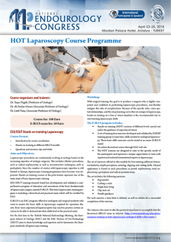
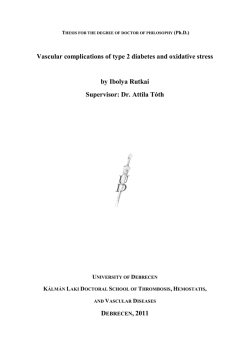
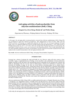
Ex-vivo tolerogenic F4/80+ antigen-presenting cells (APC) induce
efferent CD8+ regulatory T cell-dependent suppression of
experimental autoimmune uveitis
The Harvard community has made this article openly available.
Please share how this access benefits you. Your story matters.
Citation
Hsu, S-M, R Mathew, A W Taylor, and J Stein-Streilein. 2013.
“Ex-vivo tolerogenic F4/80+ antigen-presenting cells (APC)
induce efferent CD8+ regulatory T cell-dependent suppression
of experimental autoimmune uveitis.” Clinical and Experimental
Immunology 176 (1): 37-48. doi:10.1111/cei.12243.
http://dx.doi.org/10.1111/cei.12243.
Published Version
doi:10.1111/cei.12243
Accessed
February 6, 2015 10:57:27 AM EST
Citable Link
http://nrs.harvard.edu/urn-3:HUL.InstRepos:13890668
Terms of Use
This article was downloaded from Harvard University's DASH
repository, and is made available under the terms and conditions
applicable to Other Posted Material, as set forth at
http://nrs.harvard.edu/urn-3:HUL.InstRepos:dash.current.termsof-use#LAA
(Article begins on next page)
bs_bs_banner
Clinical and Experimental Immunology
O R I G I N A L A RT I C L E
doi:10.1111/cei.12243
Ex-vivo tolerogenic F4/80+ antigen-presenting cells (APC) induce
efferent CD8+ regulatory T cell-dependent suppression of
experimental autoimmune uveitis
S.-M. Hsu,*† R. Mathew,*
A. W. Taylor‡ and J. Stein-Streilein*
*Schepens Eye Research Institute, Department of
Ophthalmology, Harvard Medical School,
‡
Department of Ophthalmology, Boston
University School of Medicine, Boston, MA, USA,
and †Department of Ophthalmology, National
Cheng-Kung University Hospital, Tainan City,
Taiwan
Accepted for publication 19 November 2013
Correspondence: J. Stein-Streilein, Schepens Eye
Research Institute–Massachusetts Eye and Ear
Infirmary, Department of Ophthalmology,
Harvard Medical School, Boston, MA 02114,
USA.
E-mail: [email protected]
Summary
It is known that inoculation of antigen into the anterior chamber (a.c.) of a
mouse eye induces a.c.-associated immune deviation (ACAID), which is
mediated in part by antigen-specific local and peripheral tolerance to the
inciting antigen. ACAID can also be induced in vivo by intravenous (i.v.)
inoculation of ex-vivo-generated tolerogenic antigen-presenting cells
(TolAPC). The purpose of this study was to test if in-vitro-generated retinal
antigen-pulsed TolAPC suppressed established experimental autoimmune
uveitis (EAU). Retinal antigen-pulsed TolAPC were injected i.v. into mice 7
days post-induction of EAU. We observed that retinal antigen-pulsed
TolAPC suppressed the incidence and severity of the clinical expression of
EAU and reduced the expression of associated inflammatory cytokines.
Moreover, extract of whole retina efficiently replaced interphotoreceptor
retinoid-binding protein (IRBP) in the preparation of TolAPC used to
induce tolerance in EAU mice. Finally, the suppression of EAU could be
transferred to a new set of EAU mice with CD8+ but not with CD4+regulatory
T cells (Treg). Retinal antigen-pulsed TolAPC suppressed ongoing EAU by
inducing CD8+ Treg cells that, in turn, suppressed the effector activity of the
IRBP-specific T cells and altered the clinical symptoms of autoimmune
inflammation in the eye. The ability to use retinal extract for the antigen
raises the possibility that retinal extract could be used to produce autologous
TolAPC and then used as therapy in human uveitis.
Keywords: ACAID, autoimmunity, CD8+ Treg, EAU, tolerogenic APC
Introduction
The eye is the prototype for the study of immune-privileged
mechanisms. Because ocular antigens are not sequestered
from recognition by the immune system [1], multiple layers
of immune regulation exist, both locally and peripherally,
to preserve the visual axis. Ocular-induced regulation of
immune responses suppress inflammation and the adaptive
immune response, in part, by generating antigen-specific
regulatory T cells (Treg) that contribute to both local and
peripheral tolerance [2]. In addition, activated T cells specific for ocular antigens that are able to cross the structural
barriers of the eye meet multiple immunosuppressive
mechanisms to prevent them from finding their targets and
inducing inflammation within the eye [2–5].
In spite of all the overlapping immunoregulatory mechanisms that exist, uveitis occurs in approximately 0·2% of
the US population [6], with autoimmunity contributing to
approximately 50% of the aetiology. While 0·2% of the
population is not classified as an orphan disease, the
National Institute of Health (NIH) considers it a rare
disease. Autoimmune uveitis is a sight-threatening inflammatory disorder that affects all ages, and is a significant
cause of visual loss [7]. Each year 17·6% of active uveitis
patients experience a transient or permanent loss of vision,
and 12·5% will develop glaucoma [8]. Uveitis is also associated with several systemic diseases, including arthritis [9].
The medical community predominantly treats the clinical
symptoms of uveitis with corticosteroids, with increasing
prescriptions for biologicals such as anti-tumour necrosis
factor (TNF)-α compounds [10]. A more specific treatment and restoration of the immune homeostasis would be
a welcome treatment for uveitis.
Experimental autoimmune uveitis (EAU) is a disease of
the neural retina that is induced by immunization of
rodents (mice or rats) with retinal antigens. Generally, the
© 2013 The Authors. Clinical and Experimental Immunology published by John Wiley & Sons Ltd on behalf of British Society
37
for Immunology, Clinical and Experimental Immunology, 176: 37–48
This is an open access article under the terms of the Creative Commons Attribution-NonCommercial-NoDerivs License, which permits use
and distribution in any medium, provided the original work is properly cited, the use is non-commercial and no modifications or adaptations
are made.
S-M. Hsu et al.
antigen is introduced in concert with strong adjuvant and
pertussis toxin injections to overcome natural immune
resistance [11]. Because the use of strong adjuvants would
interfere with the induction of tolerance, we used a wellestablished adoptive transfer model of EAU, which induces
EAU by transferring interphotoreceptor retinoid-binding
protein (IRBP)-primed T cells to naive recipients in the
absence of adjuvants [7]. The purpose of this study was to
determine if restoration of aspects of immune privilege in a
mouse with EAU would interfere with and suppress the
progression of the uveitis. More specifically, we wanted to
know if we could induce suppression of the immune
response to the inciting retinal antigen by inducing tolerance by the transfer of ex-vivo-generated TolAPC to EAU
mice.
Materials and methods
Animals
C57BL/6J (B6) female (8–12 weeks old) mice were purchased from Jackson Laboratory (Bar Harbor, ME, USA)
and used for all experiments. All animals were treated
humanely and in accordance with the guidelines of the NIH
office of Laboratory Animal Welfare; the protocols were
approved by the Schepens Institutional Animal Care and
Use Committee. All experiments were conducted in accordance with the Association for Research in Vision and Ophthalmology (ARVO) Statement for the Use of Animals in
Ophthalmic and Vision Research.
Reagents
Cells were cultured in serum-free medium (SFM), consisting of RPMI-1640 (Lonza, Walkersville, MD, USA), 10 mM
HEPES, 0·1 mM non-essential amino acids, 1 mM sodium
pyruvate, 100 U/ml penicillin and 100 μg/ml streptomycin
(all purchased from Life Technologies Gaithersburg, MD,
USA). Transforming growth factor (TGF)-β2 and mouse T
cell enrichment columns were purchased from R&D
Systems (Minneapolis, MN, USA). Peptide IRBP1-20
(GPTHLFQPSLVLDMAKVLLD), representing residues
1–20 of human IRBP, was synthesized by Invitrogen
(Carlsbad, CA, USA). Myelin basic protein (MBP), pertussis
toxin (PTX) and incomplete Freund’s adjuvant (IFA) were
purchased from Sigma Life Sciences (St Louis, MO, USA).
Heat-inactivated desiccated Mycobacterium tuberculosis
(H37 RA) was purchased from Difco laboratories (Detroit,
MI, USA). Tropicacyl® (tropicamide ophthalmic solution
1%) and phenylephrine hydrochloride ophthalmic solutions 2·5% were both purchased from Akorn Inc. (Lake
Forest, IL, USA). Monoclonal antibodies for flow cytometry
such as Fc block (anti-2.4G2), forkhead box protein 3
(FoxP3) fluorescein isothiocyanate (FITC) (clone-FJK-16s),
CD8 phycoerythrin (PE) (clone LY3), CD4 PE (clone GK138
5), anti CD253 TNF-related apoptosis-inducing ligand
(TRAIL) PE (clone N2B2), F4/80 FITC (clone BM8), CD40
PE (clone IC10), as well as mouse interferon (IFN)-γ and
mouse interleukin (IL)-17A enzyme-linked immunosorbent assay (ELISA) kits were purchased from eBioscience Inc.
(San Diego, CA, USA). Mouse CD8+ and CD4+ T cell isolation kits were purchased from Miltenyi Biotech (Auburn,
CA, USA).
Flow cytometry and cell sorting
Splenic cells that were analysed by flow cytometry were
stained in the presence of a saturated concentration of Fc
block (blocks FcRγ II/III). Cells (1 × 10⁶) were stained with
the monoclonal antibodies using concentrations recommended by the manufacturer. Stained cells were analysed on
a BD LSRII Flow analyser (BD Biosciences, San Diego, CA,
USA). For sorting TRAIL+ and TRAIL– populations,
enriched CD8+ T cells were passed through a MoFlo Cell
Sorter (Cytomation, Inc., Fort Collins, CO, USA).
Induction of EAU
EAU was induced by modification of methods reported [7].
Briefly, donor B6 mice were immunized subcutaneously
(s.c.) with 100 μl of an emulsion (1:1) of phosphatebuffered saline (PBS) and IFA containing 200 μg of IRBP1–20
and 500 μg of M. tuberculosis H37RA (Difco Laboratories).
A single dose of PTX (200 ng) was injected intraperitoneally (i.p.) on the same day. The lymphocytes from
draining lymph nodes and spleens of the immunized donor
mice were collected on day 12 and activated in culture with
30 μg/ml of IRBP1–20 for 48 h, after which the non-adherent
cells were collected, washed and injected [5 × 10⁶ cells/
0·1 ml PBS/intravenously (i.v.)] into recipient B6 mice to
induce EAU.
Scoring of EAU
The ocular fundus of the mouse eyes was examined by slit
lamp two times a week for clinical signs of EAU. Pupils were
dilated using Tropicacyl® and phenylephrine hydrochloride
ophthalmic solutions. The severity of inflammation was
clinically graded on a scale of 1–5, as described previously
[12,13]. In brief, a grade of 1 or less was considered as a
negative score. Briefly, 0 = no inflammation; 1 = focal vasculitis ≤5 spots or soft exudates ≤5; 2 = linear vasculitis or
spotted exudates ≤50% of the retina; 3 = linear vasculitis or
spotted exudates ≥50% of the retina; 4 = retinal haemorrhage or severe exudates and vasculitis; and 5 = exudative
retinal detachment or subretinal (or vitreous) haemorrhage.
A mouse was considered to have uveitis if at least one of its
eyes had a score of above 1 or more. The severity of uveitis
is represented as the highest clinical score achieved by either
eye in a mouse over the 25 days of the clinical disease. The
© 2013 The Authors. Clinical and Experimental Immunology published by John Wiley & Sons Ltd on behalf of British Society
for Immunology, Clinical and Experimental Immunology, 176: 37–48
Tolerogenic APC suppress EAU
(a)
(b)
10
Double isotype
1·21
0·51
103
102
102
1·02
100 97·3
100 101 102 103 104
APC no treatment
200
38·5
101
58·7
100 2·28
100 101 102 103 104
150
100
50
0
100 101 102 103
F4/80 FITC
4
10
APC treatment with
IRBP alone
0·81
46
10
4
103
103
102
102
101
50·8
100 2·42
100 101 102 103 104
clinical symptoms of EAU post-transfer of IRBP immune
cells are less severe than the clinical symptoms of EAU
induced by traditional immunization (includes CFA and
pertussis toxin).
Histopathological evaluation
Whole eyes were collected at the peak of the clinical
response (between 21–23 days after induction of EAU by
adoptive transfer of IRBP immune cells), immersed in 10%
formaldehyde and stored until processed. Fixed and dehydrated tissues were embedded in methacrylate, and 5-μm
sections were cut through the papillary–optic nerve plane
and stained with haematoxylin and eosin (H&E). The presence or absence of disease was evaluated in a blinded
fashion by examining six sections cut at different levels for
each eye.
Preparation of TolAPC
TolAPC were prepared by a modification of methods
reported [14–17]. Briefly, thioglycolate-elicited PEC was
cultured overnight in SFM with TGF-β (5 ng/ml) and
antigen [IRBP1–20 (50 μg/ml), retinal extract (100 μg/ml),
corneal extract (100 μg/ml) or MBP (100 μg/ml)]. After
incubation, the culture media was replaced with cold (4°C)
PBS for 10 min, and the APC were removed by gently scraping the Petri dish with a rubber policeman. To verify that
TolAPC were generated, the APC were analysed by flow
cytometry for expression of CD40 and F4/80. CD40, a
co-stimulatory molecule for immune activation, was down-
APC treatment with
IRBP + TGF-β
0·078
101
120
2·45
# of cells
CD40 PE
10
103
101
Fig. 1. Flow analysis of CD40 expression. (a)
Antigen-presenting cells (APC) were treated
with transforming growth factor (TGF)-β2 and
interphotoreceptor retinoid-binding protein
(IRBP) overnight to produce tolerogenic
antigen-presenting cells (TolAPC). F4/80 is
plotted on the abscissa and CD40 on the
ordinate. (b) Upper histograph of APC stained
with F4/80 after various treatments. Lower
histograph of APC gated for F4/80 fluorescein
isothiocyanate (FITC)-positive cells and
analysed for CD40 phycoerythrin (PE). TolAPC
(black); APC without treatment (blue); APC
pulsed with IRBP (red). Shaded graph
represents the isotype control. Representative of
two experiments.
0·47
4
# of cells
4
92·3
5·22
100
100 101 102 103 104
90
60
30
0
100 101 102 103
F4/80 FITC
CD40PE
regulated but F4/80, a surface marker associated with anterior chamber (a.c.)-associated immune deviation (ACAID)
TolAPC [18], was increased (Fig. 1). Recovered APC were
suspended in PBS (107 cells/ml). Each recipient mouse was
inoculated (i.v.) with 100 μl of cell suspension (106 cells) 7
days after induction of EAU.
Preparation of T cells from spleens for treatment of
EAU mice
Because, in most experimental animal groups, the EAU
peak clinical response subsided by 24 days, we collected cells
between 21 and 23 days post-initiation of EAU. Spleens
were dissociated individually into single-cell suspensions
and labelled as (i) EAU untreated or (ii) EAU-treated. Dissociated spleen cells were passed through T cell enrichment
columns (R&D Systems). The T cell samples (5 × 10⁶/100 μl
PBS) were separated further into CD8- or CD4-positive
populations, using magnetic isolation kits on fluorescenceactivated cell sorting before being injected into new EAU
mice. Cells from our donor mouse were injected (i.v.) into
one recipient EAU mouse.
In-vitro correlate for ACAID
It has been shown previously that induction of ACAID by
antigen inoculation into the a.c. could be bypassed by
injecting TolAPC i.v [16]. Furthermore, it is known that
in-vitro ACAID cultures where the TolAPC is co-cultured
(5–7 days) with spleen cells will generate both CD4+ and
CD8+ Treg cells. Thus, TolAPC generates Treg cells in vitro and
© 2013 The Authors. Clinical and Experimental Immunology published by John Wiley & Sons Ltd on behalf of British Society
for Immunology, Clinical and Experimental Immunology, 176: 37–48
39
S-M. Hsu et al.
in vivo [19]. To generate sufficient numbers of the Treg cells
we used in-vitro cultures [16]. IRBP-pulsed TolAPC were
incubated with non-adherent spleen cells for 7 days. The
non-adherent cells were harvested and enriched for CD8+ T
cells using the magnetic affinity cell sorting (MACS®) cell
separation system following the manufacturer’s instructions
(Miltenyi Biotech). The purity of the separated cells was
checked using flow cytometry PE-conjugated CD8 monoclonal antibody.
Assay of in-vitro T helper type 1 (Th1) and
Th17-related cytokine production
Spleens were removed from each group of TolAPC or
untreated APC-treated mice 22 days after induction of EAU.
Cells were seeded in flat-bottomed, 12-well tissue culture
plates at a density of 6 × 10⁶ cells per well in 2 ml of the
culture medium RPMI-1640 supplemented with 10% fetal
calf serum, 2-mM of L-glutamine, 1-mM of sodium
pyruvate and antibiotics in the presence of hIRBP1–20
peptide (50 μg/ml) and then cultured for 48 h. To assess
cytokine production, cell-free supernatants were collected
at 48 h and assayed for IFN-γ and IL-17 by using a mouse
IFN-γ and IL-17 ELISA (Ready-SET-Go! Kit; eBioscience,
Inc.).
Local adoptive transfer (LAT) assay [20]
The LAT assay is commonly used to test the efferent suppressor activity of a population of cells [21]. The concept is
to inject (intradermally) a mixture of immune cells with
antigen into the ear pinnae of a naive mouse. The mouse
serves as a test tube for the immune response; the swelling
of the ear is evidence of a delayed hypersensitivity response
induced by the injected immune cells and antigen. CD8+ Treg
cells (5 × 10⁵ cells) were mixed with spleen cells (5 × 10⁵
cells) from mice immunized with IRBP and CFA 7 days previously and antigen was injected (10 μl/injection) into the
ear pinnae of B6 mice. The ear thickness was measured with
an engineer’s micrometer before and compared to ear
thickness 24 h after injection.
Preparation of retinal extract
The eyes were enucleated from euthanized mice. The eyeballs were cut at the equator around the ora serrata, and the
posterior pole of the eyes was separated from the anterior
pole and lens. The retina, consisting of the neural retina and
the retinal pigment epithelial cells, was extracted from the
posterior pole. The extract from one retina was placed in
500 μ of RPMI on ice (1 min) and sonicated briefly three
times for 7 s at a probe intensity of 7 (MicrosonTM XL2000
Ultrasonic liquid processor; Qsonica, LLC, Newton, CT,
USA). After removal of the insoluble material by centrifugation (200 g for 5 min), the protein concentration of the
40
retinal extract was measured at 280 nm on an ND-1000
spectrophotometer, and adjusted to approximately 4 mg/ml.
The retinal extract (100 μg/ml) was used as antigen to pulse
the TolAPC.
Preparation of corneal extract
A 1·5-mm full-thickness cornea button was trephined
(under the hydration of PBS) from euthanized mouse
tissue, and sonicated in RPMI (100 μl) on ice to prepare a
homogeneous solution. The protein concentration of the
corneal extract from one corneal button was approximately
2 mg/ml. A dilution of the corneal extract (100 μg/ml) was
used to pulse the TolAPC.
Statistical analysis
All statistical analyses were performed using PRISMTM software. Statistical differences in the incidence of uveitis and
peak clinical scores between controls versus experimental
groups were determined by non-parametric Mann–
Whitney U-tests. In some experiments statistical differences
between the course of the EAU (area under the curve)
between groups were also compared using non-parametric
Mann–Whitney U-tests. Statistical differences between
cytokine production in control and treated EAU in ELISA
assays were determined using a one-tailed Student’s t-test.
Differences were considered significant at P ≤ 0·05.
Results
IRBP-pulsed TolAPC suppress pre-existing EAU
TolAPC were generated by culturing thioglycolate-elicited
peritoneal exudate cells (PEC) in serum-free medium
(SFM) in the presence of IRBP1–20 and TGF-β2. Untreated
APC were cultured in SFM only, without IRBP and TGF-β2.
The TolAPC or untreated APC were injected (i.v. 106 cells/
mouse) into EAU mice. EAU was induced by IRBPsensitized cells that were transferred adoptively, as described
in Materials and methods. Mice with EAU that were treated
with TolAPC exhibited a delay in disease onset and a significantly (P ≤ 0·001) lower peak EAU score over time
(Fig. 2a,b). The mice that received untreated APC treatment
had a mean clinical severity score of 2 ± 0·26, while the
mice that received TolAPC treatment had a mean clinical
severity score of 0·64 ± 0·13 (P ≤ 0·05) (Table 1). In addition, examination of haematoxylin and eosin (H&E)stained paraffin-fixed slides revealed that retinal sections of
eyes from EAU mice that received TolAPC showed a
reduced cell infiltration into the vitreous cavity and their
retinal layer structures lacked the retinal folds and vascular
swelling observed in the untreated mice (Fig. 2c,d). The fact
that the mice given TolAPC had peak clinical scores of 1 or
lower (mild or no uveitis) supported the postulate that the
© 2013 The Authors. Clinical and Experimental Immunology published by John Wiley & Sons Ltd on behalf of British Society
for Immunology, Clinical and Experimental Immunology, 176: 37–48
Tolerogenic APC suppress EAU
(b)
*
Untreated APC
Tolerogenic APC
3
4
Max EAU scores
Mean clinical scores
(a)
2
1
* *
0
0
* *
*
*
3
2
1
0
5
10
Tol APC
15
20
25
Day 21
Days
(c)
(d)
Untreated APC
Tolerogenic APC
*
100×
100×
GCL
INL
**
ONL
RPE
***
(e)
IL-17
*
25 000
400
0
pg/ml
pg/ml
100
*
20 000
300
200
IFN-γ
(f)
15 000
10 000
5 000
0
Fig. 2. Effect of tolerogenic antigen-presenting cells (TolAPC) on clinical course of experimental autoimmune uveitis (EAU). (a) Average clinical
score over time of EAU in mice with and without TolAPC treatment. EAU was induced by adoptive transfer of interphotoreceptor retinoid-binding
protein (IRBP)-sensitized enriched T cells into C57BL/6 mice. A week later, TolAPC (red n = 14) or untreated antigen-presenting cells (APC) (black
n = 14) were injected intravenously (i.v., 106 cells/mouse) into EAU mice. Data shown are the mean clinical score (ordinate) of each experiment
group over time (abscissa), and are the sum of two independent experiments. Comparison of (the course of the clinical symptoms) untreated EAU
mice versus TolAPC-treated mice shows a significant difference (P ≤ 0·05) and is indicated. (b) Scatterplot shows peak scores on day 21 of individual
EAU mice were given TolAPC (red) or not (black). The peak clinical scores over time of TolAPC-treated EAU mice are significantly lower than the
peak scores over time of the untreated EAU mice. *Indicates a significant difference (P ≤ 0·05). (c,d) Photomicrographs of haematoxylin and eosin
(H&E)-stained retinal tissue. Representative photomicrographs paraffin-fixed H&E stained slides of the retina of (c) EAU mice that received
untreated APC (*leucocytes in vitreous cavity; **swelling; ***retinal fold) and (d) EAU mice that received TolAPC days post-initiation of EAU.
Retinal pigment epithelium (RPE), outer nuclear layer (ONL), inner nuclear layer (INL), ganglion cell layer (GCL). (e,f) Enzyme-linked
immunosorbent assay (ELISA) analysis of inflammatory cytokines in spleen cells harvested at day 23 of EAU mice that received either TolAPC or
spleen cells were restimulated with antigen with serum-free media for 48 h prior to collecting the supernatants for analyses. Bar graphs showing
(e) interleukin (IL)-17 and (f) interferon (IFN)-γ production. Spleen cells (from three separate mice) were harvested from APC- (solid bar) or
TolAPC-treated EAU mice (three per group) co-cultured with antigen (IRBP 50 ug/ml) (open bar). Supernatants from duplicate cultures were
harvested 48 h after restimulation with IRBP for ELISA analysis. An asterisk (*) indicates a significant difference (P ≤ 0·05).
© 2013 The Authors. Clinical and Experimental Immunology published by John Wiley & Sons Ltd on behalf of British Society
for Immunology, Clinical and Experimental Immunology, 176: 37–48
41
S-M. Hsu et al.
Untreated APC
Tolerogenic APC
Incidence of mice with
clinical score ≥2
Peak disease
score ± s.e.m.
9/14
0/14*
2·0 ± 0·026
0·64 ± 0·13*
Scores are from the experimental and control mice used for Fig. 2.
*Indicates a significant difference (P ≤ 0·05) between untreated and
TolAPC-treated mice. EAU: experimental autoimmune uveitis; s.e.m.:
standard error of the mean.
TolAPC induced suppression of the clinical symptoms of
EAU. Thus, treatment of EAU mice with IRBP-pulsed
TolAPC a week after induction of the EAU delayed its onset
and suppressed the subsequent development and severity of
EAU (Table 1).
TolAPC treatment down-regulates the production of
EAU-associated inflammatory cytokines
To examine the effect of TolAPC treatment on the production of inflammatory cytokines, IFN-γ and IL-17 [11] we
collected non-adherent cells from the spleens of EAU mice
that received TolAPC or untreated APC. The spleen cells
were restimulated with IRBP antigen for 48 h in vitro and
assayed for the production of IFN-γ and IL-17. Spleen cells
derived from TolAPC-treated EAU mice produced significantly (P ≤ 0·05) less IFN-γ (Th1) and IL-17 (Th17) than
EAU mice receiving untreated APC (Fig. 2e,f). Thus,
TolAPC, but not APC, treatment resulted in suppression of
the inflammatory cytokine response in EAU mice.
3
2
*
1
0
0
5 10 15 20 25 30
Days
TolAPC
Mean clinical
scores ± s.e.m.
Treatment of
EAU mice
(b)
(a)
Mean clinical
scores ± s.e.m.
Table 1. Incidence of uveitis and peak disease score of mice after
untreated antigen-presenting cells (APC) versus tolerogenic antigenpresenting cell (TolAPC) treatment.
3
2
*
1
0
0
5 10 15 20 25 30
Days
Fig. 3. Effect of retinal antigen-pulsed tolerogenic antigen-presenting
cells (TolAPC) on clinical course of experimental autoimmune uveitis
(EAU) induced with interphotoreceptor retinoid-binding protein
(IRBP). (a) Comparison of clinical scores of EAU mice treated with
retinal extract-pulsed TolAPC (filled triangles, n = 7) or EAU mice not
treated (open squares, n = 7). Data are shown as mean clinical EAU
score ± standard error of the mean (s.e.m.) (ordinate) over time
(abscissa). The retinal extract-pulsed TolAPC-treated mice show a
significant (P ≤ 0·05) decrease in EAU clinical score over time
compared to scores of EAU in untreated mice. (b) Antigen specificity
of TolAPC-induced suppression. The EAU mice were treated with each
type of TolAPC, 7 days post-induction of EAU (Materials and
methods). Line graph of response of EAU mice to TolAPC pulsed with
indicated antigens. The transferred antigen-presenting cells (APC)
were not pulsed with antigen (black line, n = 15) or were treated with
transforming growth factor (TGF)-β and pulsed with corneal extract
(red line, n = 7), myelin basic protein (MBP) (blue line, n = 6),
IRBP(1–20) (grey line, n = 14) or retinal extract (green line, n = 16).
Data shown are mean clinical score (ordinate) over time (abscissa). An
asterisk (*) indicates a significant difference between the areas under
the curves. Statistics were performed using Prism software (Materials
and methods).
establish suppression of EAU (Fig. 3b). Therefore, it is possible to produce EAU-specific TolAPC when the TolAPC are
pulsed with retina extract.
Ability of retinal extract- versus IRBP-pulsed TolAPC
to suppress EAU
TolAPC induce Treg cells in the spleens in host
EAU mice
Although the retinal antigens that induce the EAU in the
mice are known, the target antigens in human uveitis
remain obscure. Here, we tested if retinal protein extract
(containing IRBP and other retinal antigens) would provide
the relevant antigens for producing the TolAPC that were
effective in this model of suppression. In this experiment
EAU was induced by injecting IRBP-specific cells as before,
but the TolAPC were made by incubation with TGF-β and
IRBP or mouse retinal extract. The retinal antigen-pulsed
TolAPC were then injected (i.v.) into the EAU mice, 7 days
post-induction with the IRBP-specific cells. As before, mice
were monitored and clinical symptoms were scored every
3–30 days. We observed that the retinal extract (but not
corneal extract, control)-pulsed TolAPC were as effective as
IRBP1–20-pulsed TolAPC in reducing the clinical symptoms
of EAU (Fig. 3a). Furthermore, if the TolAPC were pulsed
with the irrelevant antigen MBP, they were not able to
Having shown that TolAPC treatment suppressed the production of inflammatory cytokines and reduced the clinical
symptoms of EAU, we next analysed the cellular mechanisms that might be responsible for the suppression. It is
known that tolerance induced by antigens a.c.-inoculated or
by TolAPC-inoculated i.v. induce Treg cells that can transfer
tolerance. To test for the presence of Treg cells, T cells were
enriched from spleens harvested (day 21) from individual
EAU mice treated with TolAPC, restimulated with IRBP in
vitro for 48 h prior to transferring (5 × 10⁶, 100 μl) to
syngeneic recipients in which EAU had been induced 7 days
previously. Control groups of mice received either
restimulated enriched T cells from EAU mice that were
injected with untreated APC or were not injected with APC.
EAU mice receiving T cells from mice treated with TolAPC
exhibited a delayed onset of their EAU symptoms with less
severity compared with the EAU mice that received cells
42
© 2013 The Authors. Clinical and Experimental Immunology published by John Wiley & Sons Ltd on behalf of British Society
for Immunology, Clinical and Experimental Immunology, 176: 37–48
Tolerogenic APC suppress EAU
from the control groups of mice (P ≤ 0·05) (Fig. 4a), suggesting that the suppression of the autoimmune inflammation was mediated by Treg cells induced by the administered
TolAPC.
CD8+, but not CD4+, Treg cells transfer suppression to
EAU mice
Previous reports showed that either a.c. injection of antigen
or i.v. transfer of in-vitro-generated TolAPC induced two
types of Treg cells: afferent CD4+ Treg and efferent CD8+ Treg
cells. Both the CD4+ and the CD8+ Treg cells have been characterized [22,23]. To determine which type of Treg cells
transferred the tolerance to EAU mice, the experiments
were repeated. Spleen cells were dissociated from spleens
harvested from EAU mice at 21 days and restimulated in
vitro with IRBP. The spleen cells were collected from two
groups of mice as follows: (i) individual EAU mice that
received untreated APC (Fig. 4b) and (ii) individual EAU
mice that received TolAPC (Fig. 4c). The spleen cells from
each mouse were restimulated in vitro in separate cultures
with IRBP (48 h), after which CD4+ T cell and CD8+ T cell
populations were enriched by magnetic bead separation. We
observed that transfer of efferent CD8+, but not the afferent
CD4+ T cells, suppressed the clinical symptoms of EAU
(Fig. 4a–c).
Characterization of the Treg cell
Both CD4+ and CD8+ Treg cells that are generated after culturing spleen cells with TolAPC have been characterized by
Keino and colleagues [22,23]. We analysed if the harvested
CD8+ T cells expressed TRAIL. In brief, spleen cells were
harvested from experimental mice that received TolAPC
and had EAU scores of 1 or less. The dissociated spleen cells
were restimulated with antigen for 48 h. Post-culturing, the
non-adherent cells were collected, enriched by magnetic
bead separation for CD8+ T cells, and an aliquot of the
enriched CD8+ T cells was examined prior to transfer by
flow cytometry for expression of TRAIL. Interestingly, the
Treg cells collected from TolAPC-treated EAU mice that
transferred tolerance to a second set of EAU immunized
mice were CD8+FoxP3+TRAIL– (Fig. 4d). Analysis of the
mean fluorescence intensity (MFI) of the CD8+ T cell populations analysed by flow cytometry showed little or no shift
in TRAIL staining (Fig. 4e).
Local adoptive transfer assay
Thus far, we show that in-vitro-generated TolAPC transferred to EAU mice suppress the clinical symptoms of EAU
by generating CD8+TRAIL– Treg cells. To evaluate further the
expression TRAIL on CD8+ Treg cells, we generated CD8+ Treg
cells in vitro [24]. The suppressor function of the in-vitrogenerated Treg cells was evaluated in a local adoptive transfer
assay (Materials and methods). Others have reported that
Treg cells are generated in cultures where TolAPC have
similar characteristics to those generated in vivo [19,24–26].
After co-culturing F4/80+ TolAPC with spleen cells for 7
days, the non-adherent cells were harvested and the CD8+ T
cells sorted into TRAIL-negative and -positive populations
(Fig. 5a). The CD8+TRAIL– T cells suppressed the response
to IRBP in a LAT assay (Fig. 5b) (there were insufficient
CD8+TRAIL+ cells to test their function). Flow analysis of
the TRAIL–CD8+ Treg cells confirmed that CD8+ Treg cells
expressed CD103 (data not shown) [22]. Thus, 7-day cultures of TolAPC with spleen cells generate CD8+CD103+
FoxP3+TRAIL– Treg cells that are capable of suppressing
efferent immune responses in vivo.
Discussion
It is reasonable to think that if autoimmunity occurs in the
eye, one or more mechanisms of immune privilege,
immune regulation, must be compromised. ACAID is a
model used to study ocular immune privilege in vivo and in
vitro that generates peripheral antigen-specific Treg cells
[2,17,27,28]. As early as 1992, Streilein and colleagues
reported that ACAID induction by intracameral inoculation
of the immunizing ocular autoantigen prior to the induction of uveitis reduced the incidence of EAU in mice [29].
No ocular inflammation was observed in the group that
received the retinal antigen via the a.c. prior to the induction of EAU, while 80% of mice that were injected a.c. with
PBS developed uveitis. Here, we extended these studies
and induced tolerance by transferring in-vitro-generated
TolAPC to experimental mice after EAU was induced. APC
become tolerogenic after exposure to immunosuppressive
factors such as TGF-β2 and antigen in vitro, and have been
shown to promote negative regulation of both Th1- and
Th2-mediated inflammation in part by generating antigenspecific Treg cells [25,27,30].
The transfer of antigen-pulsed TolAPC has been shown
to induce tolerance in both naive and sensitized mice
[15,31]. Furthermore, we have reported previously that the
adoptive transfer of TolAPC successfully abrogated immune
inflammation and clinical symptoms in mouse models for
autoimmune pulmonary interstitial fibrosis [25], airway
hypersensitivity hyper-reactivity [26] and experimental
autoimmune encephalomyelitis (EAE) [32]. Here, we show
that TolAPC pulsed with retinal antigen (IRBP or extract of
retina) are capable of reducing the clinical symptoms and
inflammatory cytokines in an adoptive transfer model of
IRBP-induced EAU. Thus, depending on the frequency of
the unknown target antigen in a retinal extract, this
approach may provide a basis for a novel cell-based therapy
using autologous cells for the treatment of ocular autoimmune diseases such as uveitis.
The TolAPC associated with eye-induced tolerance were
first defined by their surface expression of F4/80 protein
© 2013 The Authors. Clinical and Experimental Immunology published by John Wiley & Sons Ltd on behalf of British Society
for Immunology, Clinical and Experimental Immunology, 176: 37–48
43
S-M. Hsu et al.
3
2
1
0
0
*
10
20
Days
(d)
30
1
0
0
10
CD8+ T Cells: no staining
104
FoxP3 FITC
*
2
0·059
0
20 30
Days
(c) Enriched CD4 and CD8 T
cell from EAU mice
3 receiving Tol APC
Mean EAU
scores ± s.e.m.
(b) Enriched CD4, CD8 T cell
from EAU mice receiving
3 untreated APC
Whole T cells
Mean EAU
scores ± s.e.m.
Mean clinical
scores ± s.e.m.
(a)
40
2
1
0
0
10
20 30
Days
40
CD8+ T cells: TRAIL staining
104
0·15
0
103
103
102
102
101
101
0
99·9
100
100 101 102 103 104
0
99·9
100 0
10 101 102 103 104
CD8+ T cells: FoxP3 Staining CD8+ T Cells: Tol APC-ADT EAU
104
0·079
104
0
6·36
0
103
103
102
102
101
101
99·9
0
100 0
10 101 102 103 104
93·6
0
100 0
10 101 102 103 104
TRAIL PE
(e)
MFI
188
131
400
300
200
100
0 0
10
101
102
103
TRAIL PE
+
104
MFI
247
99·4
500
Cell number
Cell number
500
400
300
200
100
0
100
101
102
103
104
FoxP3 FITC
+
Fig. 4. The regulatory effects of CD4 or CD8 T cells from tolerogenic antigen-presenting cells (TolAPC)-treated experimental autoimmune uveitis
(EAU) mice. The line graph represents progression of mean clinical scores of EAU over time. The ordinate = mean EAU clinical scores;
abscissa = days post-induction of EAU. Whole T cells, CD4+ or CD8+ T cells were collected from the spleens of mice that were either TolAPC or
antigen-presenting cells (APC). Enriched T cells were collected at the peak of clinical symptoms and transferred to a new group of EAU mice to test
for their suppressor function. (a) Clinical course of EAU in mice receiving no T cells (n = 14, black line); clinical course of EAU in mice receiving T
cells harvested from the EAU mice were injected with APC (n = 4, orange line); clinical course of EAU in mice receiving T cells harvested from the
EAU mice that were injected with TolAPC (n = 4, blue line). (b) Effects on EAU of mice receiving enriched T cells (CD8+ T red line; CD4+ T green
line) harvested from the EAU mice were injected with APC. (c) Effects on EAU of mice receiving enriched T cells (CD8+ T red line; CD4+ T green
line) from EAU mice that were injected with TolAPC (n = 7 per group). Each recipient mouse received one donor equivalent. *Significant difference
(P ≤ 0·05) in overall severity of the disease over time between two indicated experimental groups. (d) Representative flow cytometry analysis of
CD8+ T cells handled in the same manner as the CD8+ T cells in (c). Upper left block shows resting CD8+ T cells with no staining; upper right block
are resting CD8+ T cells stained with tumour necrosis factor (TNF)-related apoptosis-inducing ligand (TRAIL) antibody only; lower left block shows
resting CD8+ T cells stained with forkhead box protein 3 (FoxP3) antibody only; and lower right shows the CD8+ T cells harvested from the
TolAPC-treated mice used in (c) (red line) stained from FoxP3 FITC (ordinate) and TRAIL phycoerythrin (PE) (abscissa) (e) Mean fluorescence
intensity (MFI) analyses of CD8+ T cells from TolAPC-treated EAU mice (solid line) compared to staining on CD8+ T cells from EAU mice. The
abscissa represents the intensity of the fluorescence for the indicated antibody; the ordinate shows the cell number; insert box gives the actual
reading for the MFI. Dashed line (—) APC-treated EAU CD8+ T cells; solid line (—) TolAPC-treated EAU CD8+ T cells. The left block shows that
there is a little or no increase in fluorescence intensity staining for TRAIL in the two types of CD8+ T cells compared. The right block shows a major
shift in staining intensity for the MFI of FoxP3 staining and CD8+ T cells from the TolAPC-treated EAU mice.
44
© 2013 The Authors. Clinical and Experimental Immunology published by John Wiley & Sons Ltd on behalf of British Society
for Immunology, Clinical and Experimental Immunology, 176: 37–48
Tolerogenic APC suppress EAU
(a)
No· of cells
200
150
100
69·3
50
0
100 101 102 103 104
(b)
No staining
TRAIL single staining
104
104
FoxP3 FITC
3
3
10
10
102 0·77 0·14
102
101
101
0·41
0·79 0·42
1·77
100
100 101 102 103 104
100
100 101 102 103 104
FoxP3 single staining
104
FoxP3 TRAIL double
104
103
103
102
101
5·99
0·5
0·52
102
101
100
100 101 102 103 104
Δear thickness
in μm
CD8 PE
*
*
150
100
Splenocytes from
non-immunized
mice
Splenocytes from
immunized mice
TRAIL negative
CD8 cells
(n)
50
0
+
–
–
–
+
+
–
–
+
4
4
4
6·4 0·94
0·98
100
100 101 102 103 104
TRAIL PE
Fig. 5. Effect of CD8+ tumour necrosis factor (TNF)-related apoptosis-inducing ligand (TRAIL)– cells on suppression. (a) Flow cytometry gate
used for analysis. Spleen cells were cultured with antigen-presenting cells (APC) treated with transforming growth factor (TGF)-β2 and
interphotoreceptor retinoid-binding protein (IRBP). Seven days later the CD8+ T cells were enriched by magnetic beads and single-stained for CD8
(top panel). Lower panel: an aliquot of enriched CD8+ T cells was stained with TRAIL phycoerythrin (PE) (abscissa) and forkhead box protein 3
(FoxP3) (fluorescein isothiocyanate (FITC) (ordinate). (b) Local adoptive transfer assay using CD8+TRAIL– T cells. The experiment was performed
twice with similar results. *Significant difference (P ≤ 0·05).
and were therefore thought to be macrophages [14,33–37].
However, once F4/80+ macrophages are mobile their
appearance becomes dendritic [33]. Also, although F4/80
is expressed predominantly by macrophages, F4/80 is
expressed by a subset of dendritic cells (DC) that is
tolerogenic [38,39]. Importantly, F4/80 protein expression
is necessary for the tolerogenic function of TolAPC [40].
Although F4/80 may not always distinguish macrophages
from other APC, the inability to induce ACAID in op/op
mice, a B6 mouse with a spontaneous mutation in the csf
gene region resulting in a deficiency in some macrophage
but not DC populations, supports the notion that the
F4/80+ TolAPC are macrophages [41].
A variety of DC has been shown to be tolerogenic in
experimental models [42–44]. While the majority opinion
is that immature DC are tolerogenic [44–47], the use of
immature DC for therapeutic reasons has limited potential
because of the possibility that they could mature once
exposed to the immune state of the recipient [48]. Other
investigators contend that tolerogenic DC are semi-mature
[49]; other reports show that a subtype of DC
(plasmacytoid dendritic cells) has tolerogenic capabilities
[50,51]. Thus, it becomes clear that several types of APC
have the potential of becoming tolerogenic.
Our laboratory has been successful in generating TolAPC
from thioglycolate-induced PEC and bone marrow-derived
macrophage/DC [26] as well as macrophage hybridoma no.
59 [52,53]. We have also made TolAPC from enriched
human DC isolated from human peripheral blood lymphocyte samples (unpublished data). Thus, our experience supports the idea that multiple types of APC have the potential
of maturing into TolAPC if the critical components of
TGF-β and antigen activation are present together. APC
activated by antigen in the presence of TGF-β progress
through distinct regulatory pathways, and subsequently
express distinct regulatory markers. Therefore, we propose
that immature macrophages/DC have the option to mature
through multiple pathways into immune-activating or
immune-regulating cells.
CD8+ Treg cells were first identified in ACAID induction
in the 1990s [54]. The CD8+ Treg cells induced by antigen
injection into the eye express CD103 and have a novel
genetic pattern associated with their efferent suppressor
function [22]. ACAID-induced CD8+ Treg cells can suppress
© 2013 The Authors. Clinical and Experimental Immunology published by John Wiley & Sons Ltd on behalf of British Society
for Immunology, Clinical and Experimental Immunology, 176: 37–48
45
S-M. Hsu et al.
by secreting TGF-β2 [55]. One report suggests that a population of CD8+TRAIL+ Treg cells develop in an extra-ocular
environment post-antigen inoculation to the a.c. and
mediate suppression [56]. In this paper, we analysed the
CD8+ T cell population from the spleens of the TolAPCtreated and control-treated EAU mice for expressing
TRAIL. We observed that CD8+ T cells harvested from
TolAPC-treated EAU mice were able to transfer tolerance,
but they were negative for TRAIL. Thus our data suggest
that TolAPC injected into mice with EAU generate
CD8+FoxP3+TRAIL– Treg cells can mediate and transfer efferent suppression of EAU. Apropos this observation are
reports that support the idea that the CD8+ Treg cells suppress by multiple mechanisms [57,58] and that the mechanisms are strain-dependent [59]. Reported studies show
that C57BL/6 CD8+ Treg cells express less TRAIL than
BALB/c CD8+ Treg. Moreover, C57BL/6 CD8+ Treg suppression is IL-35-, IL-10-dependent and BALB/c CD8+ Treg suppression is TRAIL-dependent.
Consistent with the idea that CD8+ Treg cells from different mouse strains use different methods to suppress is the
posit that CD8+ Treg suppressive mechanisms may vary with
the substrain of mouse used. The C57BL/6 mouse is the
most well-known inbred mouse strain and provides the
genetic background for congenic and mutant mice. There
are also a number of substrains derived from the founder
B6 strain. The fact that the substrains express genetic and
phenotypic variances is not always acknowledged in
research papers. For instance, genotyping demonstrated
genetic differences in the C57BL/6J and the C57BL/6N
substrains at 11 single nucleotide polymorphism (SNP) loci
[60]. The SNP pattern for the C57BL/6 mouse from NCI
(the C57BL/6 CrSlc substrain) and the C57BL/6N substrain
were the same [60]. Moreover, Mattapallil and colleagues
identified the CRB1rd8 mutation of the retinal degeneration
phenotype in the C57BL/6N but not the C57BL/6J
substrain [61], and cautioned researchers that these mice
provide the background from many genetically modified
strains used in the study of the eye. Indeed, recent studies
with C57BL/6 mice with the Crb1rd8 mutation with a CD11c
expression of yellow fluorescent protein (eYFP) transgenic
reporter show abnormal numbers of CD11c-positive cells in
the retina of 8–10-week-old mice [62].
The studies reported here induced CD8+TRAIL– Treg cells
in the C57BL/6J substrain, while the studies that induced
the CD8+TRAIL+ Treg cells post-antigen inoculation into the
a.c. used C57 BL/6N mice homozygous for the Rd8 mutation [56]. Thus the markers expressed and the methods
used to suppress immune responses by the ‘ACAID’induced CD8+ Treg cells may depend upon not only the
strain of mouse used [59] but also the substrain used.
This is the first time that F4/80+ TolAPC has been used to
treat existing inflammation in the eye. Others have shown
that bone marrow-derived immature DC cultured in
granulocyte–macrophage colony-stimulating factor (GM46
CSF) and pulsed with antigen were able to inhibit EAU
(induced with IRBP, CFA and PTX) if given before the
induction of the uveitis [63]. Our experimental design
differs from these and previous studies using ACAID
mechanisms to suppress EAU [29], in that the antigenpulsed, TGF-β2-treated TolAPC were given a week to 10
days after adoptively transferring EAU, suppressing an
already established autoimmune response, supporting the
possibility of the development of therapy for human autoimmune uveitis.
In summary, we show that TolAPC generated ex vivo by
TGF-β2 treatment in the presence of EAU-inciting antigen
(IRBP) or retinal antigen extract were able to modulate the
clinical symptoms and inflammatory cytokines of IRBPinduced EAU in mice. Mechanistic studies showed that the
efferent suppression could be transferred with CD8+FoxP3+
TRAIL– Treg cells. Together, these observations raise the possibility that the clinical symptoms of human uveitis might
be relieved by therapy that uses target tissue extract instead
of a specific antigen (currently unknown) to generate
TolAPC from the patients’ own cells for autologous
transfer.
Acknowledgements
We appreciate the many helpful discussions we have had
with Drs K. Lucas and D. Lee during our laboratory work in
progress meetings. We are grateful for Mr Toan Phan for his
technical assistance. We thank Ms Gianna Ramirez for the
preparation, editing and submission of this manuscript.
Author contributions
S.-M. H. and R. M. performed the experiments and wrote
parts of the manuscript. A. W. T. contributed intellectually
to the project by participating in experimental discussions,
and by providing reagents and protocols. J. S.-S. designed
the study, interpreted the data and wrote the paper. The
research was supported in part by NIH grants: EY023659;
EY11983.
Disclosure
The authors report no disclosures.
References
1 Gregerson DS, Heuss ND, Lehmann U, McPherson SW. Peripheral induction of tolerance by retinal antigen expression. J
Immunol 2009; 183:814–22.
2 Streilein JW. Ocular immune privilege: therapeutic opportunities
from an experiment of nature. Nat Rev Immunol 2003; 3:878–89.
3 Taylor AW. Neuropeptides, aqueous humor, and ocular immune
privilege. In: Troger J, Kieslbach G, Bechrakis N, eds.
Neuropeptides in the eye. Kerala: Research Signpost, 2009:79–91.
© 2013 The Authors. Clinical and Experimental Immunology published by John Wiley & Sons Ltd on behalf of British Society
for Immunology, Clinical and Experimental Immunology, 176: 37–48
Tolerogenic APC suppress EAU
4 Hori J, Wang M, Miyashita M et al. B7-H1-induced apoptosis as a
mechanism of immune privilege of corneal allografts. J Immunol
2006; 177:5928–35.
5 Ferguson TA, Griffith TS. The role of Fas ligand and TNF-related
apoptosis-inducing ligand (TRAIL) in the ocular immune
response. Chem Immunol Allergy 2007; 92:140–54.
6 Knox EG, Mayho A, Cant W, Truelove P. Iridocyclitis: incidence,
distribution and health service usage. Community Med 1980;
2:97–101.
7 Shao H, Liao T, Ke Y, Shi H, Kaplan HJ, Sun D. Severe chronic
experimental autoimmune uveitis (EAU) of the C57BL/6 mouse
induced by adoptive transfer of IRBP1-20-specific T cells. Exp Eye
Res 2006; 82:323–31.
8 Gritz DC, Wong IG. Incidence and prevalence of uveitis in Northern California; the Northern California Epidemiology of Uveitis
Study. Ophthalmology 2004; 111:491–500; discussion.
9 Qian Y, Acharya NR. Juvenile idiopathic arthritis-associated
uveitis. Curr Opin Ophthalmol 2010; 21:468–72.
10 Lindstedt EW, Baarsma GS, Kuijpers RW, van Hagen PM. AntiTNF-alpha therapy for sight threatening uveitis. Br J Ophthalmol
2005; 89:533–6.
11 Caspi R. Autoimmunity in the immune privileged eye: pathogenic
and regulatory T cells. Immunol Res 2008; 42:41–50.
12 Xu H, Koch P, Chen M, Lau A, Reid DM, Forrester JV. A clinical
grading system for retinal inflammation in the chronic model of
experimental autoimmune uveoretinitis using digital fundus
images. Exp Eye Res 2008; 87:319–26.
13 Namba K, Kitaichi N, Nishida T, Taylor AW. Induction of regulatory T cells by the immunomodulating cytokines alphamelanocyte-stimulating hormone and transforming growth
factor-beta2. J Leukoc Biol 2002; 72:946–52.
14 Wilbanks GA, Streilein JW. Macrophages capable of inducing
anterior chamber associated immune deviation demonstrate
spleen-seeking migratory properties. Reg Immunol 1992; 4:
130–7.
15 Kosiewicz MM, Alard P. Tolerogenic antigen-presenting cells:
regulation of the immune response by TGF-beta-treated antigenpresenting cells. Immunol Res 2004; 30:155–70.
16 Takeuchi M, Alard P, Streilein JW. TGF-beta promotes immune
deviation by altering accessory signals of antigen-presenting cells.
J Immunol 1998; 160:1589–97.
17 Niederkorn JY. The induction of anterior chamber-associated
immune deviation. Chem Immunol Allergy 2007; 92:27–35.
18 Wilbanks GA, Mammolenti M, Streilein JW. Studies on the induction of anterior chamber-associated immune deviation (ACAID).
II. Eye-derived cells participate in generating blood-borne signals
that induce ACAID. J Immunol 1991; 146:3018–24.
19 Kezuka T, Streilein JW. In vitro generation of regulatory CD8+ T
cells similar to those found in mice with Anterior Chamber Associated Immune Deviation. Invest Ophthalmol Vis Sci 2000;
41:1803–11.
20 Sonoda KH, Stein-Streilein J. Ocular immune privilege and
CD1d-reactive natural killer T cells. Cornea 2002; 21:S33–8.
21 Kezuka T, Streilein JW. Analysis of in vivo regulatory properties of
T cells activated in vitro by TGF beta2-treated antigen presenting
cells. Invest Ophthalmol Vis Sci 2000; 41:1410–21.
22 Keino H, Masli S, Sasaki S, Streilein JW, Stein-Streilein J. CD8+T
regulatory cells use a novel genetic program that includes CD103
to suppress Th1 immunity in eye-derived tolerance. Invest
Ophthalmol Vis Sci 2006; 47:1533–43.
23 Keino H, Takeuchi M, Kezuka T et al. Induction of
eye-derived tolerance does not depend on naturally occurring
CD4+CD25+ T regulatory cells. Invest Ophthalmol Vis Sci 2006;
47:1047–55.
24 Hara Y, Caspi RR, Wiggert B, Dorf M, Streilein JW. Analysis of an
in vitro-generated signal that induces systemic immune deviation
similar to that elicited by antigen injected into the anterior
chamber of the eye. J Immunol 1992; 149:1531–8.
25 Zhang-Hoover J, Stein-Streilein J. Tolerogenic APC generate CD8+
T regulatory cells that modulate pulmonary interstitial fibrosis. J
Immunol 2004; 172:178–85.
26 Zhang-Hoover J, Finn P, Stein-Streilein J. Modulation of
ovalbumin-induced airway inflammation and hyperreactivity by
tolerogenic APC. J Immunol 2005; 175:7117–24.
27 Stein-Streilein J. Immune regulation and the eye. Trends Immunol
2008; 29:503–86.
28 Stein-Streilein J, Streilein JW. Anterior chamber associated
immune deviation (ACAID); regulation, biological relevance, and
implications for therapy. Int Rev Immunol 2002; 21:123–52.
29 Hara Y, Caspi RR, Wiggert B, Chan CC, Wilbanks GA, Streilein
JW. Suppression of experimental autoimmune uveitis in mice by
induction of anterior chamber-associated immune deviation with
interphotoreceptor retinoid-binding protein. J Immunol 1992;
148:1685–92.
30 Katagiri K, Zhang-Hoover J, Mo JS, Stein-Streilein J, Streilein JW.
Using tolerance induced via the anterior chamber of the eye to
inhibit Th2-dependent pulmonary pathology. J Immunol 2002;
169:84–9.
31 Kitaichi N, Namba K, Taylor AW. Inducible immune regulation
following autoimmune disease in the immune-privileged eye. J
Leukoc Biol 2005; 77:496–502.
32 Faunce DE, Terajewicz A, Stein-Streilein J. Cutting edge: in vitrogenerated tolerogenic APC induce CD8+ T regulatory cells that
can suppress ongoing experimental autoimmune encephalomyelitis. J Immunol 2004; 172:1991–5.
33 Faunce DE, Sonoda KH, Stein-Streilein J. MIP-2 recruits NKT
cells to the spleen during tolerance induction. J Immunol 2001;
166:313–21.
34 Faunce DE, Stein-Streilein J. NKT cell-derived RANTES
recruits APCs and CD8+ T cells to the spleen during the
generation of regulatory T cells in tolerance. J Immunol 2002;
169:31–8.
35 D’Orazio TJ, Niederkorn JY. Splenic B cells are required for
tolerogenic antigen presentation in the induction of anterior
chamber-associated immune deviation (ACAID). Immunology
1998; 95:47–55.
36 Sonoda KH, Stein-Streilein J. CD1d on antigen-transporting APC
and splenic marginal zone B cells promotes NKT cell-dependent
tolerance. Eur J Immunol 2002; 32:848–57.
37 Wilbanks GA, Mammolenti M, Streilein JW. Studies on the
induction of anterior chamber-associated immune deviation
(ACAID) III. Induction of ACAID depends upon intraocular
transforming growth factor-beta. Eur J Immunol 1992; 22:
165–73.
38 Mahnke K, Bedke T, Enk AH. Regulatory conversation between
antigen presenting cells and regulatory T cells enhance immune
suppression. Cell Immunol 2007; 250:1–13.
39 Geissmann F, Gordon S, Hume DA, Mowat AM, Randolph GJ.
Unravelling mononuclear phagocyte heterogeneity. Nat Rev
Immunol 2010; 10:453–60.
© 2013 The Authors. Clinical and Experimental Immunology published by John Wiley & Sons Ltd on behalf of British Society
for Immunology, Clinical and Experimental Immunology, 176: 37–48
47
S-M. Hsu et al.
40 Lin HH, Faunce DE, Stacey M et al. The macrophage F4/80
receptor is required for the induction of antigen-specific efferent
regulatory T cells in peripheral tolerance. J Exp Med 2005;
201:1615–25.
41 Watte CM, Nakamura T, Lau CH, Ortaldo JR, Stein-Streilein J.
Ly49 C/I-dependent NKT cell-derived IL-10 is required for
corneal graft survival and peripheral tolerance. J Leukoc Biol
2008; 83:928–35.
42 Yamazaki S, Steinman RM. Dendritic cells as controllers of
antigen-specific Foxp3+ regulatory T cells. J Dermatol Sci 2009;
54:69–75.
43 Yu X, Harden K, Gonzalez LC et al. The surface protein TIGIT
suppresses T cell activation by promoting the generation of
mature immunoregulatory dendritic cells. Nat Immunol 2009;
10:48–57.
44 Quah BJ, O’Neill HC. Maturation of function in dendritic cells for
tolerance and immunity. J Cell Mol Med 2005; 9:643–54.
45 Onishi Y, Fehervari Z, Yamaguchi T, Sakaguchi S. Foxp3+ natural
regulatory T cells preferentially form aggregates on dendritic cells
in vitro and actively inhibit their maturation. Proc Natl Acad Sci
USA 2008; 105:10113–8.
46 Lange C, Durr M, Doster H, Melms A, Bischof F. Dendritic cellregulatory T-cell interactions control self-directed immunity.
Immunol Cell Biol 2007; 85:575–81.
47 Yamazaki S, Inaba K, Tarbell KV, Steinman RM. Dendritic cells
expand antigen-specific Foxp3+ CD25+ CD4+ regulatory T cells
including suppressors of alloreactivity. Immunol Rev 2006;
212:314–29.
48 Delgado M. Generating tolerogenic dendritic cells with
neuropeptides. Hum Immunol 2009; 70:300–7.
49 Fu BM, He XS, Yu S et al. Tolerogenic semimature dendritic cells
induce effector T-cell hyporesponsiveness by the activation of
antigen-specific CD4+ CD25+ T-regulatory cells. Exp Clin Transplant 2009; 7:149–56.
50 de Heer HJ, Hammad H, Soullie T et al. Essential role of lung
plasmacytoid dendritic cells in preventing asthmatic reactions to
harmless inhaled antigen. J Exp Med 2004; 200:89–98.
51 Bilsborough J, George TC, Norment A, Viney JL. Mucosal
CD8alpha+ DC, with a plasmacytoid phenotype, induce differentiation and support function of T cells with regulatory properties.
Immunology 2003; 108:481–92.
48
52 Kawasaki H, Martin CA, Uchida T et al. Functional analysis of
cloned macrophage hybridomas V induction of suppressor T cell
responses. J Immunol 1986; 137:2145–51.
53 Masli S, Turpie B, Hecker KH, Streilein JW. Expression of
thrombospondin in TGFbeta-treated APCs and its relevance to
their immune deviation-promoting properties. J Immunol 2002;
168:2264–73.
54 Wilbanks GA, Streilein JW. Characterization of suppressor cells in
anterior chamber-associated immune deviation (ACAID) induced
by soluble antigen. Evidence of two functionally and phenotypically distinct T-suppressor cell populations. Immunology 1990;
71:383–9.
55 Jiang L, He H, Yang P et al. Splenic CD8+ T cells secrete TGFbeta1 to exert suppression in mice with anterior chamberassociated immune deviation. Graefes Arch Clin Exp Ophthalmol
2009; 247:87–92.
56 Griffith TS, Brincks EL, Gurung P, Kucaba TA, Ferguson TA.
Systemic immunological tolerance to ocular antigens is
mediated by TRAIL-expressing CD8+ T cells. J Immunol 2011;
186:791–8.
57 Vignali DA, Collison LW, Workman CJ. How regulatory T cells
work. Nat Rev Immunol 2008; 8:523–32.
58 Vignali D. How many mechanisms do regulatory T cells need? Eur
J Immunol 2008; 38:908–11.
59 Pillai MR, Collison LW, Wang X et al. The plasticity of regulatory
T cell function. J Immunol 2011; 187:4987–97.
60 Mekada K, Abe K, Murakami A et al. Genetic differences among
C57BL/6 substrains. Exp Anim 2009; 58:141–9.
61 Mattapallil MJ, Wawrousek EF, Chan CC et al. The Rd8
mutation of the Crb1 gene is present in vendor lines of
C57BL/6N mice and embryonic stem cells, and confounds ocular
induced mutant phenotypes. Invest Ophthalmol Vis Sci 2012;
53:2921–7.
62 Chen X, Kezic J, Bernard C, McMenamin PG. Rd8 mutation in the
Crb1 gene of CD11c-eYFP transgenic reporter mice results in
abnormal numbers of CD11c-positive cells in the retina. J
Neuropathol Exp Neurol 2013; 72:782–90.
63 Jiang HR, Muckersie E, Robertson M, Forrester JV. Antigenspecific inhibition of experimental autoimmune uveoretinitis
by bone marrow-derived immature dendritic cells. Invest
Ophthalmol Vis Sci 2003; 44:1598–607.
© 2013 The Authors. Clinical and Experimental Immunology published by John Wiley & Sons Ltd on behalf of British Society
for Immunology, Clinical and Experimental Immunology, 176: 37–48


© Copyright 2026