
Download Full Text - Harvard University
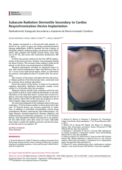
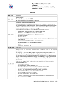
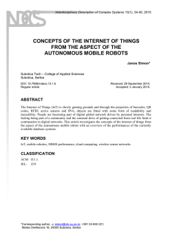
Evoked itch perception is associated with changes in functional
brain connectivity
The Harvard community has made this article openly available.
Please share how this access benefits you. Your story matters.
Citation
Desbordes, G., A. Li, M. L. Loggia, J. Kim, P. C. Schalock, E.
Lerner, T. N. Tran, et al. 2014. “Evoked itch perception is
associated with changes in functional brain connectivity.”
NeuroImage : Clinical 7 (1): 213-221.
doi:10.1016/j.nicl.2014.12.002.
http://dx.doi.org/10.1016/j.nicl.2014.12.002.
Published Version
doi:10.1016/j.nicl.2014.12.002
Accessed
February 6, 2015 10:54:03 AM EST
Citable Link
http://nrs.harvard.edu/urn-3:HUL.InstRepos:13890591
Terms of Use
This article was downloaded from Harvard University's DASH
repository, and is made available under the terms and conditions
applicable to Other Posted Material, as set forth at
http://nrs.harvard.edu/urn-3:HUL.InstRepos:dash.current.termsof-use#LAA
(Article begins on next page)
NeuroImage: Clinical 7 (2015) 213–221
Contents lists available at ScienceDirect
NeuroImage: Clinical
journal homepage: www.elsevier.com/locate/ynicl
Evoked itch perception is associated with changes in functional
brain connectivity
Gaëlle Desbordes a,⁎, Ang Li a, Marco L. Loggia a, Jieun Kim a, Peter C. Schalock c, Ethan Lerner c, Thanh N. Tran c,
Johannes Ringe, Bruce R. Rosen a, Ted J. Kaptchukd, Florian Pfab a,e,f,1, Vitaly Napadow a,b,g,1
a
Athinoula A. Martinos Center for Biomedical Imaging, Massachusetts General Hospital, Harvard Medical School, Boston, MA, USA
Department of Radiology, Logan College of Chiropractic, Chesterfield, MO, USA
Department of Dermatology, Massachusetts General Hospital, Harvard Medical School, Boston, MA, USA
d
Department of Internal Medicine, Beth Israel Deaconess Medical Center, Harvard Medical School, Boston, MA, USA
e
Department of Dermatology and Allergy, Technische Universität München, Munich, Germany
f
Department of Prevention and Sports Medicine, Technische Universität München, Munich, Germany
g
Department of Biomedical Engineering, Kyunghee University, Yongin, Korea
b
c
a r t i c l e
i n f o
Article history:
Received 21 July 2014
Received in revised form 28 October 2014
Accepted 1 December 2014
Available online 3 December 2014
Keywords:
Atopic dermatitis
Eczema
Insula
Pruritus
Putamen
a b s t r a c t
Chronic itch, a highly debilitating condition, has received relatively little attention in the neuroimaging literature.
Recent studies suggest that brain regions supporting itch in chronic itch patients encompass sensorimotor and
salience networks, and corticostriatal circuits involved in motor preparation for scratching. However, how
these different brain areas interact with one another in the context of itch is still unknown. We acquired BOLD
fMRI scans in 14 atopic dermatitis patients to investigate resting-state functional connectivity before and after
allergen-induced itch exacerbated the clinical itch perception in these patients. A seed-based analysis revealed
decreased functional connectivity from baseline resting state to the evoked-itch state between several itchrelated brain regions, particularly the insular and cingulate cortices and basal ganglia, where decreased connectivity was significantly correlated with increased levels of perceived itch. In contrast, evoked itch increased connectivity between key nodes of the frontoparietal control network (superior parietal lobule and dorsolateral
prefrontal cortex), where higher increase in connectivity was correlated with a lesser increase in perceived
itch, suggesting that greater interaction between nodes of this executive attention network serves to limit itch
sensation via enhanced top-down regulation. Overall, our results provide the first evidence of itch-dependent
changes in functional connectivity across multiple brain regions.
© 2014 The Authors. Published by Elsevier Inc. This is an open access article under the CC BY-NC-ND license
(http://creativecommons.org/licenses/by-nc-nd/3.0/).
1. Introduction
The brain circuitry supporting itch sensation is an active topic of research. While numerous studies have investigated and described painrelated brain mechanisms (Apkarian et al., 2005; Henry et al., 2011),
Abbreviations: aMCC, anterior mid-cingulate cortex; AD, atopic dermatitis; ASL, arterial
spinlabeling; BA, Brodmann area;BOLD,blood-oxygen-level dependent; dlPFC, dorsolateral
prefrontal cortex; DMN, default mode network; ECG,electrocardiography; fcMRI, functional
connectivity magnetic resonance imaging; fMRI, functional magnetic resonance imaging;
GLM, general linear model; ITCH, evoked itch resting-state scan; L, left; MNI, Montreal
Neurological Institute; MR, magnetic resonance; PCC, posterior cingulate cortex; PET, positron emission tomography; PMC,premotor cortex; pMCC, posterior mid-cingulate cortex; R,
right; REST, baseline resting-state scan; S1/M1, primary sensorimotor cortex; SCORAD,
SCORing atopic dermatitis scale; SPL, Superior parietal lobule; VAS, visual analog scale;
vlPFC, ventrolateral prefrontal cortex.
⁎ Correspondence author at: Department of Radiology, Athinoula A. Martinos Center for
Biomedical Imaging, Massachusetts General Hospital, Suite 2301, 149 Thirteenth Street,
Boston, MA 02129. Tel.: +1 617 643 2467; fax: +1 617 726 7422.
E-mail address: [email protected] (G. Desbordes).
1
Authors contributed equally to this manuscript.
much less is known about the brain circuitry involved with itch in
spite of the clinical relevance of this highly debilitating symptom,
which is present across multiple chronic itch conditions (Yosipovitch
et al., 2003). Moreover, recent studies suggest that pain perception is associated with altered resting-state functional brain connectivity, which
refers to stereotypical patterns of co-activation across multiple brain regions that show both trait and pain-state specificity (e.g., Napadow
et al., 2010; Napadow et al., 2012; Loggia et al., 2013; reviewed in
Fomberstein et al., 2013). However, itch-related functional brain connectivity, especially in chronic itch patients, has never been reported.
Atopic dermatitis (AD), also known as atopic eczema, is a pruritic
chronic inflammatory disease of the skin, affecting approximately 17.8
million persons in the United States, in which patients experience highly debilitating itch (Finlay, 2001; Lapidus, 2001; Yosipovitch et al., 2003;
Berke et al., 2012). Previous studies indicate that AD involves not only
peripheral sensitization for itch (Lee and Yu, 2011; Rahman et al.,
2011) but also central sensitization, in an analogous fashion as central
sensitization for pain in chronic pain patients (Koltzenburg, 2000),
such that stimuli that are perceived as painful in healthy subjects are
http://dx.doi.org/10.1016/j.nicl.2014.12.002
2213-1582/© 2014 The Authors. Published by Elsevier Inc. This is an open access article under the CC BY-NC-ND license (http://creativecommons.org/licenses/by-nc-nd/3.0/).
214
G. Desbordes et al. / NeuroImage: Clinical 7 (2015) 213–221
experienced as itching in AD patients (Ikoma et al., 2004; Ikoma et al.,
2006; Schmelz, 2010). Not only specific allergens, but also stress and
psychosocial factors, can exacerbate AD symptoms (Gil and Sampson,
1989; Koblenzer, 1999; Rahman et al., 2011). This complex symptomatology, and its bidirectional relationship to stress and psychosocial factors, which can be both a cause and a consequence of itching and
scratching (Gil and Sampson, 1989; Lapidus, 2001; Arck and Paus,
2006; Chida et al., 2008; Mizawa et al., 2013; Yaghmaie et al., 2013;
Schut et al., 2014), point to an essential role of the central nervous system in AD (Ikoma et al., 2004; Misery, 2011; Pfab et al., 2012a; Darlenski
et al., 2014).
In healthy subjects, a number of studies indicate that experimentallyinduced itch activates a brain network which includes pre-motor and
supplementary motor area, thalamus, and cingulate, insular, inferior parietal, and prefrontal cortices (reviewed in Pfab et al., 2012a). However,
few studies have investigated the brain circuitry supporting itch in AD.
Studies from our group and others suggest that brain response to experimentally induced itch in AD differs from healthy subjects, consistent
with abnormal itch sensitivity in AD (Ikoma et al., 2004; Schneider
et al., 2008; Ishiuji et al., 2009; Pfab et al., 2010; Pfab et al., 2011; Pfab
et al., 2012b; Napadow et al., 2014). In a recent functional MRI (fMRI)
study, we evoked clinically-relevant itch in AD patients by using
individually-matched allergens and our validated temperaturemodulated itch induction procedure (Napadow et al., 2014). In this procedure, a thermode is used to modulate skin temperature between 32 °C
(warm) and 25 °C (cool), in 20-s blocks (Pfab et al., 2006). This procedure
has reliably been used to experimentally modulate itch, such that cooling
the skin gradually increases itch sensation over the first 10 s, followed by
a phase in which itch sensation reaches a peak plateau (Pfab et al., 2006;
Pfab et al., 2010). We found that different brain regions were activated
during these different phases of itch induction (i.e., increasing itch and
peak itch plateau). During the increasing itch phase, the right anterior
insula, right anterior middle cingulate cortex, bilateral striatum (putamen and caudate), right globus pallidus, and right ventrolateral prefrontal cortex were activated. Conversely, during the peak itch plateau phase
the right dorsolateral prefrontal cortex, bilateral premotor cortex, and
left superior parietal lobule were activated. Notably, reduction of itch
after non-pharmacological (acupuncture) therapy was associated with
reduced activation in the putamen and right anterior insula (Napadow
et al., 2014), further highlighting the important role of these regions in
itch processing.
In the present study, we investigated functional brain connectivity
before and after experimentally inducing (or exacerbating) itch, using
allergen skin prick in nonlesional skin while AD patients were resting
in the MRI scanner. We used functional connectivity magnetic resonance imaging (fcMRI) with seed-based correlation methods to evaluate changes in functional brain connectivity associated with itch. To
our knowledge, this is the first study to investigate how functional
brain connectivity is modulated by itch, particularly the clinicallyrelevant itch state — a common cause of suffering in AD patients.
2. Materials and methods
2.1. Study participants
Patients aged 18–60 with a clinical diagnosis of AD and a score greater than 18 on the SCORing Atopic Dermatitis (SCORAD) scale (European
Task Force on Atopic Dermatitis 1993; Schmitt et al., 2013) were enrolled. Recruitment was by print and e-mail advertisement as well as referrals from physician colleagues in the Department of Dermatology at
the Massachusetts General Hospital. Volunteers who met eligibility
criteria via phone screening were invited to a preliminary session in
the laboratory during which a licensed dermatologist (F.P.) assessed
their eligibility to the study via medical examination and focused history. In addition, eligible participants had to show type-I sensitivity to
grass pollen and/or dust mites, as demonstrated by wheal and flare
formation upon skin prick testing. For each participant, the most effective itch-inducing allergen was determined among Timothy grass pollen (100,000 bioequivalent allergy units per milliliter) and the two
most common types of house dust mites in North America and Europe
(Dermatophagoides pteronyssinus,10,000 allergy units per milliliter,
and Dermatophagoides farinae,10,000 allergy units per milliliter, Allergy
Laboratories, Oklahoma City, OK). Participants also completed the Edinburgh Inventory for handedness (Oldfield, 1971). The study protocol
was approved by the Human Research Committee of Massachusetts
General Hospital and all study participants gave informed consent. Participants were required to stop all immunosuppressive medications at
least 10 days prior to the study to avoid potential suppression of itch
perception.
2.2. Data acquisition
As part of a previously published study of the anti-pruritic effects of
acupuncture (Napadow et al., 2014), each patient took part in an initial
clinical screening session followed by two separate fMRI sessions (at
least 1 week apart) during which resting-state scans were performed
before and after allergen itch induction (but before any verum or placebo treatment was applied), with corresponding self-report ratings of
perceived itch. The baseline resting-state scan (REST) occurred at the
beginning of the session. The allergen-evoked itch resting-state scan
(ITCH) was performed 15–17 min after allergen skin prick, within the
period of time when allergen induced itch sensation is known to persist
after skin prick (Pfab et al., 2010). A single drop of the most pruritogenic
allergen (as determined during the clinical screening session) was applied on a non-lesion site on the left volar forearm. The skin was then
punctured with a plastic MR-compatible lancet (Duotip Test II, Lincoln
Diagnostics, Decatur, IL) such that the allergen solution was deposited
at the dermal–epidermal junction, where the terminals of itch-related
C-fibers are located (Shelley and Arthur, 1957). Using this procedure,
an itch sensation develops with a median latency of 35 s and persists
more than 15 min after application (Pfab et al., 2010). An absorbent
gauze pad was then used to carefully remove the drop of allergen solution 120 s after application. With this method, the skin puncture is sufficiently shallow to avoid sustained pain, as previously reported (Pfab
et al., 2006), and can be considered an induction of pure itch rather
than a combination of itch and pain.
Brain imaging was performed on a Siemens Trio 3 Tesla MRI scanner
(Siemens AG, Erlangen, Germany) with vendor-supplied 32-channel
multi-array head coil. A high-resolution T1-weighted anatomical scan
was collected using an isotropic multi-echo MPRAGE pulse sequence
(TR/TE1/TI = 2530/1.64/1200 ms, 256 × 256 matrix, 256 mm field-ofview (FOV), 7° flip angle) (van der Kouwe et al., 2008). Functional imaging (BOLD fMRI) was performed using a gradient echo T2*-weighted
pulse sequence (TR/TE = 2 s/30 ms, 32 anterior commissure–posterior
commissure (AC−PC) aligned slices, slice thickness 3.6 mm, 64 × 64
matrix, 200 mm FOV, 90° flip angle). Each resting-state fMRI scan had
a duration of 6 min (180 time points). Electrocardiography (ECG) and
respiratory activity were simultaneously recorded throughout the
scans using a Powerlab system (ML880, ADInstruments, Colorado
Springs, CO) at a 400 Hz sampling rate. ECG data were acquired and filtered using an MR-compatible physiological monitor (Magnitude 3150
MRI Patient Monitor, In vivo, Gainesville, Florida) designed to minimize
radio frequency and gradient switching artifacts generated during the
MRI scan. Respiratory data were collected using a custom-built system
based on that devised by Binks et al. (2007) which consists of two
MR-compatible pneumobelts placed around the chest and abdomen
and connected to air pressure transducers (PX138-0.3D5V, Omegadyne,
Inc., Sunbury, Ohio).
Immediately before the baseline scan and immediately after the
induced-itch scan, subjects were asked to rate the intensity of experienced itch on a visual analog scale (VAS) from 0 (no itch) to 100
(most intense itch imaginable), with 33 corresponding to an “urge to
G. Desbordes et al. / NeuroImage: Clinical 7 (2015) 213–221
scratch” threshold, as in our previous studies (Pfab et al., 2005; Pfab
et al., 2006; Valet et al., 2008; Pfab et al., 2010; Pfab et al., 2011). It
should be noted that subjects were instructed to refrain from scratching
even if the perceived itch was above this threshold, and we confirmed
by observation that subjects did not engage in scratching during the
scans.
2.3. Data analysis
2.3.1. Preprocessing
BOLD data preprocessing and analysis were performed using tools
from the FMRIB Software Library (FSL) (http://www.fmrib.ox.ac.uk/
fsl) (Smith et al., 2004; Woolrich et al., 2009), the FreeSurfer (v. 5.2)
suite (http://surfer.nmr.mgh.harvard.edu/) (Dale et al., 1999; Fischl
et al., 1999; Fischl et al., 2002; Fischl et al., 2004), AFNI (Cox, 1996;
Cox and Hyde, 1997), and RETROICOR (Glover et al., 2000) for retrospective correction of physiological motion artifacts using our peripheral measures of cardiac and respiratory activities as independent
assessments of physiological noise in the BOLD signal.
Preprocessing consisted of: (1) B0 fieldmap correction (dewarping);
(2) physiological noise correction with RETROICOR; (3) slice timing correction (‘slicetimer’, FSL); (4) head motion correction (MCFLIRT, FSL
(Jenkinson et al., 2002)); (5) skull stripping (BET, FSL (Smith, 2002));
(6) nonlinear registration to the Montreal Neurological Institute
(MNI) template (FLIRT/FNIRT, FSL); and (7) 0.008–0.1 Hz band-pass
temporal filtering (‘1dBandpass’, AFNI). The translation parameters
resulting from the motion correction step were then used to compute
the relative mean motion during each scan. Relative mean motion was
defined as the average over time of the absolute displacement of each
brain volume as compared to the previous volume in time, where the
absolute displacement at each time point was computed as the rootmean-square of the x, y, and z translation parameters (Van Dijk et al.,
2012).
2.3.2. Seed-based functional connectivity analyses
Functional connectivity was computed using a conventional seedbased correlation analysis method. This method consists in (1) choosing
a specific voxel or cluster of voxels in the brain, referred to as the seed,
(2) extracting the time series of the BOLD signal in this seed region, and
215
(3) using this time series as a regressor in a general linear model (GLM)
analysis to calculate whole-brain, voxel-wise functional connectivity
maps of covariance with this seed region (Biswal et al., 1995; Cole
et al., 2010). Our seed regions of interest were chosen as the clusters significantly activated during the “increasing itch” or “peak itch plateau”
phase from an evoked, allergen-induced itch block fMRI scan in the
same AD subjects, as previously reported (Napadow et al., 2014). Importantly, activation clusters came from different fMRI runs than those
investigated with our resting fcMRI approach, assuring independence
between the seed fMRI time series during rest and that during
temperature-modulated evoked itch scanning. The brain regions from
which ROI seeds were chosen were the right anterior insula (MNI coordinates in the Talairach–Tournoux Atlas: X = 30 mm, Y = 18 mm, Z =
2 mm), right anterior mid-cingulate cortex (aMCC) (X = 8, Y = 34, Z =
32), right putamen (X = 24, Y = 18, Z = 4), left putamen (X = −22,
Y = 10, Z = 10), right caudate (X = 20, Y = 18, Z = 8), left caudate
(X = −20, Y = 10, Z = 16), right globus pallidus (X = 18, Y = 4,
Z = 4), right ventrolateral prefrontal cortex (vlPFC) (X = 34, Y = 40,
Z = 6), right dorsolateral prefrontal cortex (dlPFC) (X = 36, Y = 20,
Z = 52), right premotor cortex (PMC) (X = 22, Y = −4, Z = 62), left
PMC (X = −20, Y = −20, Z = 62), and left superior parietal lobule
(SPL) (X = −28, Y = −44, Z = 64) (see Table 1). For each seed region,
the BOLD signal was averaged across a 4-mm radius sphere centered on
each cluster3s peak voxel. The resulting time series was then used as a
regressor in a standard GLM analysis using FSL, along with 10 regressors
of no interest: 6 motion correction parameters, the average time series
from cerebrospinal fluid voxels in the lateral ventricles, the average
time series from white matter voxels in deep parietal white matter,
and cardiac and respiratory time series obtained by convolving the cardiac and respiratory signals with hemodynamic response functions
from the ‘RVHR’ model (Chang et al., 2009). The output of this GLM analysis for each seed region of interest was a 3D volume (“brain map”) in
which each voxel value represented the strength of functional connectivity between this seed and every other voxel in the brain.
2.3.3. ITCH–REST difference map for each subject
For each subject, a difference map was computed by calculating the
difference in parameter estimates for connectivity taken from REST
Table 1
Seed-based functional connectivity results. For each seed, statistically significant clusters (with a cluster-forming threshold of Z N 2.3 and a cluster significance level of p b 0.05) are listed.
For each cluster, the Z-value, MNI coordinates, and atlas label of the peak voxel are listed, along with Pearson3s correlation coefficient r and corresponding p-value of the correlation with
level of perceived itch.
Seed region
Z-value
X
Y
Z
Atlas label
Correlation with perceived itch
R premotor cortex
3.56
3.16
3.92
3.81
3.61
3.62
3.38
3.78
3.95
3.68
4.01
3.35
3.92
3.69
3.61
4.24
4.32
4.04
4.03
4.39
3.86
3.98
4.01
−9
8
−30
−18
−12
−52
−4
−60
−61
−2
−0.5
−11
−53
−41.5
43.5
29.5
34
28.5
58
−10.5
−51.5
−5
35.5
−64.5
−48
−44
−36
−60
−66
−18
−20.5
−17.5
−22
−35
−41.5
−24.5
29
−10.5
−14
−21.5
−12.5
−26.5
−73
19.5
−25.5
27
66
61
−14
−2
14
6
40
22.5
11
48
41
29.5
32
27.5
15
11.5
10
9.5
22
23.5
−4.5
44
49
L precuneus
R superior parietal lobule (5L)
L fusiform gyrus
L parahippocampal gyrus
L posterior cingulate cortex
L middle temporal gyrus
L mid-cingulate cortex
Left postcentral gyrus
L transverse (or superior) gyrus, BA42
L medial frontal gyrus, BA6 or L paracentral lobule
L/R mid-cingulate cortex
L posterior cingulate cortex
L inferior parietal lobule
L dlPFC (Middle frontal gyrus)
R posterior insula
R putamen (not the same locus as the R putamen seed)
R posterior insula
R putamen
R inferior parietal lobule
L cuneus, BA18
L inferior frontal gyrus (pars orbitalis)
L mid-cingulate cortex or paracentral lobule
R superior/middle frontal gyrus (dlPFC)
n.s.
n.s.
n.s.
r = –0.39, p = 0.045
r = –0.42, p = 0.025
n.s.
n.s.
n.s.
n.s.
n.s.
n.s.
n.s.
R insula
R putamen
L superior parietal lobule
R anterior mid-cingulate cortex
R caudate
R globus pallidus
r = –0.40, p = 0.036
n.s.
n.s.
n.s.
r = –0.40, p = 0.036
n.s.
n.s.
n.s.
r = –0.37, p = 0.050
n.s.
216
G. Desbordes et al. / NeuroImage: Clinical 7 (2015) 213–221
versus ITCH scan runs using ‘fslmaths’ (FSL). The variance map for this
difference map was estimated by adding the parameter estimate variances computed from the REST and ITCH scan runs. These maps, calculated from all subjects, were then passed up to a group level analysis
using FSL, as described below.
was no significant correlation between perceived itch and age across
subjects, neither during REST (r = 0.14, p = 0.63), nor during ITCH
(r = 0.18, p = 0.54).
2.3.4. Group-level analysis of main effect of itch on functional connectivity
A group-level analysis yielded a paired difference map between
REST and ITCH across subjects and therefore indicated the main effect
of itch induction on functional connectivity. Multiple comparisons correction was performed with cluster correction, with a cluster-forming
threshold of Z N 2.3 and a cluster significance level of p b 0.05. Finally,
for visualization purposes, the resulting brain maps were spatially
smoothed using the cubic resampling function in AFNI and overlaid on
the 0.5 mm MNI152 template.
Since subject motion during the scan can significantly affect measures of functional connectivity, it is important to measure and report
head motion in any study of functional connectivity (Power et al.,
2012; Van Dijk et al., 2012). Here we verified that mean motion ranged
within previously published values in a healthy population (Van Dijk
et al., 2012) and did not significantly differ between REST (0.100 ±
0.043 mm, mean ± standard deviation) and ITCH (0.097 ±
0.037 mm; paired t-test, p = 0.9). We further verified that gross translational motion never exceeded 3 mm on any axis. We also verified that
the participants3 breathing rates did not change between the REST condition (0.35 Hz ± 0.11, mean ± standard deviation) and the ITCH condition (0.34 Hz ± 0.10, paired t-test, p = 0.19).
2.3.5. Correlation between perceived itch and level of functional
connectivity
Using only the brain regions in which a main effect was found, we
then performed a linear regression analysis between the average main
effect within the region and the itch perception score difference between the REST and ITCH conditions. This analysis specifically indicated
which functional connectivity changes between REST and ITCH were
correlated with perceived change in itch, thus linking changes in functional connectivity with itch more directly than the main effect analysis
alone, and taking advantage of the intersubject variability in itch responses. Therefore, in this final analysis, changes in functional connectivity were deemed related to itch only in brain regions where there
was both (i) a main effect of itch and (ii) a significant correlation across
subjects between change in functional connectivity and change in perceived itch (from REST to ITCH).
3. Results
Fourteen AD patients (8 females, 13 right-handed, age: 25.4 ±
9.1 years old, SCORAD: 38.7 ± 14.9, mean ± standard deviation) participated in the study, with itch induction performed using the most
pruritogenic allergen for each patient among Timothy grass pollen
(N = 8), D. pteronyssinus house dust mite (N = 3), or D. farinae house
dust mite (N = 3).
3.1. Itch rating levels
Perceived itch increased significantly after allergen induction (REST:
6.4 ± 11.7; ITCH: 32.0 ± 16.6, mean ± standard deviation on a 0–100
visual analog scale; paired t-test, p b 10−7; Fig. 1), as expected. There
Fig. 1. Change in perceived itch intensity from the baseline resting state (‘REST’) to the allergen-evoked itch state (‘ITCH’), on a 0–100 scale, for all experimental sessions (N = 14,
with 2 sessions per subject). The dashed line indicates the “urge to scratch” threshold (defined at the 33 level). The thicker, black line represents the mean across subjects and sessions. The asterisk indicates that the ratings during ‘ITCH’ were significantly different from
those during ‘REST’ (paired t-test, p b 10−7).
3.2. Head motion and respiration during the scan
3.3. Correlation between perceived itch and level of functional connectivity
While functional connectivity changed significantly from REST to
ITCH for several of the seed regions of interest (see Table 1), the change
in functional connectivity was significantly correlated with change in
perceived itch for a smaller subset of regions. Among these regions,
functional connectivity decreased during ITCH, compared to REST, in
most cases. Specifically, connectivity was decreased and negatively correlated with change in itch for connectivity between the right anterior
insula and left hippocampal formation (MNI coordinates of the
peak voxel: X = −18, Y = −36, Z = –2, r = –0.39, p = 0.045)
and left posterior cingulate cortex (PCC, X = −12, Y = −60, Z = 14,
r = –0.42, p = 0.025), between the right aMCC and right putamen
(X = 28.5, Y = −12.5, Z = 9.5, r = –0.40, p = 0.036), and between
the right globus pallidus and left posterior mid-cingulate cortex
(pMCC, X = −5, Y = −25.5, Z = 44, r = –0.37, p = 0.050; Fig. 2 and
Table 1). Thus, for these connectivity changes, a higher increase in perceived itch was associated with a greater decrease in connectivity. The
exception to this pattern was for connectivity between the left SPL
and left dlPFC (X = −41.5, Y = 29, Z = 27.5), which demonstrated an
increase during ITCH, and a negative correlation between change in
connectivity and change in itch (r = –0.40, p = 0.036; Fig. 3 and
Table 1). In other words, a higher increase in perceived itch was associated with a lower increase in SPL–dlPFC connectivity.
4. Discussion
In this fcMRI study of allergen-induced itch in AD patients, we found
that an increase in perceived itch was significantly correlated with a decrease in functional connectivity between brain regions previously
demonstrated to activate in response to phasic itch provocation
(Napadow et al., 2014). These brain regions included the insula, cingulate cortex, and basal ganglia. Specifically, greater itch was associated
with reduced connectivity between the right anterior insula and both
hippocampal formation and posterior cingulate cortex. Similarly, itch
was associated with reduced connectivity between the anterior midcingulate cortex and putamen, and between the globus pallidus and
posterior mid-cingulate cortex. While itch was mostly associated with
reduced functional connectivity, increased connectivity between the superior parietal lobule and dorsolateral prefrontal cortex was associated
with an increase in perceived itch. Moreover, a negative correlation was
found between SPL–dlPFC connectivity and itch rating; that is, subjects
showing greater increase in SPL–dlPFC connectivity also reported lower
increase in itch sensation. This result suggests that greater interaction
between the SPL and dlPFC serves to limit itch sensation via enhanced
top-down regulation. Our study clearly demonstrates for the first time
that “resting” functional connectivity is altered when AD patients
G. Desbordes et al. / NeuroImage: Clinical 7 (2015) 213–221
217
Fig. 2. Changes in functional connectivity from the baseline resting state (‘REST’) to the allergen-evoked itch state (‘ITCH’). A. Seed-based functional connectivity from the right anterior
insula (MNI coordinates: [30, 18, 1]). Top: Significant cluster in the left hippocampal formation (peak voxel coordinates: [−30, −44, −14] in the fusiform gyrus). Bottom: Significant
cluster in the left posterior cingulate cortex (PCC) (peak voxel coordinates: [−12, −60, 14]). In each of the two panels, the graph on the left shows z-score levels of functional connectivity
(main effect), in the resting state (‘REST’; white bar) and 15–17 min after allergen-based itch induction (‘ITCH’; black bar). The graph on the right shows for each scan session (N = 28) the
difference in connectivity from ‘REST’ to ‘ITCH’ as a function of the difference in perceived itch intensity from ‘REST’ to ‘ITCH’, as reported by participants; linear regression across subjects is
shown (red line) along with corresponding r and p values. B. Seed-based functional connectivity from the right anterior mid-cingulate cortex (coordinates: [8, 34, 28]), showing a significant cluster in the right putamen (peak voxel coordinates: [28, −12, 9]). C. Seed-based functional connectivity from the right globus pallidus (coordinates: [18, 4, 3]), showing a significant
cluster in the left posterior mid-cingulate cortex (peak voxel coordinates: [−5, −25, 44]).
218
G. Desbordes et al. / NeuroImage: Clinical 7 (2015) 213–221
Fig. 3. Seed-based functional connectivity from the left superior parietal lobule (coordinates: [−28, −40, 61]), showing a significant cluster in the left rostral middle frontal gyrus, part of
left dorsolateral prefrontal cortex (peak voxel coordinates: [−41, 29, 27]). Same graph conventions as in Fig. 2.
experience itch, and these state-specific alterations can be directly
linked with the intensity of perceived itch.
Overall, we found that a sustained itch state altered functional connectivity for nodes known to be activated by evoked itch. For instance,
we found that functional connectivity between the basal ganglia (caudate and globus pallidus) and cingulate cortex, normally present at
rest (Postuma and Dagher, 2006), decreased with increased itch (see
Fig. 2B, 2C). This finding is reminiscent of our recent pain neuroimaging
study in which we found that experimentally-induced sustained
myofascial pain in healthy adults reduced functional connectivity between the somatotopic cortical representation within the primary sensorimotor (S1/M1) area contralateral to the pain stimulus site and other
regions of the primary sensorimotor network (Kim et al., 2013). Hence,
in both studies, brain areas activated by an evoked stimulus (whether
itch or pain) showed decreased connectivity within their intrinsic connectivity network.
Evoked itch in our AD patients was also associated with a decrease in
functional connectivity between the right anterior insula and both left
hippocampal formation and left PCC (see Fig. 2A), both of which are
nodes of the default mode network (DMN) (Buckner et al., 2008). Interestingly, while the anterior insula is not usually connected with DMN
regions, chronic pain patients show greater DMN–insula connectivity
at rest than healthy controls (Napadow et al., 2010; Loggia et al.,
2013), and even in healthy subjects, DMN–insula connectivity increases
with state anxiety (Dennis et al., 2011). In our AD subjects, functional
connectivity between the right anterior insula and these two DMN
nodes (left hippocampal formation and left PCC) was present during a
resting state and decreased after itch induction, and this decrease was
significantly correlated with an increase in perceived itch. The
allergen-induced itch may therefore have disrupted the resting connectivity patterns that support chronic (versus evoked) itch perception in
these patients, perhaps by shifting attention to an evoked stimulus. Together, these results reinforce the notion that unpleasant interoceptive
experiences such as itch or pain may decrease functional connectivity
between brain regions that are functionally connected at rest.
In contrast, while connectivity between itch-processing brain areas
was mostly decreased during the sustained itch state, increased connectivity was noted between SPL and dlPFC. In fact, we found a negative
correlation between itch intensity and the change in connectivity between the SPL and dlPFC. In other words, a higher increase in functional
connectivity between SPL and dlPFC was associated with a lower increase in perceived itch after allergen itch induction (see Fig. 3). This
suggests that increased SPL–dlPFC connectivity may be a protective
mechanism, limiting perceived itch severity after allergen itch induction
in AD. Both the SPL and dlPFC sub-regions in our results are key nodes of
the frontoparietal control network, which supports executive processing of cognitive control (Vincent et al., 2008). Hence, our finding suggests that subjects able to regulate increased information exchange
within this network were better able to limit the severity of sustained
itch sensation after allergen itch induction. Interestingly, we previously
found that SPL and dlPFC were activated by evoked allergen itch during
the “peak itch” plateau phase (Napadow et al., 2014), an experimental
condition likely to be more closely related to sustained itch than the
preceding “increasing itch” phase, when itch sensation was steadily increasing. Of note, the brain regions that were activated during the “increasing itch” phase (anterior insula, cingulate, putamen) instead
showed decreased connectivity in response to sustained evoked itch.
This suggests that different brain networks become engaged when
itch sensation first develops versus when itch is a steadily ongoing
perception.
The continuous, sustained itch state induced in our study was associated with altered functional connectivity between several brain regions which previous studies have reported as being activated by
evoked itch stimuli. In healthy subjects, brain activation during itch
has been investigated by performing functional brain imaging – with either positron emission tomography (PET), BOLD fMRI, or arterial spin
labeling (ASL) fMRI – while inducing itch experimentally using various
means such as histamine prick, allergen prick, cowhage spicule application, or electrical stimulation (Walter et al., 2005; Herde et al., 2007;
Leknes et al., 2007; Mochizuki et al., 2007; Pfab et al., 2008; Valet
et al., 2008; Yosipovitch et al., 2008; Mochizuki et al., 2009; Vierow
et al., 2009; Jeffry et al., 2011; Kleyn et al., 2012; Papoiu et al., 2012;
Pfab et al., 2012a). These studies, most of them conducted with healthy
subjects rather than chronic itch patients, highlight the involvement of a
network of brain regions which includes ipsilateral pre-motor and supplementary motor areas and contralateral cingulate cortex, insular cortex, thalamus, inferior parietal cortex, and dorsolateral prefrontal cortex
(reviewed in Pfab et al., 2012a). In addition, the above studies found
that itch stimuli activate habit-encoding motor planning areas, including the putamen, probably in relation to the motor act of scratching
which is strongly associated with itch (Yosipovitch et al., 2008;
Vierow et al., 2009). In the present study, itch perception was associated
with reduced connectivity between the insula, cingulate cortex, and
basal ganglia (i.e., putamen, globus pallidus) — regions whose activation
G. Desbordes et al. / NeuroImage: Clinical 7 (2015) 213–221
was strongly associated with itch in previous evoked-itch neuroimaging
studies.
Notably, functional connectivity for basal ganglia areas such as the
putamen and globus pallidus was significantly altered during sustained
itch. These areas are critical in regulating both voluntary and involuntary movements, and may play an important role in the scratch response to itch. Putamen activation has been reported not only during
scratching, with greater activation in the presence of itch than in its absence, but also during itching without scratching, suggesting that putamen activation may represent the urge and/or intention to scratch
(Vierow et al., 2009). We previously reported that putamen activation
was maximal during an increasing itch phase in AD patients subjected
to allergen itch (Napadow et al., 2014). The putamen is also a key structure within the striato-thalamo-cortical circuitry implicated in action
motivation and initiation and in habitual, repetitive behavior (Graybiel,
2008). This circuit is dysfunctional in various psychopathologies, including addiction (Koob and Volkow, 2010), which is consistent with its implication in the “urge” to scratch (Leknes et al., 2007; Vierow et al., 2009).
Here we found that functional connectivity between the basal ganglia
(caudate and globus pallidus) and cingulate cortex decreased with increased itch (see Fig. 2B, 2C). The cingulate cortex is part of the salience
network and is activated during itch (Pfab et al., 2012a), which is a highly
salient stimulus, particularly to AD patients. Therefore, since the basal
ganglia as well as the cingulate cortex are activated during itch, the observed decrease in functional connectivity between these regions during
experimentally-induced itch may be another case of diminished connectivity between regions known to be activated by an evoked noxious
stimulus, as noted above (Kim et al., 2013).
Limitations to our study should also be noted. For instance, the ITCH
scan took place 15–17 min after allergen itch induction, a time at which
persisting itch intensity was no longer maximal. While perceived itch
was still robust enough to alter functional brain connectivity, future
studies should acquire fMRI data closer in time to itch induction,
allowing for greater power to evaluate changes in brain connectivity
supporting a persistent itch state. On the other hand, the long delay
after initial itch induction allowed for any early transients in itch perception and physiological brain response to subside, thus improving a
functional connectivity assessment based on 6 min of scanning. Additionally, while we found other significant changes in functional connectivity after itch induction (see Table 1), these changes were not
significantly correlated with changes in perceived itch, indicating that
they may be only indirectly related to itch perception or may have
been caused by order effects (since the REST scan always preceded the
ITCH scan). Hence, the results reported here account for any potential
order effects as we focused on functional connectivity changes that
were also significantly correlated with the level of perceived itch.
In conclusion, the present results shed new light on our recent findings of the neural correlates of allergen-induced itch in AD, which involved a broad network of brain regions associated with emotional
processing (anterior insula, anterior cingulate cortex), salience (insula,
striatum), executive function (ventrolateral and dorsolateral prefrontal
cortex, cingulate cortex), sensory-motor integration (superior parietal
lobule), and motor planning (globus pallidus, premotor cortex, striatum) (Napadow et al., 2014). As increased SPL–dlPFC connectivity
may reduce itch perception in AD, future studies should explore how
cognitive and attentional psychological interventions, such as cognitive
behavioral therapy and meditation training (Chida et al., 2007;
Rosenkranz et al., 2013; Schut et al., 2013), might modulate
frontoparietal control network connectivity in response to an upregulated itch state, and thereby reduce itch perception via top-down
regulation. Since longitudinal studies have found functional as well as
structural changes in the brain after such interventions (Linden, 2006;
Felmingham et al., 2007; Hölzel et al., 2011; Desbordes et al., 2012;
Hölzel et al., 2013), there is cautious optimism that these types of intervention may “rewire” dysfunctional brain networks in chronic itch patient populations, such as AD.
219
Funding
This work was supported by the National Institutes of Health
(V.N.: K01AT002166, R01AT004714; G.D.: K01AT008225; B.R.R:
P01AT002048; T.K.: K24AT004095; E.L.: R01AR057744). This research was carried out in part at the Athinoula A. Martinos Center
for Biomedical Imaging at the Massachusetts General Hospital,
using resources provided by the Center for Functional Neuroimaging
Technologies, P41EB015896, a P41 Biotechnology Resource Grant
supported by the National Institute of Biomedical Imaging and Bioengineering (NIBIB), National Institutes of Health. This work also
involved the use of instrumentation supported by the NIH Shared
Instrumentation Grant Program S10RR021110, S10OD010364, and
S10RR023401. This study was also partly funded by a grant from
the German Research Foundation (pf 690/2-1; http://www.dfg.de/
en/research_funding/), and the Christine Kühne Center of Allergy
and Education (CK-Care; http://www.ck-care.ch/index.php?id=
2659&L=4).
References
Apkarian, A.V., Bushnell, M.C., Treede, R.-D., Zubieta, J.-K., 2005. Human brain mechanisms
of pain perception and regulation in health and disease. Eur. J. Pain 9 (4), 463–484.
http://dx.doi.org/10.1016/j.ejpain.2004.11.00115979027.
Arck, P., Paus, R., 2006. From the brain–skin connection: the neuroendocrine–immune
misalliance of stress and itch. Neuroimmunomodulation 13 (5–6), 347–356. http://
dx.doi.org/10.1159/00010486317709957.
Berke, R., Singh, A., Guralnick, M., 2012. Atopic dermatitis: an overview. Am. Fam. Physician 86 (1), 35–4222962911.
Binks, A.P., Banzett, R.B., Duvivier, C., 2007. An inexpensive, MRI compatible device to
measure tidal volume from chest-wall circumference. Physiol. Meas. 28 (2),
149–159. http://dx.doi.org/10.1088/0967-3334/28/2/00417237587.
Biswal, B., Yetkin, F.Z., Haughton, V.M., Hyde, J.S., 1995. Functional connectivity in the
motor cortex of resting human brain using echo-planar MRI. Magn. Reson. Med. 34
(4), 537–541. http://dx.doi.org/10.1002/mrm.19103404098524021.
Buckner, R.L., Andrews-Hanna, J.R., Schacter, D.L., 2008. The brain3s default network: anatomy, function, and relevance to disease. Ann. N. Y. Acad. Sci. 1124, 1–38. http://dx.
doi.org/10.1196/annals.1440.01118400922.
Chang, C., Cunningham, J.P., Glover, G.H., 2009. Influence of heart rate on the BOLD signal:
the cardiac response function. Neuroimage 44 (3), 857–869. http://dx.doi.org/10.
1016/j.neuroimage.2008.09.02918951982.
Chida, Y., Hamer, M., Steptoe, A., 2008. A bidirectional relationship between psychosocial factors and atopic disorders: a systematic review and meta-analysis.
Psychosom. Med. 70 (1), 102–116. http://dx.doi.org/10.1097/PSY.
0b013e31815c1b7118158379.
Chida, Y., Steptoe, A., Hirakawa, N., Sudo, N., Kubo, C., 2007. The effects of psychological
intervention on atopic dermatitis. A systematic review and meta-analysis. Int. Arch.
Allergy Immunol. 144 (1), 1–9. http://dx.doi.org/10.1159/00010194017449959.
Cole, D.M., Smith, S.M., Beckmann, C.F., 2010. Advances and pitfalls in the analysis and interpretation of resting-state fMRI data. Front. Syst. Neurosci. 4, 8. http://dx.doi.org/10.
3389/fnsys.2010.0000820407579.
Cox, R.W., Hyde, J.S., 1997. Software tools for analysis and visualization of fMRI data. N.M.R.
Biomed. 10 (4–5), 171–178. http://dx.doi.org/10.1002/(SICI)1099-1492(199706/08)
10:4/5b171::AID-NBM453N3.0.CO;2-L9430344.
Cox, R.W., 1996. AFNI: software for analysis and visualization of functional magnetic resonance neuroimages. Comput. Biomed. Res. 29 (3), 162–1738812068.
Dale, A.M., Fischl, B., Sereno, M.I., 1999. Cortical surface-based analysis. I. Segmentation
and surface reconstruction. Neuroimage 9 (2), 179–194. http://dx.doi.org/10.1006/
nimg.1998.03959931268.
Darlenski, R., Kazandjieva, J., Hristakieva, E., Fluhr, J.W., 2014. Atopic dermatitis as a
systemic disease. Clin. Dermatol. 32 (3), 409–413. http://dx.doi.org/10.1016/j.
clindermatol.2013.11.00724767188.
Dennis, E.L., Gotlib, I.H., Thompson, P.M., Thomason, M.E., 2011. Anxiety modulates insula
recruitment in resting-state functional magnetic resonance imaging in youth
and adults. Brain Connect. 1 (3), 245–254. http://dx.doi.org/10.1089/brain.2011.
003022433052.
Desbordes, G., Negi, L.T., Pace, T.W., Wallace, B.A., Raison, C.L., Schwartz, E.L., 2012. Effects
of mindful-attention and compassion meditation training on amygdala response to
emotional stimuli in an ordinary, non-meditative state. Front. Hum. Neurosci. 6,
292. http://dx.doi.org/10.3389/fnhum.2012.0029223125828.
European Task Force on Atopic Dermatitis, 1993. Severity scoring of atopic dermatitis: the
SCORAD index. Consensus report of the European Task Force on Atopic Dermatitis.
Dermatology 186 (1), 23–318435513.
Felmingham, K., Kemp, A., Williams, L., Das, P., Hughes, G., Peduto, A., Bryant, R., 2007.
Changes in anterior cingulate and amygdala after cognitive behavior therapy of posttraumatic stress disorder. Psychol. Sci. 18 (2), 127–129. http://dx.doi.org/10.1111/j.
1467-9280.2007.01860.x17425531.
Finlay, A.Y., 2001. Quality of life in atopic dermatitis. J. Am. Acad. Dermatol. 45 (1 Suppl),
S64–S66. http://dx.doi.org/10.1067/mjd.2001.11701011423879.
220
G. Desbordes et al. / NeuroImage: Clinical 7 (2015) 213–221
Fischl, B., Salat, D.H., Busa, E., Albert, M., Dieterich, M., Haselgrove, C., van der Kouwe, A.,
Killiany, R., Kennedy, D., Klaveness, S., et al., 2002. Whole brain segmentation: automated labeling of neuroanatomical structures in the human brain. Neuron 33 (3),
341–355. http://dx.doi.org/10.1016/S0896-6273(02)00569-X11832223.
Fischl, B., Salat, D.H., van der Kouwe, A.J., Makris, N., Ségonne, F., Quinn, B.T., Dale, A.M.,
2004. Sequence-independent segmentation of magnetic resonance images.
Neuroimage 23 (Suppl. 1), S69–S84. http://dx.doi.org/10.1016/j.neuroimage.2004.
07.01615501102.
Fischl, B., Sereno, M.I., Dale, A.M., 1999. Cortical surface-based analysis. II: Inflation, flattening, and a surface-based coordinate system. Neuroimage 9 (2), 195–207. http://
dx.doi.org/10.1006/nimg.1998.03969931269.
Fomberstein, K., Qadri, S., Ramani, R., 2013. Functional MRI and pain. Curr. Opin.
Anaesthesiol. 26 (5), 588–593. http://dx.doi.org/10.1097/01.aco.0000433060.59939.
fe ISSN: 0952-7907.
Gil, K.M., Sampson, H.A., 1989. Psychological and social factors of atopic dermatitis. Allergy
44 (Suppl. 9), 84–89. http://dx.doi.org/10.1111/j.1398-9995.1989.tb04322.x2683846.
Glover, G.H., Li, T.Q., Ress, D., 2000. Image-based method for retrospective correction of
physiological motion effects in fMRI: RETROICOR. Magn. Reson. Med. 44 (1),
162–16710893535.
Graybiel, A.M., 2008. Habits, rituals, and the evaluative brain. Annu. Rev. Neurosci. 31,
359–387. http://dx.doi.org/10.1146/annurev.neuro.29.051605.11285118558860.
Henry, D.E., Chiodo, A.E., Yang, W., 2011. Central nervous system reorganization in a variety of chronic pain states: a review. PM&R 3 (12), 1116–1125. http://dx.doi.org/
10.1016/j.pmrj.2011.05.018.
Herde, L., Forster, C., Strupf, M., Handwerker, H.O., 2007. Itch induced by a novel method
leads to limbic deactivations – a functional MRI study. J. Neurophysiol. 98 (4),
2347–2356. http://dx.doi.org/10.1152/jn.00475.200717715198.
Hölzel, B.K., Carmody, J., Vangel, M., Congleton, C., Yerramsetti, S.M., Gard, T., Lazar, S.W.,
2011. Mindfulness practice leads to increases in regional brain gray matter density.
Psychiatry Res. 191 (1), 36–43. http://dx.doi.org/10.1016/j.pscychresns.2010.08.
00621071182.
Hölzel, B.K., Hoge, E.A., Greve, D.N., Gard, T., Creswell, J.D., Brown, K.W., Barrett, L.F.,
Schwartz, C., Vaitl, D., Lazar, S.W., 2013. Neural mechanisms of symptom improvements in generalized anxiety disorder following mindfulness training. Neuroimage
Clin. 2, 448–458. http://dx.doi.org/10.1016/j.nicl.2013.03.01124179799.
Ikoma, A., Fartasch, M., Heyer, G., Miyachi, Y., Handwerker, H., Schmelz, M., 2004. Painful
stimuli evoke itch in patients with chronic pruritus: central sensitization for itch.
Neurology 62 (2), 212–217. http://dx.doi.org/10.1212/WNL.62.2.21214745056.
Ikoma, A., Steinhoff, M., Ständer, S., Yosipovitch, G., Schmelz, M., 2006. The
neurobiology of itch. Nat. Rev. Neurosci. 7 (7), 535–547. http://dx.doi.org/10.1038/
nrn195016791143.
Ishiuji, Y., Coghill, R.C., Patel, T.S., Oshiro, Y., Kraft, R.A., Yosipovitch, G., 2009.
Distinct patterns of brain activity evoked by histamine-induced itch reveal
an association with itch intensity and disease severity in atopic dermatitis.
Br. J. Dermatol. 161 (5), 1072–1080. http://dx.doi.org/10.1111/j.1365-2133.
2009.09308.x19663870.
Jeffry, J., Kim, S., Chen, Z.-F., 2011. Itch signaling in the nervous system. Physiology (Bethesda) 26 (4), 286–292. http://dx.doi.org/10.1152/physiol.00007.201121841076.
Jenkinson, M., Bannister, P., Brady, M., Smith, S., 2002. Improved optimization for the robust and accurate linear registration and motion correction of brain images.
Neuroimage 17 (2), 825–841. http://dx.doi.org/10.1006/nimg.2002.113212377157.
Kim, J., Loggia, M.L., Edwards, R.R., Wasan, A.D., Gollub, R.L., Napadow, V., 2013. Sustained
deep-tissue pain alters functional brain connectivity. Pain 154 (8), 1343–1351. http://
dx.doi.org/10.1016/j.pain.2013.04.01623718988.
Kleyn, C.E., McKie, S., Ross, A., Elliott, R., Griffiths, C.E., 2012. A temporal analysis of the
central neural processing of itch. Br. J. Dermatol. 166 (5), 994–1001. http://dx.doi.
org/10.1111/j.1365-2133.2012.10849.x22283926.
Koblenzer, C.S., 1999. Itching and the atopic skin. J. Allergy Clin. Immunol. 104 (3 2),
S109–S113. http://dx.doi.org/10.1016/S0091-6749(99)70052-710482861.
Koltzenburg, M., 2000. Neural mechanisms of cutaneous nociceptive pain. Clin. J. Pain 16 (3
Suppl), S131–S138. http://dx.doi.org/10.1097/00002508-200009001-0000411014457.
Koob, G.F., Volkow, N.D., 2010. Neurocircuitry of addiction. Neuropsychopharmacology 35
(1), 217–238. http://dx.doi.org/10.1038/npp.2009.11019710631.
Lapidus, C.S., 2001. Role of social factors in atopic dermatitis: the US perspective. J. Am.
Acad. Dermatol. 45 (1 Suppl), S41–S43. http://dx.doi.org/10.1067/mjd.2001.
11701711423872.
Lee, C.-H., Yu, H.-S., 2011. Biomarkers for itch and disease severity in atopic dermatitis.
Curr. Probl. Dermatol. 41, 136–148. http://dx.doi.org/10.1159/00032330721576954.
Leknes, S.G., Bantick, S., Willis, C.M., Wilkinson, J.D., Wise, R.G., Tracey, I., 2007. Itch and
motivation to scratch: an investigation of the central and peripheral correlates of
allergen- and histamine-induced itch in humans. J. Neurophysiol. 97 (1), 415–422.
http://dx.doi.org/10.1152/jn.00070.200616914620.
Linden, D.E., 2006. How psychotherapy changes the brain — the contribution of functional
neuroimaging. Mol. Psychiatry 11 (6), 528–538. http://dx.doi.org/10.1038/sj.mp.
400181616520823.
Loggia, M.L., Kim, J., Gollub, R.L., Vangel, M.G., Kirsch, I., Kong, J., Wasan, A.D., Napadow, V.,
2013. Default mode network connectivity encodes clinical pain: an arterial spin labeling study. Pain 154 (1), 24–33. http://dx.doi.org/10.1016/j.pain.2012.07.
02923111164.
Misery, L., 2011. Atopic dermatitis and the nervous system. Clin. Rev. Allergy Immunol. 41
(3), 259–266. http://dx.doi.org/10.1007/s12016-010-8225-z21181506.
Mizawa, M., Yamaguchi, M., Ueda, C., Makino, T., Shimizu, T., 2013. Stress evaluation in
adult patients with atopic dermatitis using salivary cortisol. BioMed Res. Int. 2013,
138027. http://dx.doi.org/10.1155/2013/13802723971022.
Mochizuki, H., Inui, K., Tanabe, H.C., Akiyama, L.F., Otsuru, N., Yamashiro, K., Sasaki, A.,
Nakata, H., Sadato, N., Kakigi, R., 2009. Time course of activity in itch-related brain
regions: a combined MEG-fMRI study. J. Neurophysiol. 102 (5), 2657–2666. http://
dx.doi.org/10.1152/jn.00460.200919710378.
Mochizuki, H., Sadato, N., Saito, D.N., Toyoda, H., Tashiro, M., Okamura, N., Yanai, K., 2007.
Neural correlates of perceptual difference between itching and pain: a human fMRI
study. Neuroimage 36 (3), 706–717. http://dx.doi.org/10.1016/j.neuroimage.2007.
04.00317524669.
Napadow, V., Kim, J., Clauw, D.J., Harris, R.E., 2012. Decreased intrinsic brain connectivity
is associated with reduced clinical pain in fibromyalgia. Arthritis Rheum. 64 (7),
2398–2403. http://dx.doi.org/10.1002/art.3441222294427.
Napadow, V., LaCount, L., Park, K., As-Sanie, S., Clauw, D.J., Harris, R.E., 2010. Intrinsic brain connectivity in fibromyalgia is associated with chronic pain intensity. Arthritis Rheum. 62 (8), 2545–2555. http://dx.doi.org/10.1002/art.
2749720506181.
Napadow, V., Li, A., Loggia, M.L., Kim, J., Schalock, P.C., Lerner, E., Tran, T.-N., Ring, J., Rosen,
B.R., Kaptchuk, T.J., et al., 2014. The brain circuitry mediating antipruritic effects of
acupuncture. Cereb. Cortex 24 (4), 873–882. http://dx.doi.org/10.1093/cercor/
bhs36323222890.
Oldfield, R.C., 1971. The assessment and analysis of handedness: the Edinburgh inventory.
Neuropsychologia 9 (1), 97–113. http://dx.doi.org/10.1016/0028-3932(71)9006745146491.
Papoiu, A.D., Coghill, R.C., Kraft, R.A., Wang, H., Yosipovitch, G., 2012. A tale of two itches.
Common features and notable differences in brain activation evoked by cowhage and
histamine induced itch. Neuroimage 59 (4), 3611–3623. http://dx.doi.org/10.1016/j.
neuroimage.2011.10.09922100770.
Pfab, F., Athanasiadis, G.I., Huss-Marp, J., Fuqin, J., Heuser, B., Cifuentes, L., Brockow, K.,
Schober, W., Konstantinow, A., Irnich, D., et al., 2011. Effect of acupuncture on
allergen-induced basophil activation in patients with atopic eczema: a pilot trial. J.
Altern. Complement. Med. 17 (4), 309–314. http://dx.doi.org/10.1089/acm.2009.
068421443446.
Pfab, F., Hammes, M., Bäcker, M., Huss-Marp, J., Athanasiadis, G.I., Tölle, T.R., Behrendt, H.,
Ring, J., Darsow, U., 2005. Preventive effect of acupuncture on histamine-induced itch:
a blinded, randomized, placebo-controlled, crossover trial. J. Allergy Clin. Immunol.
116 (6), 1386–1388. http://dx.doi.org/10.1016/j.jaci.2005.08.05516337477.
Pfab, F., Kirchner, M.-T., Huss-Marp, J., Schuster, T., Schalock, P.C., Fuqin, J., Athanasiadis,
G.I., Behrendt, H., Ring, J., Darsow, U., et al., 2012b. Acupuncture compared with
oral antihistamine for type I hypersensitivity itch and skin response in adults with
atopic dermatitis: a patient- and examiner-blinded, randomized, placebocontrolled, crossover trial. Allergy 67 (4), 566–573. http://dx.doi.org/10.1111/j.
1398-9995.2012.02789.x22313287.
Pfab, F., Valet, M., Napadow, V., Tölle, T.R., Behrendt, H., Ring, J., Darsow, U., 2012a. Itch
and the brain. Chem. Immunol. Allergy 98, 253–265. http://dx.doi.org/10.1159/
00033652922767068.
Pfab, F., Valet, M., Sprenger, T., Huss-Marp, J., Athanasiadis, G.I., Baurecht, H.J.,
Konstantinow, A., Zimmer, C., Behrendt, H., Ring, J., et al., 2010. Temperature modulated histamine-itch in lesional and nonlesional skin in atopic eczema — a combined
psychophysical and neuroimaging study. Allergy 65 (1), 84–94. http://dx.doi.org/10.
1111/j.1398-9995.2009.02163.x19804445.
Pfab, F., Valet, M., Sprenger, T., Toelle, T.R., Athanasiadis, G.I., Behrendt, H., Ring, J., Darsow,
U., 2006. Short-term alternating temperature enhances histamine-induced itch: a biphasic stimulus model. J. Invest. Dermatol. 126 (12), 2673–2678. http://dx.doi.org/10.
1038/sj.jid.570057717008877.
Pfab, F., Valet, M., Tölle, T., Behrendt, H., Ring, J., Darsow, U., 2008. Recent progress in
unraveling central nervous system processing of itch sensation. World Allergy Organ.
J. 1 (10), 168–173. http://dx.doi.org/10.1097/WOX.0b013e318187ff7023282675.
Postuma, R.B., Dagher, A., 2006. Basal ganglia functional connectivity based on a metaanalysis of 126 positron emission tomography and functional magnetic resonance
imaging publications. Cereb. Cortex 16 (10), 1508–1521. http://dx.doi.org/10.1093/
cercor/bhj08816373457.
Power, J.D., Barnes, K.A., Snyder, A.Z., Schlaggar, B.L., Petersen, S.E., 2012. Spurious but systematic correlations in functional connectivity MRI networks arise from subject motion. Neuroimage 59 (3), 2142–2154. http://dx.doi.org/10.1016/j.neuroimage.2011.
10.01822019881.
Rahman, S., Collins, M., Williams, C.M., Ma, H.-L., 2011. The pathology and immunology of
atopic dermatitis. Inflamm. Allergy Drug Targets. 10 (6), 486–496. http://dx.doi.org/
10.2174/18715281179810493521864272.
Rosenkranz, M.A., Davidson, R.J., MacCoon, D.G., Sheridan, J.F., Kalin, N.H., Lutz, A., 2013. A
comparison of mindfulness-based stress reduction and an active control in modulation of neurogenic inflammation. Brain Behav. Immun. 27 (1), 174–184. http://dx.
doi.org/10.1016/j.bbi.2012.10.01323092711.
Schmelz, M., 2010. Itch and pain. Neurosci. Biobehav. Rev. 34 (2), 171–176. http://dx.doi.
org/10.1016/j.neubiorev.2008.12.00419146873.
Schmitt, J., Langan, S., Deckert, S., Svensson, A., von Kobyletzki, L., Thomas, K., Spuls, P.,
Harmonising Outcome Measures for Atopic Dermatitis (HOME) Initiative, 2013. Assessment of clinical signs of atopic dermatitis: a systematic review and recommendation. J. Allergy Clin. Immunol. 132 (6), 1337–1347. http://dx.doi.org/10.1016/j.jaci.
2013.07.00824035157.
Schneider, G., Ständer, S., Burgmer, M., Driesch, G., Heuft, G., Weckesser, M., 2008. Significant differences in central imaging of histamine-induced itch between atopic dermatitis and healthy subjects. Eur. J. Pain 12 (7), 834–841. http://dx.doi.org/10.1016/j.
ejpain.2007.12.00318203636.
Schut, C., Bosbach, S., Gieler, U., Kupfer, J., 2014. Personality traits, depression and
itch in patients with atopic dermatitis in an experimental setting: a regression analysis. Acta Derm. Venereol. 94 (1), 20–25. http://dx.doi.org/10.2340/00015555163423756579.
Schut, C., Weik, U., Tews, N., Gieler, U., Deinzer, R., Kupfer, J., 2013. Psychophysiological effects of stress management in patients with atopic dermatitis: a randomized
G. Desbordes et al. / NeuroImage: Clinical 7 (2015) 213–221
controlled trial. Acta Derm. Venereol. 93 (1), 57–61. http://dx.doi.org/10.2340/
00015555-141522983681.
Shelley, W.B., Arthur, R.P., 1957. The neurohistology and neurophysiology of the itch sensation in man. AMA Arch. Derm. 76 (3), 296–323. http://dx.doi.org/10.1001/
archderm.1957.0155021002000413457411.
Smith, S.M., Jenkinson, M., Woolrich, M.W., Beckmann, C.F., Behrens, T.E., Johansen-Berg,
H., Bannister, P.R., De Luca, M., Drobnjak, I., Flitney, D.E., et al., 2004. Advances
in functional and structural MR image analysis and implementation as FSL.
Neuroimage 23 (Suppl. 1), S208–S219. http://dx.doi.org/10.1016/j.neuroimage.
2004.07.05115501092.
Smith, S.M., 2002. Fast robust automated brain extraction. Hum. Brain Mapp. 17 (3),
143–155. http://dx.doi.org/10.1002/hbm.1006212391568.
Valet, M., Pfab, F., Sprenger, T., Wöller, A., Zimmer, C., Behrendt, H., Ring, J., Darsow, U.,
Tölle, T.R., 2008. Cerebral processing of histamine-induced itch using short-term alternating temperature modulation — an fMRI study. J. Invest. Dermatol. 128 (2),
426–433. http://dx.doi.org/10.1038/sj.jid.570100217657239.
Van der Kouwe, A.J., Benner, T., Salat, D.H., Fischl, B., 2008. Brain morphometry with
multiecho MPRAGE. Neuroimage 40 (2), 559–569. http://dx.doi.org/10.1016/j.
neuroimage.2007.12.02518242102.
Van Dijk, K.R., Sabuncu, M.R., Buckner, R.L., 2012. The influence of head motion on intrinsic functional connectivity MRI. Neuroimage 59 (1), 431–438. http://dx.doi.org/10.
1016/j.neuroimage.2011.07.04421810475.
221
Vierow, V., Fukuoka, M., Ikoma, A., Dörfler, A., Handwerker, H.O., Forster, C., 2009. Cerebral representation of the relief of itch by scratching. J. Neurophysiol. 102 (6),
3216–3224. http://dx.doi.org/10.1152/jn.00207.200919776365.
Vincent, J.L., Kahn, I., Snyder, A.Z., Raichle, M.E., Buckner, R.L., 2008. Evidence
for a frontoparietal control system revealed by intrinsic functional connectivity.
J. Neurophysiol. 100 (6), 3328–3342. http://dx.doi.org/10.1152/jn.90355.
200818799601.
Walter, B., Sadlo, M.N., Kupfer, J., Niemeier, V., Brosig, B., Stark, R., Vaitl, D., Gieler, U., 2005.
Brain activation by histamine prick test-induced itch. J. Invest. Dermatol. 125 (2),
380–382. http://dx.doi.org/10.1111/j.0022-202X.2005.23817.x16098050.
Woolrich, M.W., Jbabdi, S., Patenaude, B., Chappell, M., Makni, S., Behrens, T., Beckmann,
C., Jenkinson, M., Smith, S.M., 2009. Bayesian analysis of neuroimaging data in FSL.
Neuroimage 45 (1 Suppl), S173–S186. http://dx.doi.org/10.1016/j.neuroimage.2008.
10.05519059349.
Yaghmaie, P., Koudelka, C.W., Simpson, E.L., 2013. Mental health comorbidity in patients
with atopic dermatitis. J. Allergy Clin. Immunol. 131 (2), 428–433. http://dx.doi.org/
10.1016/j.jaci.2012.10.04123245818.
Yosipovitch, G., Greaves, M.W., Schmelz, M., 2003. Itch. Lancet 361 (9358), 690–694.
http://dx.doi.org/10.1016/S0140-6736(03)12570-612606187.
Yosipovitch, G., Ishiuji, Y., Patel, T.S., Hicks, M.I., Oshiro, Y., Kraft, R.A., Winnicki, E., Coghill,
R.C., 2008. The brain processing of scratching. J. Invest. Dermatol. 128 (7), 1806–1811.
http://dx.doi.org/10.1038/jid.2008.318239615.

© Copyright 2026