
Reduced Expression of Vascular Cell Adhesion Molecule-l
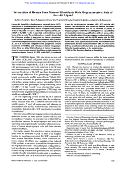
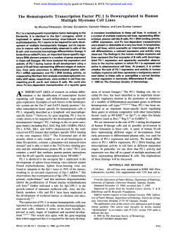
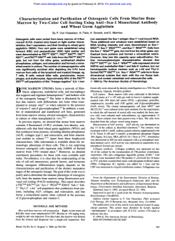
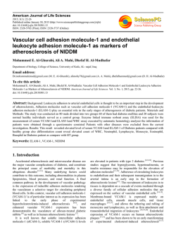
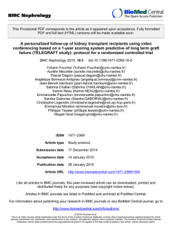
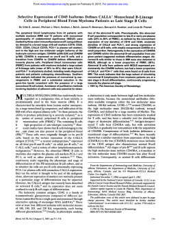
From www.bloodjournal.org by guest on February 6, 2015. For personal use only.
Reduced Expression of Vascular Cell Adhesion Molecule-l on Bone Marrow
Stromal Cells Isolated From Marrow Transplant Recipients Correlates With
a Reduced Capacity to Support Human B Lymphopoiesis In Vitro
By Bonnie N. Dittel and Tucker W. LeBien
A common sequela t o allogeneic or autologous bone marrow transplantation (BMT) is a delay in the reconstitution
of a functional B-cell immune response. Therefore,we examined whether the posttransplant BM microenvironment is
deficient in supporting the proliferation and/or differentiation of B-cell precursors.BM stromal cell cultures were established from patients who received allogeneic orautologousBMT for acute lymphoblastic leukemia,Hodgkin‘s
disease, or non-Hodgkin’s lymphoma. These cuttures were
then compared with normal donor BM stromal cell cultures
for expression of adhesion molecules and the capacity t o
support the adhesion and interleukin-7 (IL7)-dependent
growth of normal B-cell precursors. Analysisof BM stromal
cell cultures established from 28 BMT recipients showed a
significantly reduced expressionof cell surface vascular cell
adhesion molecule-l (VCAM-l/CDlOG), comparedwith nor-
mal donorBM stromal cells.TransplantBM stromal cell
CD106 expression was responsiveto regulatory cytokines in
a manner qualitatively comparable with normal donor BM
stromal cells. Thelevel of B-cell precursor adhesion
to transplant BM stromal cells correlated with the level of CD106
expression. Of 19 evaluable transplant BM stromal cell cultures, eight exhibited a reduced capacity to support the
growth of CD19+/lightchain- normal B-cell precursors. The
capacity of transplant BM stromal cells t o support B-cell
precursor growth correlated with thelevel of CD106 expression, and the level of B-cell precursor adhesion. Our collective results may provide new mechanistic insight into why
B-cell recoveryis delayed post-BMT and underscorethe importance of VCAM-1ICDlO6 in regulating B lymphopoiesis.
0 1995 by The American Society of Hematology.
B
was motivated by the well-known B-cell immune deficiency
that is frequently present in recipients of allogeneic and autologous BMTt4 (and references therein). The kinetics of
immune reconstitution post-BMT exhibit a general pattern:
CD8’ cytotoxic T cells, monocytes, and neutrophils reach
normal numbers in peripheral blood within 1 month, whereas
CD4’ helper T cells and B cells are more delayed in appearance.I4B-cell dysfunction in the early (3 months) post-BMT
period has been attributed to increased T-cell suppression,
decreased T-cell help, or failure to recapitulate B-cell ontogeny because of chronic graft-versus-host d i ~ e a s e . Not~~-~~
withstanding the insight provided by these studies, the mechanism underlying the profound reduction in circulating B
cells during the early post-BMT period has not been elucidated. The results in the current study define heretofore unidentified deficiencies in the capacity of BM stromal cells
derived from BMT recipients to support the adhesion and
growth of B-cell precursors.
LYMPHOPOIESIS occurs exclusively in the bone marrow (BM) of adult mammals and requires the regulated
processing of developmental signals from both cellular and
extracellular matrix components in the BM microenvironment. BM stromal cells, a complex array of nonhematopoietic fixed tissue cells in the medullary cavity, play a crucial
role in murine B-cell development.’” The development of a
long-term murine BM culture that supports the growth of
early B-lineage cells has facilitated analyses of stromal cell
f ~ n c t i o n We
. ~ have developed an in vitro BM stromal cell
culture that supports the interleukin-7 (IL-7)-dependent
growth of normal human B-cell precursor^.^ Recent studies
have shown that CD19+/CD34+ pro-B cells are the 1L-7responsive cells in our BM stromal cell culture, whereas
CD19+/CD34- pre-B cells are completely nonresponsive to
IL-7.’ Our human BM culture, and similar cultures developed by
all
exhibit an absolute dependency Bof
cell precursor growth on an intact BM stromal cell microenvironment.
Several studies have found that the primary adhesive interaction between B-cell precursors and BM stromal cells is
mediated byvery late antigen-4 (VLA-4, also designated
CD49dKD29) and vascular cell adhesion molecule-l
(VCAM-I, also designated CD106), re~pectively.~-”
Miyake
et a1’0.’2 reported that monoclonal antibody (MoAb) to
CD106 and CD49dCD29 inhibit the outgrowth of murine
lymphoid cells in long-term BM cultures. Mouse MoAb to
the CD49d and CD29 subunits of human VLA-4 inhibit the
growth of terminal transferase+ human lymphoid progenit o r ~ .However,
~
Kaisho et all3 reported that anti-CD106
failed to inhibit the growth of the murine pre-B cell line
DW34 on human SV40 transformed BM stromal cell lines
known to express CD106.
Given the importance of an intact BM stromal cell microenvironment for sustaining normal murine B lymphopoiesis,*.*we were interested in developing a strategy to evaluate
the role of BM stromal cells during human B-cell ontogeny.
TO achieve this goal, we characterized the B-lymphopoietic
supportive capabilities ofBM stromal cells isolated from
patients undergoing BM transplantation (BMT). Our study
Blood, Vol 86, No 7 (October 1). 1995:pp 2833-2841
MATERIALS AND METHODS
Cells. BM from normal 18- to 21-week-old human fetuses, normal adult donors, and BMT recipients was obtained in accordance
From the Department of Laboratory Medicine/Pathology and the
Bone Marrow Transplant Program, University of Minnesota Medical
School, Minneapolis.
Submitted October 28, 1994; accepted May 26, 1995.
Supported by Grants No. POI CA-21737 and R01 CA-31685from
the National Institutes of Health and theMinnesota Medical Foundation. B.N.D. was the recipient of a predoctoralfellowship from National Institutes of Health Immunology Training Grant No. T32 AI07313.
Address reprint requests to Tucker W. LeBien, PhD, Box 609
UMHC, Department of Laboratory MedicindPathology, University
of Minnesota Medical School, Minneapolis, MN 55455.
The publication costs of this article were defrayed in part by page
charge payment. This article must therefore be hereby marked
“advertisement” in accordance with 18 U.S.C. section 1734 solety to
indicate this fact.
0 1995 by The American Society of Hematology.
0006-4971/95/8607-0017$3.00/0
2833
From www.bloodjournal.org by guest on February 6, 2015. For personal use only.
2834
with the guidelines of the University of Minnesota Committee on
the Use of Human Subjects in Research. CD10f/CD19'Aight chainB-cell precursors were isolated from human fetal BM using magnetic
bead depletion, as previously described."
Adult BM stromal cells were established from normal donors."
Briefly, normal adult BM was centrifuged on Ficoll-Hypaque gradients (Sigma Chemical CO, St Louis, MO). Then, 10 to 40 x lo6
interface cells were cultured in individual T75 tissue culture flasks
containing 10 mL of EX-CELL 320 (JRH Biosciences, Lenexa,
KS) supplemented with 10% fetal calf serum (FCS), 100 units/mL
penicillin, and 100 pglmL streptomycin. The adherent stromal cells
reached confluence in 7 to 14 days and were passaged twice in EXCELL 320/10% FCS. At this point, we had typically generated four
to nine T75 flasks of normal donor BM stromal cells in approximately 40 days. Normal donor BM stromal cells were used at second
or third passage.
Transplant BM stromal cell cultures were initiated from BM aspirates (2 to 10mL) obtained from individuals receiving autologous or
allogeneic BMT for B-cell precursor-acute lymphoblastic leukemia
(BCP-ALL), Hodgkin's disease (HD), or non-Hodgkin's lymphoma
(NHL). The conditioning regimens and posttransplant therapies administered to these patients are described elsewhere.22.z3BM aspirates were obtained at 21 or 28 days post-BMT from all patients.
Recent analysis of the cellularity of these day 2l/day 28 post-BMT
marrows indicates that they contain from 1% to 100% of the nucleated cell concentration of normal donor BM. BM was washed twice
inRPM1 1640/2% FCS (Hyclone, Ogden, UT), and the cellular
components were plated into a T25 tissue culture flask containing
EX-CELL 320/20% FCS. Nonadherent cells were washed off 1 to
3 days later. Adherent cells were maintained in EX-CELL 320/
20% FCS and monitored for growth by light microscopy. Cultures
exhibiting growth initially appeared as foci of adventitial reticular
cells that underwent expansion tonear confluence within 7 to 14
days. The adherent cell contents of T25 flasks were then expanded
into individual T75 flasks containing EX-CELL 320120% FCS. Once
the stromal cells reached confluence (7 to 14 additional days), they
were passaged twice using txypsiniEDTA. At this point, we had
typically generated four to nine 'I75 flasks of transplant BM stromal
cell cultures in approximately 54 days. Successfully established second or third passage transplant BM stromal cell cultures were maintained in EX-CELL 320/0% FCS without morphological changes or
loss of viability, for greater than 6 months.
Stromal cell cultures were successfully established from 27 BMT
patients, referred to by unique patient numbers (UPN 1-27). Of the
27 cultures, 15 were from patients with BCP-ALL (UPN 1-15), 8
were from NHL (UPN 16-23), and 4 were from HD (UPN 24-27).
Eleven BCP-ALL patients received allogeneic BMT and 4 received
autologous BMT, all 8 NHL patients received autologous BMT, and
1 HD patient received allogeneic BMT and 3 received autologous
BMT. All 27 BMT stromal cell cultures were evaluated for expression of VCAM-I/CD106. However, logistical problems precluded
evaluation of all 27 for the capacity to support adhesion and growth
of B-cell precursors.
Anribodiesand cytokines. The adhesion molecules recognized
by MoAb in the current study have several different designations. For
purposes of continuity, we will use the CD nomenclature throughout.
Therefore, PECAM-1 = CD31, H-CAM = CD44, JCAM-I = CD54,
VCAM-1 = CD106, the a subunit of LFA-I = CDlla, the 8 2
subunit of LFA-I = CD18, the a1 subunit of VLA-1 = CD49a. the
a 2 subunit of VLA-2 = CD49b, the a 3 subunit of VLA-3 = CD49c.
the a 4 subunit of VLA-4 = CD49d, the a 5 subunit of VLA-5 =
CD49e, the a 6 subunit of VLA-6 = CD49f, the @ lsubunit of VLA1-6 = CD29 and the a subunit of the vitronectin receptor = CD51.
The VLA-4, VLA-S, and LFA-I heterodimers will be referred to as
CD49dCD29, CD49e/CD29, and CD1 laICD18, respectively.
4B9/anti-CD10624and LB2/anti-CD54" were gifts from Drs John
DITTELANDLEBIEN
Harlan and Ed Clark (both from the University of Washington,
Seattle). PSDZ/anti-CD29, P2G12/anti-CD31, P3H9-3- llanti-CD44,
TS217/anti-CD49a, PlHS/anti-CD49b, PlBS/anti-CD49c, P4C2/
anti-CD49d. PlD6/anti-CD49e, GOH3/anti-CD49f, P3G8/anti-CD5I
were gifts from Dr Elizabeth Wayner (Fred Hutchinson Cancer ReP8B1-2/anti-CD106 and P4G11-2/
search Center, Seattle, WA).26-Z*
anti-CD54 were produced by immunizing mice with IL-18-induced
adult BM stromal cells. The specificity of the latter two MoAbs was
confirmed by immunoprecipitation. GH12 and ED11 are CD106
specific MoAb were kindly provided by Dr Laurelee Osbom (Biogen, Cambridge, MA)?9 Hybridoma cells secreting W6/32/antiHLA-A, B, C, and L243/anti/MHC class I1 were obtained from the
American Type Culture Collection (Rockville, MD). JS/anti-CDlO
was purchased from Coulter Immunology (Hialeah, FL). Hybridoma
cells secreting 25Cllanti-CD19 were kindly provided by Dr Stephen
Peiper (University of Louisville School of Medicine, Louisville,
KY). 25C1 was conjugated to phycoerythrin (PE) using standard
methods.3"HPCA-Uanti-CD34 and Leu-IZ-PE/anti-CD19 were purchased from Becton Dickinson Immunocytometry Systems (San
Jose, CA). IgG1, IgG2a, and IgG2b control mouse myeloma proteins
were purchased from Organon Teknika-Cappel (Durham, NC).
F(ab'), goat antimouse Ig-FITC was purchased from Tago, Inc
(Burlingame, CA).
Recombinant human IL-18, recombinant human IL-4, and porcine
transforming growth factor-8 (TGF-8) were obtained from R & D
Systems (Minneapolis, MN). Recombinant human IL-7 was obtained
from PeproTech Inc (Rocky Hill, NJ).
Flow cytometry. Expression of cell surface molecules onBM
stromal cells was analyzed by indirect immunofluorescent staining
andflow cytometry, as previously described." BM stromal cells
from normal donors and transplant recipients were dissociated from
tissue culture flasks using Cell Dissociation Solution (Sigma Chemical CO).This nonenzymatic solution does not disrupt epitopes recognized by the MoAb used to characterize the stromal cells. Stromal
cells were stained for the expression of CD10, CD29, CD31, CD34,
CD44, CD49a-f, CD5 1, CD54, CD106, Class I major histocompatibility complex (MHC) and class 11 MHC using saturating concentrations of MoAb, counterstained with F(ab'), goat antimouse
IgG-FITC, and fixed in 1% paraformaldehyde before analysis on a
FACScan (Becton Dickinson, San Jose, CA) using CONSORT 30
software. Negative controls included mouse IgG,, IgGZ., or IgGZh
myeloma proteins, Data are reported as percent positive cells or
mean channel fluorescence (MCF) of 5,000 cells analyzed on a log
scale. The influence of cytokines on BM stromal cell CD106 and
CD54 expression was conducted as previously described."
Adhesion assay. Adhesion of CDlO'ilight chain- B-cell precursors to normal and transplant BM stromal cells was performed as
previously described." Briefly, 50,000 Naz "CrO, radiolabeled
CDlO'Aight chain- cells were incubated with normal or transplant
BM stromal cells in the presence or absence of blocking MoAb for
2 hours at 37°C. After two washes to remove nonadherent cells, the
stromal cell adherent B-cell precursors were quantitated on a y counter. Each experimental variable was tested in replicates of eight.
Percent B-cell precursor adherence was calculated as follows:
% Adherence =
Mean Adherent cpm
x 100
Total Input cpm
B-cell precursor growth assay. B-cell precursors were cultured
in vitro as previously described: with the following modifications.
Normal or transplant BM stromal cells were seeded onto 96-well
flat-bottom microtiter plates at 3,000 cells per wellin EX-CELL
320/10% FCS. After 3 days, the medium was switched to X-VIVO
1010% FCS and maintained until further use ( 2 to 4 days). Normal
B-cell precursors were seeded onto normal or transplant BM stromal
cells at approximately 20,000 to 40,000 cells per well in X-VIVO
10/0% FCS containing 10 ng/mL IL-7. The cultures were fed every
From www.bloodjournal.org by guest on February 6, 2015. For personal use only.
2835
BONE MARROW STROMAL CELL VCAM-1
5 days with fresh X-VIVO 10/0% FCS containing 10 ndmL IL-7.
CD19+ B-cell precursors were quantitated on days 0, 7, 14, and 21
by harvesting the cellular contents of the microtiter wells with Cell
Dissociation Solution (Sigma) and stained for expression of CD19
using Leu-12-PE or 25C1-PE. For each condition, the cellular contents from 12 microtiter wells were pooled in groups of three wells,
to provide four replicate data points. Polystyrene6.0 micron microspheres (Polysciences, Inc. Warrington, PA) were used to quantify
cell numbers using the F A C S C ~ ~ ? . ~ '
Statistics. Statisticalsignificancewasdetermined
bytheunpaired Student's t-test using Statworks (Cricket Software, Philadelphia, PA). Linear regression analysis and Pearson's correlation
coefficient were determined using Cricket Graph (Cricket Software).
RESULTS
Transplant BM stromal cells express lower levels of
CD104 compared with normal BM stromal cells. B-cell Stroma
precursor/BM stromal cell adhesive interactions mediated by
30
CD49dCD29 and CD106 regulate the in vitro adhesion and
B
growth of normal B-cell precursors in human^'^^^" and
g 25~ c e . ~ n . Therefore,
~ 2
we reasoned that an alteration in the
BMT Recipient
Normal
Stroma
U
'C
v)
expression and/or function of CD106 on BM stromal cells
v)
Q)
20after BMT may help explain the delayed appearance of peL
n
0
A
ripheral blood B cells so frequently observed posttransG 15BM aspirates from 73 BMT recipients were colt
lected on day +21 or +28 posttransplant, and cultured as
v)
n
described in Materials and Methods. Stromal cell cultures
u
were successfully established from 27 patients (see Materials
e
Q
and Methods). No correlation was observed between the
Q)
success or failure in establishing transplant BM stromal cell
I:
cultures relative to the hematopoietic disorder, type of transplant (allogeneic or autologous), age of the recipient, or
BMT Recipient
Normal
number of remissions patients had experienced at the time
Stroma
Stroma
their marrows were cultured.
We compared the CD106 fluorescence intensity on BM
Fig 1. Expression of CD106 and CD54 on normal and transplant
stromal cells established from 12 normal donors and 27BMT
stromal cells. BM stromal cells from 27 transplant recipients IO) and
recipients. As shown in Fig lA, the mean CD106 MCF on
12 normal donors (A) were analyzed for the expression of CD106 (A)
and CD54(B1 by indirect immunofluorescence on a FACScan. The
normalBM stromal cells was 23.4 9.8 (range, 5 to 36),
individual data points represent the CD106 or CD54 M W minus the
whereas the mean CD106 MCF on transplant BM stromal
MCF of the isotype control. If an individualtransplant or normal strocells was 14.8 5 8.7 (range, 2 to 38). This difference was
mal cell culture was analyzed more thanonce, the value represented
significant (P < .Ol). In contrast, Fig 1B shows that the
in (A) and (B) is the mean of all observations.
CD54 MCF on normal BM stromal cells was6.7 2 4.7
(range, 2 to 18), whereas the CD54 MCF on transplant BM
stromal cells was 6.8 t 5.7 (range, 0 to 26). Even though
stained with MoAb specific for CD106 domain 1 (MoAb
the transplant BM stromal cells analyzed had a statistically
4B9), the domain 1-2 interface (P8B1-2), and domain 4
significant reduction in the intensity of CD106 expression,
(GH12). The results in Fig 2 clearly demonstrate thatall
their expression of CD10, CD29, CD44, CD49a, CD49b,
three anti-CD106 MoAb stain both normal and transplant
CD49c, CD49e, CD51, and class I MHC was indistinguishBM stromal cells. Furthermore, normal and transplant BM
stromal cells express the 7 Ig form of CD106, as indicated by
able from normal BM stromal cells (data not shown).
CD106 cell surface expression on normalBM stromal
positive staining with the CD106 domain 4-specific MoAb
cells exhibited minor fluctuation as a function of time in
GH12. A second MoAb specific for CD106 domain 4 (ED1 1)
culture, but expression was essentially unchanged for periods
stained normal and transplant BM stromal cells in an identiup to 4 months in serum-free medium (data not shown).
cal manner to MoAb GH12 (data not shown). The low-level
CD106 staining in Fig 2 (similar to our previous study")
Expression of the 7 Ig form of CD104 on normal and
transplant BM stromal cells. Because the 4th Ig domain
does not preclude the possibility that CD106 negative stroin the 7 Ig domain form of CD106 can be alternatively
mal cells exist. However, they would be present in normal
spliced to yield a 6 Ig domain form of CD106,32.33 we
examand transplant BM stromal cell cultures.
ined BM stromal cells obtained from both normal donors
Cytokine regulation of CD104 on normal and transplant
and transplant recipients for the expression of these two
BM stromal cells. In previous studies we showed that cell
CD106 isoforms. BM stromal cells cultured from one normal
surface expression of CD106 on normal BM stromal cells
donor and from transplant recipients UPN 25 and 26 were
is upregulated by the cytokines L-lg and IL-4, and down-
From www.bloodjournal.org by guest on February 6, 2015. For personal use only.
DIlTEL AND LEBIEN
2836
1
NormalStroma
MCF=55
UPN 26
UPN 25
1
MCF=161
Domain 1
1
MCF=43
MCF=14
Domain
Domain4
FluorescenceIntensity
Fig 2. Expression of the 7-19 isoform of CD106 on BM stromal cells from a normal donor and Hodgkin's disease transplant recipients UPN
25 and 26. B M stromal cells were stained by indirect immunofluorescence with anti-CD106 MoAb 489. P8B1-2, and GH12 at 10 pg/mL to
detect CD106 "19-like" domains 1,1/2 interface, and domain 4, respectively, and analyzed on a FACScan. Expression of CD106 isrepresented
by solid lines and the isotype control is represented by the dotted lines in the top panel of histograms. The CD106 MCF is indicated in the
upper righthand corner of each individual histogram.
regulated by TGF-D." The cytokine regulated increase or
decrease in CD106 led to a corresponding increase or decrease in the adhesion ofB-cell precursors.'' The data in
Table I show that treatment of normal and transplant BM
stromal cells with IL-ID and IL-4 leads to an increase in
CD106 expression, and an increase in B-cell precursor adhesion. Conversely. incubation of normal BM stromal cells
with TGF-/3 leads to a reduction in CD106 expression and
a corresponding decrease in B-cell precursor adhesion. Evaluation of the effect of TGF-Don transplant BM stromal
cells was compromised by the decreased CD106 expression
present on the majorityof transplant BM stromal cells (Table
1 and Fig 1 ).
Using MoAb toa panel of adhesion molecules, we showed
that B-cell precursor adhesion to transplant BM stromal cells
was mediated by CD49d/CD29 and CD106 (data not shown),
comparable to our earlier studies with normal BM stromal
cells."
Trurlsplant BM s t r o n d cells from n subset of patients
.show CI reduced cnpacih t o support tire in vitro proliferation
of B-cell precursors. We usedanIL-7/BM
stromal cell
culture to examine the capacity of transplant BM stromal
cells to support the in vitro growth of normal B-cell precur-
sors. As shown in Table 2,8 of the 19 transplant BM stromal
cell cultures examined exhibited a reduced capacity to supportB-cell precursor proliferation (<90% of control BM
stromal cells at all three timepoints, and a mean < 80%).
The remaining 1 1 transplant BM stromal cell cultures supportedthe in vitro growth of B-cell precursors similar to
normal stromal cells (>90% of control stroma for at least
one time point, and a mean > 80%). Statistical comparison
of the 8 cultures with reduced supportive capacity (mean ?
SD = 66.2% ? 6.8%) to the 1 1 cultures with normal supportive capacity (mean ? SD = 89.6% ? 5.9%) showed a P
value of < .OOl (unpaired t-test). The reduced support of Bcell precursor growth observed with some transplant BM
stromal cells could notbe explained by simple differences
in plating densities of the BM stromal cells. When normal
BM stromal cells were seeded at densities of 6,000, 3,000,
1,500, or 750 cells/well, similar numbers of CD19' cells
were recovered fromeach culture 14 days later (data not
shown).
Adhesion of B-cell precur.sors is correlated to the expression of CDl06, but not CD54, on transplant BM stromal
cells. We have previously reported" and demonstrated in
Table l , that B-cell precursor adhesion to normal and trans-
ROMAL
From www.bloodjournal.org by guest on February 6, 2015. For personal use only.
BONE MARROW
CELL VCAM-1
2837
Table 1. Adhesion of B-Cell Precursorsto Normal and BMT Stromal Cells CorrelatesWith the Expression of CD106
Normal Stroma
UPN 9
CD106'
Medium alone
IL-lp (100 pg/mL)
IL-4 (10 ng/mL)
TGF-8 (1 ng/mL)
CD106
CD106
Fluorescence
Condition
UPN 21
Fluorescence
% Adhesion*
23 -c 1.2
31 I
1.0
39 2 1.1
19 2 1.0
26
36
34
11
19
40
18
33
34
14
% Adhesion
14
-t 0.8
33 2 0.8
2 1.8
19 2 0.7
28
Fluorescence
% Adhesion
8
16
15
2 0.5
21 2 0.6
2 1.2
11 i 0.6
6
~
Numbers represent the CD106 MCF value of a single stromal cell culture measured on a FACScan.
t Values represent the mean t SE of eight replicates.
plant BM stromal cells parallels the cytokine regulated expression of CD106. Linear regression analysis comparing
the level of cell surface CD106 expression by transplant BM
stromal cells with the ability of the same stromal cells to
support the adhesion of B-cell precursors showed a linear
relationship (Fig 3A, R = .638, P = .006). In contrast, no
linear relationship was observed between B-cell precursor
adhesion and CD54 expression (Fig 3B, R = .152, P = S73).
Expression of CD106 on transplant BM stromal cells is
correlated to the level of IL-7-induced B-cell precursor
Table 2. A Subset of BMT Recipient Stromal Cells Exhibits a
Reduced Capacityto Support the In Vitro Growthof Normal
B-Cell Precursors
B-Cell Precursor Growth*
UPN
Diseasenransplanr
Day7
Day 14
Day21
Mean
6
7
8
9
10
11
12
14
16
17
18
19
20
21
22
23
25
26
27
ALL-ALL0
71t
56t
95
86
81t
80t
102
84
69t
92
102
58
66t
82t
84t
92
72
48
98
68t
59t
95
92
69t
60
82
78
52t
100
93
100
80t
82t
49 t
123
98
98
106
56t
57t
93
68
53t
55t
64
102
63t
65t
57t
94
82
68t
65t
83
88
61t
97
99
88
72t
79t
63t
94
84
84
93
ALL-AUTO
NHL-AUTO
HD-AUTO
100
101
106
71t
73t
56t
68
83
105
75
Abbreviations: ALL-acute lymphoblastic leukemia; NHL, non-Hodgkin's lymphoma; HD, Hodgkin's disease; ALLO-allogeneic, AUTO-autologous.
Values represent the percentage of CD19' B-lineage cells recovered from transplant BM stromalcells compared with the number of
CD19' B-lineage cells recovered from normal BM stromal cells. The
percentage was derived by dividing theabsolute CD19' cell number
recovered on transplant stromal cells bythe absolute CD19'cell number recovered on normal stromalcells. The mean value is theaverage
of the three time points.
t Values defined as having a reduced capacity to support the in
CD19' B-cell precursors (<go% of control stromal
cells
vitro growth of
at all three timepoints and a mean of <EO%).
proliferation. Because the in vitro proliferation of normal
human B-cell precursors is absolutely dependent on BM
stromal cells: linear regression analysis was used to compare
transplant BM stromal cell CD106 expression with the ability to support B-cell precursor proliferation. As shown in
Fig 4A, the expression of CD106 was linearly related to the
recovery of CD19+ cells on day 7 ( R = .459, P = .07).
However, this relationship was lost by day 14 (Fig 4B, R =
.l18, P = .66). Likewise, no linear relationship existed on
day 21 ( R = .145, P = .58)(data not shown). When CD54
expression by transplant BM stromal cells was compared
with the recovery of CD19' B-cell precursors on days 7 (Fig
4C) and 14 (Fig 4D), no linear relationship was observed.
Adhesion of B-cell precursors to transplant BM stromal
cells is correlated to the proliferation of B-cell precursors.
Because CD106 expression was correlated to B-cell precursor adhesion (Fig 3A) and proliferation (Fig 4A), we analyzed the relationship between the ability of transplant BM
stromal cells to support the adhesion and proliferation of Bcell precursors. The data in Fig 5 show a linear relationship
between adhesion of B-cell precursors and the maximum
recovery of CD19+ cells on either day 7, 14, or 21 ( R =
S38, P = .03).Weaker correlations between adhesion and
proliferation of B-cell precursors were observed on days 7
( R = .263, P = .14), 14 ( R = .377, P = .12), and 21 ( R =
,322, P = .16).
DISCUSSION
Studies of human B-cell ontogeny have been facilitated
by the development of BM stromal cell-dependent cultures
that support the in vitro growth of normal B-cell precursors?6'8 Adhesion of normal B-cell precursors to BM
stromal cells is primarily mediated by CD49dCD29 and
CD106.9-LL
MoAb to murine CD49dCD29" and CD106"
block B lymphopoiesis in Whitlock-Witte cultures, and
MoAb to the CD49d and CD29 subunits of human VLA-4
inhibit the growth of TdT+ lymphoid progenitors? However,
evidence implicating CD106 in functional events beyond the
initial adhesion of human B-cell precursors to BM stromal
cells has not been reported. The delay in B-cell reconstitution
after BMT has been known for some time.I4 Given the lack
of a unifying mechanistic model to explain this delay, we
hypothesized that an abnormality in BM stromal celVB-cell
precursor interactions may be involved. The rationale underlying this hypothesis rests partially on the large body of
evidence identifying the toxic effects of infectious agents,
chemical agents, and ionizing irradiation on the BM micro-
0
From www.bloodjournal.org by guest on February 6, 2015. For personal use only.
2838
DllTEL AND LEBEN
A
C
.-0
150
v)
v)
P)
L.
n
x
W
100
(D
0
F
50
P
V
1
l
L
o !
75
50
B
-=
.
,
.
II
m
,
.
,
.
,
.
I
25
B Cell PrecursorAdhesion
B
A
400
C
.-0
v)
v)
300 4
x
W
d
v)
P
V
A A '
25
50
75
A
100
125
150
B Cell PrecursorAdhesion
Fig 3. Relationship between CD106 expression and CD54 expression on transplant BM stromal cells and their capacity to support
the adhesion of B-cell precursors. B-cell precursor adhesion on the
horizontal axis is represented as the adherent cpm of B-cell precursors to transplant BM stromal cells, divided by the adherent cpm of
B-cellprecursors to normal BM stromal cells, multiplied by 100.
CD106 (A) or CD54 (B) expression onthe vertical axis is represented
as the CD106 or CD54 MCF (minus the isotype control MCF) of transplant BM stromal cells, divided by the CD106 or CD54 MCF (minus
the isotype control MCF) of normal BM stromal cells, multiplied by
100. Eachindividualdata point represents CD106 expression
(A) plotted against B-cell precursor adhesion forUPN 6-12, 14, 16, 17,20-23,
and 25-27, or CD54 expression (B) plotted against B-cell precursor
adhesion for UPN 6-12, 14, 16, 17, 20-22, and 25-27. If an individual
transplant BM stromal cell culture was analyzed more than once,
the value represented is the mean of all observations. Analysis of
transplant and normal BM stromal cells for CD106and CD54 expression and adhesion of B-cell precursorswas conducted on the same
day for each individual data point.
e n v i r ~ n m e n tTherefore,
.~~
we reasoned that one consequence
of this BM damage might be an alteration in the capacity of
BM stromal cells to support the adhesion and growth of Bcell precursors. This could, in turn, delay the appearance of
normal B-cell numbers in peripheral blood.
Although normal human B-cell precursors can be grown
on BM stromal
the technology has not developed
to the point where limiting numbers (ie, the number of Bcell precursors that would be present in BM aspirates at
day 28 (post-BMT) can be routinely grown. We therefore
compared the functional integrity of BM stromal cells established from BMT recipients and normal donors for their
adhesion molecule expression and the capacity to support
the adhesion and growth of normal B-cell precursors. The
strategy we employed was to establish BM stromal cell CUItures from patients who had received allogeneic or autologous BM transplants for BCP-ALL, NHL, or HD. We SUCcessfully established BM stromal cells from 27 of 73
patients, but observed no relationship between successful
establishment and type of transplant, underlying disease, or
recipient age. One explanation for the failure to establish
confluent BM stromal cell cultures may have been damage
from irradiation andor chemotherapy, resulting in extreme
hypocellularity in the BM aspirates used to initiate the cultures (see below).
Although the hypocellularity of recipient marrowat 21
to 28 days post-BMT resulted in a consistent delay in the
establishment of transplant BM stromal cell cultures compared withnormal donor BM, the cell surface phenotype
was indistinguishable, with the exception of CD106. The
level of CD106 was significantly lower on BMT recipient
stromal cell cultures compared with normal donors (Fig 1 ),
buttherewasno
apparent abnormality in CD106 isoform
expression (Fig 2 ) or regulated response to cytokine stimulation (Table 1). The reduced CD106 expression has several
possible explanations including: outgrowth of a subpopulation of BM stromal cells expressing low CD106, senescence
of transplant BM stromal cells, or inheritable changes in
BM stromal cell gene expression secondary to DNA damage
caused by irradiation or chemotherapy. The former possibility seems unlikely for several reasons. First, 16 distinct cell
surface antigens identified by MoAb were identical in expression on normal and transplant BM stromal cells, with
the exception of CD106. In addition, cytokine responsiveness based onregulated expression of CD106 was essentially the same (Table l), andbothnormaland
transplant
BMstromal cells expressed the 7 Igdomain isoform of
CD106 (Fig 2). Thus, by the criteria of constitutive expression of many distinct gene products, and cytokine-regulated
expression of CD106, the predominant cellular component
of normal and transplant BM stromal cell cultures was the
same. We cannot rule out the possibility that small numbers
of macrophages were presentin some of the stromal cell
cultures, and macrophage-derived cytokines (eg, IL- 1) can
regulate CD106 expression. Although we didnot monitor
macrophage content in all the BM stromal cell cultures used
in the current study, our previous studies have shown that
CD 1 1b+/HLA-DR+macrophages are very low in number or
undetectable in second passage BM stromal cells."
Transplant BM stromal cells deficient in the capacity to
support the adhesion and growth of B-cell precursors may
have been senescent, due to a potentially increased number
of doublings required to generate confluent monolayers. We
tried to control for this possibility by classifying our BM
stromal cell cultures using criteria previously used to categorize aging fibroblasts in vitro.?' We only used normal and
transplant BM stromal cell cultures that displayed morphologic characteristics of mitotic and postmitotic fibroblasts;
that is, fibroblasts able to adhere, spread, and form confluent
monolayers? Transplant and normal BM stromal cell cul-
From www.bloodjournal.org by guest on February 6, 2015. For personal use only.
2839
BONE MARROWSTROMALCELLVCAM-1
200
A
W
-
(11
B
n 100
nl
75
50
25
100
.
.D .
,
D
.
,
.
,
.
125
B CellPrecursorGrowth
B CellPrecursorGrowth
A
4000
m
P
W"
P
.
A
AA
200200
v)
n
P
A A
100100
A
O
25
.
.
A
.
50
A
A
.
.
75
.
U
'
100
P
A
"
A
A
n
125
B CellPrecursorGrowth
B CellPrecursorGrowth
Fig 4. Relationship between CD106 expression andCD54 expression on transplant BM stromal cells and their cqpacity to support the IL7-induced proliieration of Bsell precursors. B-cell precursorproliieration on the horizontal axis is represented as absolute numbersof CD19+
cells recoveredfrom transplant BM stromal cells, divided by the absolute numbers of CD19+ cells recoveredfrom normal BM stromal cells,
multiplied by 100. CD106 (A, B) or CD54 IC, D) expression on the vertical axis is representedas the CD106 or CD54 MCF (minus the irotype
control MCF) of transplant stromal cells divided by the CD106 or CD54 MCF(minusthe isatype control MCFI of normal stromal cells, multiplied
by 100. Individual data points represent CD106 expression plotted against B a l l precursor proliieretion on day 7 (AI and day 14 (B) for UPN
6-12,14,16,17,20-23, and 25-27, or CD54 expression plotted against B e l l precursor proliferation on day 7 (C) and day 14 (D) for UPN 6-12,
14, 16, 17,20-22, and 25-27. If an individual tranrplant BM stromal cell was analyzed morethan once, the value representedis the mean of a11
observations. Analysis of transplant and normal BM stromal cells for CD106 and CD54 expression was conducted the same day the Bsell
precursor proliferation assay was initiated.
tures were both used at second or third passage and displayed
indistinguishable morphological characteristics and growth
kinetics. The optimal approach for initiation and establishment of stromal cell cultures would employ positive selection (perhaps using anti-CD106 MoAb) of BM stromal cell
precursors (ie, the adventitial reticular cells that adhere to
tissue culture flasks and undergo expansion), and subsequent
plating of equal numbers in culture on day 0.
Transplant BM stromal cell cultures expressing reduced
levels of CD106 may have acquired genetic changes induced
by irradiation or chemotherapy. The reduced expression of
cell surface CD106 (Fig 1) could reflect alterations in transcription, translation, or protein stability. CD106 expression
is regulated differently in individual cell types. For example,
CD106 is only expressed on endothelial cells following cytokine ~timulation.~~*~'
Alternatively, BM stromal cellsg"'
and follicular dendritic cells3' constitutively express CD106,
which can be increased by specific cytokines. Although transcriptional regulation ofBM stromal cell CD106 has not
been characterized, transcription of CD106 in endothelial
cells is known to be regulated by two NF-kB sites within
the CD106 promoter.3941Independent of the mechanism by
which CD106 expression is regulated following cytokine
stimulation of BM stromal cells, this process does not appear
to be grossly altered in stromal cells established from transplant recipients (Table 2). Thus, the mechanism by which
constitutive CD106 expression is reduced in BMT recipient
stromal cells remains to be determined.
Although not investigated in the current study, alterations
in cytokine production could also be involved. TGF-P downregulates CD106 expression in normal BM stromal cells,"
and CD106 is constitutively expressed by BM stromal cells
established under Dexter culture conditi0ns.4~~~
Thus, overexpression of stromal cell TGF-P could lead to reduced levels of CD106 expression, in addition to exerting a general
antiproliferative effect on a variety of lymphohematopoietic
cells. It is noteworthy that increased levels of TGF-P production by BM stromal cells from patients with chronic lymphocytic leukemia have been reported.&
Our inability to establish transplant BM stromal cell cultures from -60% of BM aspirates may reflect the intrinsic
hypocellularity of recipient BM in the first several weeks
posttransplant. Indeed, some BM aspirates gave rise to adherent cells with only a limited capacity to grow, thereby
yielding partially confluent cultures that could not be expanded for functional studies. The toxic effects of ionizing
From www.bloodjournal.org by guest on February 6, 2015. For personal use only.
2840
DITTEL AND LEBIEN
150
to support B-cell precursor adhesion and growth. The reduced CD106 expression could result in a lower threshold of
cell signaling events delivered to B-cell precursors through
CD49dCD29, leading to a decrease in B-cell precursor proliferation. However, given the array of toxic effects exerted
by irradiation and chemotherapy on the BM microenviron~ n e n tit, ~
is ~almost certain that additional abnormalities exist
that compromise the B lymphopoietic supportive capacity
of BM stromal cells.
c
.-0
v)
Q
c
125
2
100
75
ACKNOWLEDGMENT
50
m
1
50
75
100
125
150
B Cell Precursor Growth
Fig 5. Relationship between the capacity of transplant BM stromal cells to support B-cell precursor adhesion and
117-induced proliferation of Bsell precursors. B-cell precursor proliferation on the
horizontalaxis is represented asthe absolute number ofCDl9+ cells
recovered from transplant BM stromal cells, dividedby the absolute
number of CD19+cellsrecovered from normal BM stromal cells,
multiplied by 100. B-cell precursor adhesion on the vertical axis is
represented as the adherent cpm of Bsell precursors to transplant
BM stromal cells, divided by the adherent cpm of B-cell precursors
to normal BM stromal cells, multiplied by 100. Each individual data
point represents the maximum recovery of CD19* cells on day
7,14,
or 21 plotted against B a l l precursor adhesion forUPN 6-12, 14, 16,
17,ZO-23, and 25-27. If an individual transplantBM stromal cell was
analyzed more than once, the value represented is the mean of all
observations. Analysis oftransplant and normal BM stromel cells for
edhesion of &cell precursorswas conducted the same day the B-cell
precursor proliferation assay was initiated.
irradiation on the BM microenvironment are l e g i ~ n . ~Ex~.~'
periments in the mouse have noted damage to BM stromal
cells after irradiation or chemotherapy, manifested as morphologically altered cytoplasmic organelles andplasma
membranes, cytoplasmic vacuoles, and nuclear damage.34
Our difficulty in establishing confluent monolayers might
find explanation in earlier murine studies. Whereas highdose-irradiated marrow sites showed no evidence of structural damage in situ, significant cell kill was observed after
one to two traversals of the cell cycle in
High-doseirradiated sites left undisturbed for 6 months showed significant repair of potentially lethal irradiation da1nage.4~Extrapolating these findings to our study, it is conceivable that
some of our transplant BM stromal cell culture failures could
reflect an inability to repair irradiation damage that culminated in premature stromal cell death in vitro. Other studies
have noted heterogeneity in the radiosensitivity of human
BM stromal cell lines (established from a single donor) to
low-dose-rate irradiation." Therefore, it would seem highly
possible, and even probable, that radiobiologic differences
would exist in radiosensitivity and repair capacity of BM
stromal cells from different transplant recipients.
In conclusion, our results further illuminate the functional
importance of CD106 expression on human BM stromal cells
that support the IL-7-dependent growth of human B-cell
precursors. We have shown that BM stromal cells isolated
from transplant recipients have a reduced expression of
CD 106, and this reduction correlates with a reduced capacity
We thank Joanne Howard and the bone marrow transplant physicians for their generous assistance in providing bone marrow aspirates and Dr Michael Steinbuch for providing information on the
clinical characteristics of the patients. Drs John Kersey and Daniel
Weisdorf provided helpful comments on the manuscript. We thank
Karen Nelson for assistance with word processing. Dr Laurelee Osborn (Biogen Corp. Cambridge, MA) kindly provided the GH12 and
ED11 anti-CD106 Ab. We particularly thank Dr Steve Rich for his
advice on statistical analyses.
REFERENCES
1. Kincade PW, Lee G, Watanabe T, Sun L, Scheid MP: Cells
and molecules that regulate B lymphopoiesis in bone marrow. Annu
Rev Immunol 7:111, 1989
2. Dorshkind K: Regulation of hemopoiesis by bone marrow stromal cells and their products. Annu Rev Immunol 8:1l l , 1990
3. Whitlock CA, Witte ON: Long-term culture of B lymphocytes
and their precursors from murine bone marrow. Roc Natl Acad Sci
USA 79:3608, 1982
4. Wolf ML, Buckley JA, Goldfarb A, Law C-L, LeBien TW:
Development of a bone marrow culture for maintenance and growth
of normal human B cell precursors. J Immunol 147:3324, 1991
5. Dittel BN, LeBien TW: The growth response to IL-7 during
normal human B cell ontogeny is restricted to B-lineage cells expressing CD34. J Immunol 154:58, 1995
6. McGinnes K, Quesniaux V, Hitzler J, and Paige CJ: Human
B-lymphopoiesis is supported by bone marrow-derived stromal cells.
Exp Hematol 19:294, 1991
7. Ryan DH, Nuccie BL, Abboud CN: Inhibition of human bone
marrow lymphoid progenitor colonies by antibodies to VLA integrins. J Immunol 149:3759, 1992
8. Moreau I, Duvert V, Banchereau J, Saeland S : Culture of human fetal B-cell precursors on bone marrow stroma maintains highly
proliferative CD20"" cells. Blood 81:1170, 1993
9. Ryan DH, Nuccie BL, Abboud CN, Winslow JM: Vascular
cell adhesion molecule-l and the integrin VLA-4 mediate adhesion
of human B cell precursors to cultured bone marrow adherent cells.
J Clin Invest 88:995, 1991
IO. Miyake K, Medina K, Ishihara K, Kimoto M, Auerbach R,
Kincade PW: A VCAM-like adhesion molecule on murine bone
marrow stromal cells mediates binding of lymphocyte precursors in
culture. J Cell Biol 114:557, 1991
11. Dittel BN, McCarthy JB, Wayner EA, LeBien TW: Regulation of human B-cell precursor adhesion to bone marrow stromal
cells by cytokines that exert opposing effects on the expression of
vascular cell adhesion molecule-l (VCAM-1). Blood 81:2272, 1993
12. Miyake K, Weissman L,Greenberger JS, Kincade PW: Evidence for a role of the integrin VLA-4 in lympho-hemopoiesis. J
Exp Med 173:599, 1991
13. Kaisho T, Oritani K, Ishikawa J, Tanabe M, Muraoka 0, Ochi
T, Hirano T: Human bone marrow stromal cell lines from myeloma
and rheumatoid arthritis that can support murine pre-B cell growth.
J Immunol 149:4088, 1992
14. Lum LG: The kinetics of immune reconstitution after human
marrow transplantation. Blood 69:369, 1987
From www.bloodjournal.org by guest on February 6, 2015. For personal use only.
BONE MARROW STROMAL CELL VCAM-1
15. Wimperis JZ, Brenner MK, Prentice HG, Thompson EJ, Hoffbrand AV: B cell development and regulation after T cell-depleted
marrow transplantation. J Immunol 138:2445, 1987
16. Kiesel S, Pezzutto A, Moldenhauer G, Rainer R, Korbling
M, Hunstein W, Dorken B: B-cell proliferative and differentiative
responses after autologous peripheral blood stem cell or bone marrow transplantation. Blood 72:672, 1988
17. Bengtsson M, Smedmyr B, Festin R, Oberg G, Simonsson B,
Tottennan TH: B lymphocyte regeneration in marrow and blood
after autologous bone marrow transplantation: Increased numbers of
B cells carrying activation and progression markers. Leuk Res
13:791, 1989
18. Kagan JM, Champlin RE, Saxon A: B-cell dysfunction following human bone marrow transplantation: Functional-phenotypic
dissociation in the early posttransplant period. Blood 74777, 1989
19. Small T N , Keever CA, Weiner-Fedus S, Heller G, O’Reilly
RJ, Flomenberg N: B-cell differentiation following autologous, conventional, or T-cell depleted bone marrow transplantation: A recapitulation of normal B-cell ontogeny. Blood 76:1647, 1990
20. Storek J, Ferrara S , Ku N, Giorgi JV, Champlin RE, Saxon
A: B cell reconstitution after human bone marrow transplantation:
Recapitulation of ontogeny? Bone Marrow Transplant 12:387, 1993
21. Villablanca JG, Anderson JM, Moseley M, Law C-L, Elstrom
RL, LeBien T W : Differentiation of normal human pre-B cells in
vitro. J Exp Med 172:325, 1990
22. Weisdorf DJ, Woods WC, Nesbit ME, Uckun F, Dusenbery
K, Kim T, Haake R, Thomas W, Kersey JH, Ramsay NKC: Allogeneic bone marrow transplantation for acute lymphoblastic leukaemia:
Risk factors and clinical outcome. Br J Haematol 86:62, 1993
23. Weisdorf D, Katsanis E, Verfaillie C, Ramsay NKC, Haake
R, Garrison L, Blazar BR IL-laadministered after autologous transplantation: A phase I/II clinical trial. Blood 84:2044, 1994
24. Carlos TM, Schwartz BR, Kovach NL, Yee E, Rosso M,
Osbom L, Chi-Ross0 G, Newman B, Lobb R, Harlan JM: Vascular
cell adhesion molecule-l mediates lymphocyte adherence to cytokine-activated cultured human endothelial cells. Blood 76:965, 1990
25. Clark EA, Yokochi Y: Human B cell and B cell blast-associated surface molecules defined with monoclonal antibodies, In Bernard, A, Bournsell L, Dausset J, Milstein C, Scholossman SF (eds):
Leukocyte Typing I. Human Leukocyte Differentiation Antigens.
Berlin, Germany, Springer, 1984, p 339
26. Wayner EA, Garcia-Pardo A, Humphries MJ, McDonald JA,
Carter WC: Identification and characterization of the T lymphocyte
adhesion receptor for an alternative cell attachment domain (CS-l)
in plasma fibronectin. J Cell Biol 109:1321, 1989
27. Wayner EA, Carter WC: Identification of multiple cell adhesion receptors for collagen and fibronectin in human fibrosarcoma
cells possessing unique a and common p subunits. J Cell Biol
105:1873, 1987
28. Wayner EA, Carter WC, Piotrowicz RS, Kunicki TJ: The
function of multiple extracellular matrix receptors in mediating cell
adhesion to extracellular matrix: Preparation of monoclonal antibodies to the fibronectin receptor that specifically inhibit cell adhesion
to fibronectin and react with platelet glycoproteins Ic-IIa. J Cell Biol
107:1881, 1988
29. Osbom L, Vassallo C, Benjamin CD: Activated endothelium binds
lymphocytes though a novel bmding site in the alternately spliced domain of vascular cell adhesion molecule-l. J Exp Med 17699,1992
30. Coligm E,Kruisbeek AM, Margulies DH, ShevachEM, Stmber
W: Current Protocols in Immunology. Greene Publishing
Associates and
Wiley-IntersEience, New York, N Y , 5.3.5 Supplement 3, 1992
31. Larson AW, LeBien Tw: Cross-linking CD40 on human B cell
precursors inhibitsor enhancesgrowthdepending on the stage of development and the interleukin CO-stimulus. J Immunol 153:584, 1 9 9 4
32. Cybulsky MI, Fries JWU, Williams AJ, Sultan P, Davis VM,
Gimbrone MA Jr, Collins T Alternative splicing of human VCAM1 in activated vascular endothelium. Am J Pathol 138:815, 1991
2841
33. Hession C, Tizard R, Vassallo C, Schiffer SB, Goff D, Moy
P, Chi-Rosso G, Luhowskyj S, Lobb R, Osbom L: Cloning of an
alternate form of vascular cell adhesion molecule-l (VCAM-l). J
Biol Chem 266:6682, 1991
34. Greenberger JS: Toxic effects on the hematopoietic microenvironment. Exp Hematol 19:1101, 1991
35. Bayreuther K, Rodemann HP, Hommel R, Dittmann K, AIbiez M, Francz PI: Human skin fibroblasts in vitro differentiate along
a terminal cell lineage. Proc Natl Acad Sci USA 8551 12, 1988
36. Rice GR, Munro JM, Bevilacqua M P Inducible cell adhesion
molecule 110 (INCAM-110) is an endothelial receptor for lymphocytes. A CD1 IKD18-independent adhesion mechanism. J Exp Med
171:1369,1990
37. Osborn L, Hession C, Tizard R, Vassallo C, Luhowskyj S ,
Chi-Rosso G, Lobb R: Direct expression cloning of vascular cell
adhesion molecule I, a cytokine-induced endothelial protein that
binds to lymphocytes. Cell 59:1203, 1989
38. Clark EA, Grabstein KH, Shu CL: Cultured human follicular
dendritic cells. Growth characteristics and interactions with B lymphocytes. J Immunol 148:3327, 1992
39. Iademarco MR, McQuillan JJ, Rosen CD, Dean DC: Characterization of the promoter for vascular cell adhesion molecule-l
(VCAM-1). J Biol Chem 267:16323, 1992
40. Iademarco M F , McQuillan JJ, Dean DC: Vascular cell adhesion molecule 1: Contrasting transcriptional control mechanisms in
muscle and endothelium. Proc Natl Acad Sci USA 90:3943, 1993
41. Shu H B , Agranoff AB, Nabel EG, Leung K, Duckett CS,
Neish AS, Collins T, Nabel GJ: Differential regulation of vascular
cell adhesion molecule 1 gene expression by specific NF-KBsubunits
in endothelial and epithelial cells. Mol Cell Biol 13:6283, 1993
42. Eaves C J , Cashman JD, Kay RI, Dougherty T, Otsuka T,
Gaboury LA, Hogge DE, Lansdorp PM, Eaves AC, Humphries RK:
Mechanisms that regulate the cell cycle status of very primitive
hematopoietic cells in long-term human marrow cultures. 11. Analysis of positive and negative regulators produced by stromal cells
within the adherent layer. Blood 78:110, 1991
43. Teixido J, Hemler M E , Greenberger JS, Anklesaria P: Role
of B, and B2 integrins in the adhesion of human CD34histem cells
to bone marrow stroma. J Clin Invest 90:358, 1992
44. Simmons PJ, Masinovsky B, Longenecker BM, Berenson R,
Torok-Storb B, Gallatin WM: Vascular cell adhesion molecule-l
expressed by bone marrow stromal cells mediates the binding of
hematopoietic progenitor cells. Blood 80:388, 1992
45. Liesveld JL, Abboud CN: Marrow stromal cell exposure to
hematopoietic progenitors results in increased production of TGFp. Exp Hematol 22:769, 1994 (abstr)
46. Lagneaux L, Delforge A, Dorval C, Bron D, Stryckmans P:
Excessive production of transforming growth factor-p by bone marrow stromal cells in B-cell chronic lymphocytic leukemia inhibits
growth of hematopoietic precursors and interleukin-6 production.
Blood 82:2379, 1993
47. Bierkens JG, Hendry JH, Testa NG: The radiation response
and recovery of bone marrow stroma with particular reference to
long-term bone marrow cultures. Eur J Haematol 43:95, 1989
48. Greenberger JS, Otten JA, Eckner RJ, Tennant RW: In vitro
quantitation of lethal and leukemogenic effects of gamma irradiation
on stromal and hematopoietic stem cells in continuous mouse bone
marrow culture. Int J Radiat Oncol Biol Phys 8:1155, 1982
49. Gallini R, Hendry JH, Molineux G, Testa NG: The effect of
low dose rate on recovery of hemopoietic and stromal progenitor
cells in a-irradiated mouse bone marrow. Radiat Res 115:481, 1988
50. Fitzgerald TJ, Santucci MA, Harigaya K, Woda B, McKenna
M, Sakakenny MA, Pierce JH, Kase K, Holland CA, Greenberger
JS: Radiosensitivity of permanent human bone marrow stromal cell
lines: effect of dose rate. Int J Radiat Oncol Biol Phys 15:1153,
1988
From www.bloodjournal.org by guest on February 6, 2015. For personal use only.
1995 86: 2833-2841
Reduced expression of vascular cell adhesion molecule-1 on bone
marrow stromal cells isolated from marrow transplant recipients
correlates with a reduced capacity to support human B
lymphopoiesis in vitro
BN Dittel and TW LeBien
Updated information and services can be found at:
http://www.bloodjournal.org/content/86/7/2833.full.html
Articles on similar topics can be found in the following Blood collections
Information about reproducing this article in parts or in its entirety may be found online at:
http://www.bloodjournal.org/site/misc/rights.xhtml#repub_requests
Information about ordering reprints may be found online at:
http://www.bloodjournal.org/site/misc/rights.xhtml#reprints
Information about subscriptions and ASH membership may be found online at:
http://www.bloodjournal.org/site/subscriptions/index.xhtml
Blood (print ISSN 0006-4971, online ISSN 1528-0020), is published weekly by the American
Society of Hematology, 2021 L St, NW, Suite 900, Washington DC 20036.
Copyright 2011 by The American Society of Hematology; all rights reserved.


© Copyright 2026