
Impact of partial sleep deprivation on Immune markers
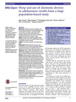
Sleep Medicine 14 (2013) 1031–1034
Contents lists available at ScienceDirect
Sleep Medicine
journal homepage: www.elsevier.com/locate/sleep
Original Article
Impact of partial sleep deprivation on immune markers
A. Wilder-Smith a,b,⇑, F.B. Mustafa c, A. Earnest d, L. Gen c, P.A. MacAry c
a
Department of Medicine, National University Singapore, Yong Loo Lin School of Medicine, Singapore
Lee Kong Chian School of Medicine, Nanyang Technological University, Singapore
c
Department of Microbiology, Immunology Programme, National University of Singapore, Yong Loo Lin School of Medicine, Singapore
d
Centre for Quantitative Medicine, Duke-NUS Graduate Medical School, Singapore
b
a r t i c l e
i n f o
Article history:
Received 23 January 2013
Received in revised form 9 June 2013
Accepted 3 July 2013
Available online 28 August 2013
Keywords:
Sleep deprivation
Immune markers
Immune impairment
Lymphocyte proliferation assays
Common cold
Respiratory infections
a b s t r a c t
Background: Sleep quality is considered to be an important predictor of immunity. Lack of sleep therefore
may reduce immunity, thereby increasing the susceptibility to respiratory pathogens. A previous study
showed that reduced sleep duration was associated with an increased likelihood of the common cold.
It is important to understand the role of sleep in altering immune responses to understand how sleep
deprivation leads to an increased susceptibility to the common cold or other respiratory infections.
Objective: We sought to examine the impact of partial sleep deprivation on various immune markers.
Patients and methods: Fifty-two healthy volunteers were partially sleep deprived for one night. We took
blood samples before the sleep deprivation, immediately after, and 4 and 7 days after sleep deprivation.
We measured various immune markers and used a generalized estimating equation (GEE) to examine the
differences in the repeated measures.
Results: CD4, CD8, CD14, and CD16 all showed significant time-dependent changes, but CD3 did not. The
most striking time-dependent change was observed for the mitogen proliferation assay and for HLA-DR.
There was a significant decrease in the mitogen proliferation values and HLA-DR immediately after the
sleep deprivation experiment, which started to rise again on day 4 and normalized by day 7.
Conclusions: The transiently impaired mitogen proliferation, the decreased HLA-DR, the upregulated
CD14, and the variations in CD4 and CD8 that we observed in temporal relationship with partial sleep
deprivation could be one possible explanation for the increased susceptibility to respiratory infections
reported after reduced sleep duration.
Ó 2013 Elsevier B.V. All rights reserved.
1. Introduction
Sleep is commonly considered a restorative process with
supportive influences on immune functions [1]. A modest amount
of sleep loss alters molecular processes that drive cellular immune
activation and induce inflammatory cytokines and inflammatory
markers [2,3]. Experimental studies have demonstrated that sleep
deprivation results in poorer immune function (i.e., reduced
natural killer cell activity, suppressed IL-2 production) [2,4,5], as
well as increased circulating levels of inflammatory markers (i.e.,
IL-6, tumor necrosis factor a [TNF-a], C-reactive protein) [4,6,7].
Sleep loss induces a functional alteration of the monocyte proinflammatory cytokine response [2,4]. Sleep deprivation also has
been found to attenuate antibody response to immunizations [8].
Because sleep quality is considered to be an important predictor
of immunity, in turn it also may increase one’s susceptibility to the
⇑ Corresponding author. Address: Lee Kong Chian School of Medicine, Nanyang Technological University, 11 Mandalay Road, Singapore 308232, Singapore.
Tel.: +65 6592 3829.
E-mail address: [email protected] (A. Wilder-Smith).
1389-9457/$ - see front matter Ó 2013 Elsevier B.V. All rights reserved.
http://dx.doi.org/10.1016/j.sleep.2013.07.001
common cold [9]. A graded average sleep duration was associated
with an increased likelihood of a cold [9]. The common cold is the
most frequent illness managed in general practice [10], with an
enormous economic impact [11,12].
It is important to understand the role of sleep in altering
immune responses to understand how sleep deprivation leads to
an increased susceptibility to the common cold or other respiratory
infections. In our study, we sought to examine the impact of partial
sleep deprivation on various immune markers.
2. Methods
2.1. Participants
Healthy volunteers between the age of 21 and 65 years not
involved in shift work or night duties were recruited by local
advertisement from the general population in Singapore. Equal
numbers of men and women were selected. Specific exclusion criteria included sleep disorders, use of sleeping tablets within the
past 8 weeks, long-haul flight within the past 8 weeks, any current
1032
A. Wilder-Smith et al. / Sleep Medicine 14 (2013) 1031–1034
illness (i.e., current respiratory infections, current or history of
psychiatric disease), alcohol abuse, autoimmune diseases or any
diseases associated with immunosuppression, immunosuppressive
therapy (i.e., steroids, inhaled steroids), and any endocrine disorders. Written informed consent was provided by all subjects. The
study was approved by the institutional review board of Singapore.
2.2. Test site and conditions
The overnight partial sleep deprivation experiment started at
9:00 pm and lasted for 10 h, until 7:00 am. All subjects stayed in
one room without beds and remained seated throughout most of
the night. Subjects were given a light meal and snacks and drinks.
Sleeping tablets and alcohol- and caffeine-containing beverages
were not permitted. The study coordinator stayed in the room
for continuous supervision, and the subjects played card games
or talked for most of the night. If subjects did fall asleep, the study
coordinator recorded the time and disrupted their sleep after a
maximum of 2 h. A total sleeping time of a maximum of 3 h was
allowed. The total sleeping time was recorded for every subject.
Subjects recorded their subjective stress and fatigue ratings on a
Likert scale (values 1–4 with 4 being most stressed or tired, respectively) at baseline, day 1, and day 7. Venous samples were taken
before and after the night with sleep deprivation and again on days
4 and 7. To exclude circadian variation, blood samples were taken
at the same time of the day at 11:00 am.
to stimulate lymphocyte proliferation and levels of proliferation
quantitated by a 3H-thymidine incorporation assay. Table 1
summarizes the markers and their main functions.
2.4. Statistical analysis
We used the generalized estimating equation (GEE) to examine
the difference in parameter estimates at days 1, 4, and 7 with baseline. The advantage of GEE models is that they allow for the correlation within each subject across the follow-up periods [16].
Because each subject was measured several times in the course
of our study, such a correlation should not be ignored; therefore,
GEE is the most appropriate statistical tool. Because the outcome
is continuous, we specified a Gaussian distribution for the family,
along with an identity link for the model. We used the exchangeable correlation matrix in the model. Data analysis was performed
in Stata V10.2 (StataCorp, College Station, TX, USA) and level of
significance was set at 5%.
3. Results
Fifty-two subjects (26 men and 26 women) with a mean age of
31 years (range, 21–65 y) were included in our study. The median
sleeping time was 2 h (range, 0–3 h). The mean stress ratings on
the Likert scale at baseline and days 1 and 7 were 2.12, 2.98, and
2.19. The mean ratings for fatigue at baseline and on days 1 and
7 were: 2.53, 3.82, and 2.57. Study subjects therefore showed the
2.3. Laboratory analyses
Venous samples were analyzed for complete blood cell count,
immunologic markers, HLA-DR expression, and the mitogen proliferation assay. White blood cells were separated from erythrocytes
using the hemolysis method [13]. Flow cytometry analysis was
performed using the staining protocol, as previously described
[14]. CD3, CD4, CD8, CD14, and CD16 lymphocyte blood cell counts
were measured. In addition, we examined the expression of HLADR on monocytes [15].
2.3.1. Mitogen assay
Lymphocyte proliferation in response to the nonspecific mitogen phytohemagglutinin was determined by 3H-thymidine incorporation. Peripheral blood mononuclear cells were isolated by
the Ficoll-Paque (specific gravity, 1.077) gradient density method.
The peripheral blood sample was centrifuged at 600 g at 18 °C
for 20 min. The peripheral blood mononuclear cell layer was
collected and washed with phosphate buffer saline solution containing 0.4% sodium citrate to remove platelets. The cells were
resuspended in HyClone RPMI 1640 media supplemented with
10% fetal calf serum (PAA Laboratories), 100 U/mL penicillin, and
100 lg/mL streptomycin (Gibco) at a final concentration of
1 106/mL. The mitogen phytohemagglutinin (Sigma–Aldrich,
Singapore) was added at a final concentration of 2 lg/mL for 48 h
Table 1
Immune markers and their main functions.
Marker
Description
CD3
CD4
CD8
CD14
CD16
HLA-DR
Lineage marker: T cells
Lineage marker: helper T cells
Lineage marker: cytotoxic T cells
Lineage marker: monocytes
Lineage marker: neutrophils
Major histocompatibility complex class II
molecule
Functional evaluation of the immune system
Mitogen proliferation
assay
Table 2
Generalized estimating equations showing the differences on days 1, 4, and 7 after a
night of partial sleep deprivation compared to the day before sleep deprivation
(baseline).
Parameter
Mean
Difference
95% Confidence interval
P value
CD3
Day 1
Day 4
Day 7
Before (mean value)
2.45
1.18
2.03
65.96
0.84
4.46
1.25
5.73
2.11
5.32
.144
.482
.225
CD4
Day 1
Day 4
Day 7
Before (mean value)
67.05
77.96
19.56
678.55
14.04
130.97
72.57
120.05
24.96
33.44
.013
.004
.469
CD8
Day 1
Day 4
Day 7
Before (mean value)
51.55
41.67
16.48
337.14
16.90
76.32
51.13
86.19
7.03
18.16
.004
.018
.351
CD14
Day 1
Day 4
Day 7
Before (mean value)
1.14
1.15
0.12
4.65
0.39
1.91
0.64
1.90
0.40
0.87
.003
.003
.765
CD16
Day 1
Day 4
Day 7
Before (mean value)
4.69
7.91
0.71
76.59
9.57
3.03
4.17
0.20
12.79
5.59
.06
.001
.775
HLA-DR
Day 1
Day 4
Day 7
Before (mean value)
7.34
4.85
0.40
27.20
10.07
7.57
2.32
4.62
2.12
3.13
<.001
<.001
.771
Mitogen
Day 1
Day 4
Day 7
Before (mean value)
39996.85
15403.24
4072.50
82173.13
48281.91
23688.30
12357.55
31711.80
7118.19
4212.56
<.001
<.001
.335
A. Wilder-Smith et al. / Sleep Medicine 14 (2013) 1031–1034
Fig. 1. Lymphocyte proliferation with stimulation from phytohemagglutinin on
days 1, 4, and 7 after a partially sleep-deprived night in comparison to the baseline
(day 0).
highest extent of fatigue and stress immediately after the partially
sleep-deprived night.
The results of the complete blood cell count for all subjects were
within reference range at baseline, and there were no significant
changes in the absolute values and proportion of leucocytes, lymphocytes, and eosinophils immediately after the partial sleep
deprivation or after 4 or 7 days (data not shown). Partial sleep
deprivation over one night also did not result in any changes in
the T-cell marker CD3. However, there were statistically significant
changes for CD4 and CD8, which showed an increase immediately
after the experiment but then decreased by day 4 and returned to
baseline levels by day 7 (Table 2). CD14 increased on the day after
the partially sleep-deprived night and then significantly decreased
below the baseline, whereas CD16 had changes in the opposite
direction (Table 2).
The most striking time dependent change was observed for the
mitogen proliferation assay and for HLA-DR. There was a significant decrease in the mitogen proliferation values immediately
after the sleep-deprivation experiment, which started to rise again
on day 4 and normalized by day 7 (Table 2; Fig. 1). HLA-DR significantly decreased on days 1 and 4 after the sleep-deprived night
and normalized to baseline values by day 7 (Table 2).
4. Discussion
We studied the effect of a single night with partial sleep deprivation (sleep duration <3 h) on various immune markers and on
functional leucocyte proliferation assays. Because partial sleep
deprivation in daily life is more common than total sleep deprivation, we decided to only measure the effect of partial sleep deprivation. All measured markers showed statistically significant
changes, except for CD3. CD4 and CD8 both immediately increased
after the sleep deprivation then decreased below the baseline on
day 4 and normalized to baseline values by day 7. Variations in
healthy volunteers over time in CD4 counts have been described
[17]; however, the initial activation of both CD4 and CD8 on day
1 and subsequent suppression of both markers by day 4 were
statistically different to the baseline and both normalized after
7 days to baseline values. Therefore, these changes are unlikely
to be a normal variation but appear to be the result of a
time-dependent transient effect of sleep deprivation on early
CD4/CD8 activation, followed by short-lived suppression. The
1033
clinical importance of these findings is unclear. Because the
absolute values of CD4 did not fall below clinically relevant values
of 500, the depression of CD4 on day 4 therefore does not present
true severe immune suppression but only transient mild immune
suppression.
The protein CD14 is a membrane-bound protein on the surface
of mononuclear cells which recognizes bacterial lipopolysaccharide [18]. In an animal model, CD14 was shown to be critical to initiate neutrophil recruitment into the airways and was found to be
an essential mediator of lipopolysaccharide-induced airway disease [19]. The upregulated CD14 that we found immediately after
a sleep-deprived night may act as a marker for disease activity.
CD16 can be found on the surface of natural killer cells and
neutrophil leukocytes [20]. The phenotypic changes that we saw
after sleep deprivation possibly provide a cellular basis for an
increase in natural killer cell activity, which is observed in the
common cold [21].
Furthermore, we found significant changes in HLA-DR and the
mitogen proliferation assay, with the most predominant change
on the first day after sleep deprivation, which started to increase
again by day 4 and normalized by day 7. HLA-DR is an antigen presentation molecule present on antigen-presenting cells such as
monocytes and B cells. An increased level of HLA-DR expression
is normally associated with activation in antigen-presenting cells
in response to proinflammatory signals, potentiating the cell’s ability to induce stronger T-cell proliferation and respond to maturation signals [15]. Likewise a decrease in HLA-DR expression is
associated with anti-inflammatory or immunosuppressive signals,
which can potentially lead to a decreased ability of monocytes to
induce T-cell proliferation. Low levels of HLA-DR expression on
peripheral blood monocytes have been demonstrated to correlate
with the risk for infection in surgery, trauma, and adult transplant
patients [22]. The decrease in HLA-DR expression after partial sleep
deprivation may be a potential signal of an increased susceptibility
to infection.
The lymphocyte proliferative response can be viewed as a functional evaluation of the immune system [23]. The lymphocyte
proliferation assay showed a clear linear time-dependent response
in our GEE models. There was a marked decrease following the
sleep deprivation that rose on day 4 and normalized by day 7 (similar levels compared to baseline).
We are unsure of the clinical implications of this transient
decrease for HLA-DR and mitogen proliferation. However, both
indicate a certain degree of immunosuppression and decreased
ability to induce T-cell proliferation. Impaired immune function
in healthy individuals possibly represents a suboptimal level of
immune functioning that should not be compared with depressed
immune status, which may occur in individuals who are
immunosuppressed.
Our study showed an inherent variability in the parameters
studied. Possible solutions include increasing the sample size to
even out the noise or adding studies to determine the intraclass
coefficient. However, the method that we chose was the GEE,
which is a robust statistical approach used to longitudinally analyze how subjects are measured at different points in time. The
advantage of GEE models is that they allow for the correlation
within each subject across the follow-up periods [16], and the
comparisons therefore are in line with the baseline of each subject,
which should reduce the possibility of spurious results.
In summary, although our study does not answer the question
of whether or not these changes result in clinically relevant
outcomes, we do postulate that the transiently impaired mitogen
proliferation, the decreased HLA-DR, the upregulated CD14, and
the variations in CD4 and CD8 that we observed in temporal relationship with partial sleep deprivation could be one possible explanation for the increased susceptibility to respiratory infections
1034
A. Wilder-Smith et al. / Sleep Medicine 14 (2013) 1031–1034
reported after reduced sleep duration. Further studies are needed
to investigate if the transient immune impairment observed in
our study indeed translates to a higher risk for acquiring respiratory infections.
Funding sources
The study was funded by the National University of Singapore
(Start-up grant: PI: AWS).
Conflict of interest
The ICMJE Uniform Disclosure Form for Potential Conflicts of
Interest associated with this article can be viewed by clicking on
the following link: http://dx.doi.org/10.1016/j.sleep.2013.07.001.
References
[1] Hamer M, Wolvers D, Albers R. Using stress models to evaluate immunomodulating effects of nutritional intervention in healthy individuals. J Am Coll
Nutr 2004;23:637–46.
[2] Irwin MR, Wang M, Campomayor CO, Collado-Hidalgo A, Cole S. Sleep
deprivation and activation of morning levels of cellular and genomic
markers of inflammation. Arch Intern Med 2006;166:1756–62.
[3] Irwin M. Effects of sleep and sleep loss on immunity and cytokines. Brain
Behav Immun 2002;16:503–12.
[4] Vgontzas AN, Zoumakis E, Bixler EO, Lin HM, Follett H, Kales A, et al. Adverse
effects of modest sleep restriction on sleepiness, performance, and
inflammatory cytokines. J Clin Endocrinol Metab 2004;89:2119–26.
[5] Irwin M, Mascovich A, Gillin JC, Willoughby R, Pike J, Smith TL. Partial sleep
deprivation reduces natural killer cell activity in humans. Psychosom Med
1994;56:493–8.
[6] Vgontzas AN, Zoumakis M, Papanicolaou DA, Bixler EO, Prolo P, Lin HM, et al.
Chronic insomnia is associated with a shift of interleukin-6 and tumor necrosis
factor secretion from nighttime to daytime. Metabolism 2002;51:887–92.
[7] Shearer WT, Reuben JM, Mullington JM, Price NJ, Lee BN, Smith EO, et al.
Soluble TNF-alpha receptor 1 and IL-6 plasma levels in humans subjected to
the sleep deprivation model of spaceflight. J Allergy Clin Immunol
2001;107:165–70.
[8] Spiegel K, Sheridan JF, Van Cauter E. Effect of sleep deprivation on response to
immunization. JAMA 2002;288:1471–2.
[9] Cohen S, Doyle WJ, Alper CM, Janicki-Deverts D, Turner RB. Sleep habits and
susceptibility to the common cold. Arch Intern Med 2009;169:62–7.
[10] Mossad SB. Treatment of the common cold. BMJ 1998;317:33–6.
[11] Gwaltney Jr JM, Hendley JO, Simon G, Jordan Jr WS. Rhinovirus infections in an
industrial population. I. The occurrence of illness. N Engl J Med
1966;275:1261–8.
[12] Fendrick AM, Monto AS, Nightengale B, Sarnes M. The economic burden of
non-influenza-related viral respiratory tract infection in the United States.
Arch Intern Med 2003;163:487–94.
[13] Lundahl J, Hallden G, Hallgren M, Skold CM, Hed J. Altered expression of
CD11b/CD18 and CD62L on human monocytes after cell preparation
procedures. J Immunol Methods 1995;180:93–100.
[14] Mustafa FB, Ng FS, Nguyen TH, Lim LH. Honeybee venom secretory
phospholipase A2 induces leukotriene production but not histamine release
from human basophils. Clin Exp Immunol 2008;151:94–100.
[15] Sztein MB, Steeg PS, Johnson HM, Oppenheim JJ. Regulation of human
peripheral blood monocyte DR antigen expression in vitro by lymphokines
and recombinant interferons. J Clin Invest 1984;73:556–65.
[16] Zeger SL, Liang KY, Albert PS. Models for longitudinal data: a generalized
estimating equation approach. Biometrics 1988;44:1049–60.
[17] Carmichael KF, Abayomi A. Analysis of diurnal variation of lymphocyte subsets
in healthy subjects in the Caribbean, and its implication in HIV monitoring and
treatment. Afr J Med Med Sci 2006;35:53–7.
[18] Werners AH, Bull S, Vendrig JC, Smyth T, Bosch RR, Fink-Gremmels J, et al.
Genotyping of Toll-like receptor 4, myeloid differentiation factor 2 and CD-14
in the horse: an investigation into the influence of genetic polymorphisms on
the LPS induced TNF-alpha response in equine whole blood. Vet Immunol
Immunopathol 2006;111:165–73.
[19] Brass DM, Hollingsworth JW, McElvania-Tekippe E, Garantziotis S, Hossain I,
Schwartz DA. CD14 is an essential mediator of LPS-induced airway disease. Am
J Physiol Lung Cell Mol Physiol 2007;293:L77–83.
[20] Krishnaraj R, Blandford G. Age-associated alterations in human natural killer
cells. 2. Increased frequency of selective NK subsets. Cell Immunol
1988;114:137–48.
[21] Hsia J, Sztein MB, Naylor PH, Simon GL, Goldstein AL, Hayden FG. Modulation
of thymosin alpha 1 and thymosin beta 4 levels and peripheral blood
mononuclear cell subsets during experimental rhinovirus colds. Lymphokine
Res 1989;8:383–91.
[22] Hoffman JA, Weinberg KI, Azen CG, Horn MV, Dukes L, Starnes VA, et al.
Human leukocyte antigen-DR expression on peripheral blood monocytes and
the risk of pneumonia in pediatric lung transplant recipients. Transpl Infect Dis
2004;6:147–55.
[23] Nielsen HB, Pedersen BK. Lymphocyte proliferation in response to exercise. Eur
J Appl Physiol Occup Physiol 1997;75:375–9.


© Copyright 2026