
Hereditary Antithrombin Deficiency: Heterogeneity of the
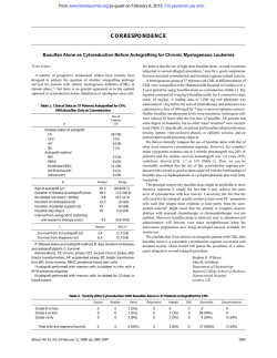
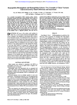
From www.bloodjournal.org by guest on February 6, 2015. For personal use only.
Hereditary Antithrombin Deficiency: Heterogeneity of the Molecular Basis
and Mortality in Dutch Families
By H.H. van Boven, R.J. Olds, S.-L. Thein, P.H. Reitsma, D.A. Lane, E. Briet, J.P. Vandenbroucke, and F.R. Rosendaal
We studied the molecular basis and genetic heterogeneity
of hereditary antithrombin (111) deficiency in nine Dutch families. Polymerase chain reaction (PCR) amplification and direct sequencing of all antithrombin
gene exons and flanking
intronic regions identified mutationsin eight families. Given
the opportunity t o correlate the molecular basis with survival, we addressed the relevance of molecular defects t o
mortality in inherited antithrombin deficiency. The defects
included single nucleotide deletions (7671 del G, 7768-69
del G ) and insertions (5501 ins A, 2463 G-+TC) that lead t o
frameshifts, a single base substitution 15381 C-T (129Arg-+
stop)] leading t o a premature termination codon, and single
base substitutions resulting in amino acid substitutions
[2652 A 4 (63TyrSer), 13380 T 4 (42111etThr). and 13407
G-T (430Cys-+Phe)]. All affected individuals were heterozy-
gous for the defects. Previously we foundin Dutch families
that antithrombindeficiency did notlead t o higher mortality
compared with the general population. In accordance with
these findings, w e observed no excess mortality in the nine
families [Observed:Expected, 52:52.6; standardised mortality ratio(SMR] 1.0,95% confidence interval (Cl), 0.7-1.31. Our
findings confirmeda considerable genetic heterogeneity underlying antithrombin deficiency. We therefore concluded
that the lack of excess mortality in these families is not
caused b y a Dutch mild defect. We suggest that thelongevity is not affected by molecular defects in the antithrombin
gene and hypothesize that differences in mortality or natural
history between families most likely result from other (genetic) risk factors.
0 1994 by The American Society of Hematology.
A
thrombosis related to the age of onset was highest between
years 15 and 40, Hirsh et a18 made a strong case for prophylactic use of anticoagulants between the ages of 15 and 40
years. Similarly, if certain molecular defects can be shown
to be associated with clinically severe phenotypes, a case
could be made for prophylactic anticoagulant therapy in individuals with such genotypes. At present, no studies have
been designed to address this matter.
Previously, we studied the natural history of 10 Dutch
kindreds with regard to mortality? We found no excess mortality in comparison with the general population. From that
study we concluded that, in general, antithrombin deficiency
did not appear so severe a disease as to warrant long-term
prophylactic anticoagulation of asymptomatic gene carriers.
Although this was the first study inwhich the natural history
of antithrombin deficiency was studied in a formal design,
two issues were correctly r a i ~ e d :first,
~ whether for some
reason these Dutch families all had mildforms of the disease,
for instance due to the presence of one “Dutch” molecular
defect with mild clinical expression in these families; and
second, whether the overall mortality figures might mask
differences in mortality between individual families, related
to the underlying defect.
In the present study we investigated the molecular basis
NTITHROMBIN IS A natural anticoagulant that exerts
its action by inhibiting clotting factor IIa (thrombin),
factor IXa, and factor Xa. The gene coding for the antithrombin protein has been localized to chromosome 1q23-25, and
its complete sequence was recently elucidated.’ Basedon
structural and sequence homology, antithrombin belongs to
the serpin (serine proteinase inhibitor) family of proteins.
Hereditary antithrombin deficiency is a rare autosomal
dominant disorder. A variety of underlying molecular defects
have been identified.’ Inherited antithrombin deficiency may
be classified into two major types based on the results of
functional and immunologic assays. Type I deficiencyis
characterized by reduced functional and immunologic antithrombin levels, both at approximately 50% of normal. Type
I1 deficiency results from the presence of a variant functionally inactive protein with almost normal antigen levels, but
functionally the levels are reduced. Type I1 deficiency may
be further subclassified into variants with reactive site defects, heparin-binding defects, and variants with pleiotropic
effects?
It iswell recognized that hereditary antithrombin deficiency is associated with a risk of thromboembolic disease.
However, it is still a matter of debate whether the clinical
seventy of thromboembolic episodes and, hence, mortality
might vary between families. It has been suggested that the
underlying defect might be relevant to clinical ~everity.~
Many families have been reported since Egeberg first described antithrombin deficiency in 1965.5The type I1 variants
with reduced affinity for heparin seem to be associated with
a low frequency of thrombotic episodes, except when present
in homozygous individuals: Excluding the heparin-binding
variants, cumulative incidences of thrombosis of 15% to
100% have been suggested for inherited antithrombin deficiency.’
It is important to know if some forms of deficiency are
truly more clinically severe than others, as such information
would play a decisive role in clinical management. For example, to decide that long-term prophylactic treatment with
anticoagulants is required for (a)symptomatic carriers, the
risk of thrombosis should outweigh the disadvantages of
long-term anticoagulant treatment. Because the incidence of
Blood, Vol 84, No 12 (December 15), 1994: pp 4209-4213
From the Department of Clinical Epidemiology, and the Haemostasis and Thrombosis Research Center, University Hospital, Leiden,
The Netherlands: the Department of Haematology, Charing Cross
and Westminster Medical School, London; and the Institute of Molecular Medicine, John Radcllffe Hospital, Oxford, UK.
Submitted March 24, 1994: accepted August 18, 1994.
Supported by Ter Meulen Fund, Royal Netherlands Academy of
Arts and Sciences, and by The Wellcome Trust, UK.
Address reprint requests to F.R. Rosendaal, MD, PhD, Department of Clinical Epidemiology, Sldg 1, CO-P University Hospital,
PO Box 9600, 2300 RC Leiden, The Netherlands.
The publication costs of this article were defrayed in part by page
charge payment. This article must therefore be hereby marked
“advertisement” in accordance with 18 U.S.C. section 1734 solely to
indicate this fact.
0 1994 by The American Society of Hematology.
0006-4971/94/8412-0012$3.00/0
4209
From www.bloodjournal.org by guest on February 6, 2015. For personal use only.
VAN BOVEN ET AL
4210
of antithrombin deficiency in nine families to identify the
the possibilityof a mild
underlyingdefectsandaddress
"Dutch" defect with regard to mortality.
MATERIALS AND METHODS
Patients. Of the 10 families from the previous mortality study,"
9 families were available for further investigation. In each family,
venepuncture was performed in two individuals previously known
to be affected by plasma phenotype. The blood samples were collected from the antecubital vein in 1/10v01 of 0.11 mmol/L trisodium
citrate. Plasma was prepared by centrifugation for 10 minutes at
2,000g at 10°C and stored at -70°C until use.
Antithrombin assays. Antithrombin antigen concentration was
measured by immunoelectrophoresis according to the methodof
Laurell.gbAmidolytic heparin cofactor assays (Chromogenie, Molndal, Sweden) were used for antithrombin activity measurement. The
presence of a variant antithrombin protein was investigated by
crossed immunoelectrophoresis (CIE) in the presence of heparin."'
Southern blot analysis. Genomic DNA extracted from blood leukocytes by standard methods was digested with restriction enzymes
Pst I, EcoRI, BamHI, and HindIII, using conditions recommended
by the manufacturers (New England Biolabs [Beverly, MA]and
Promega [Madison, WI]). The digested DNAwas fragmented by
gel electrophoresis and transferred onto nylon membranes (Hybond,
Amersham, UK) by Southern blot. These membranes were subsequently hybridized to an antithrombin cDNA probe that had been
labeled with ["Pld-cytidine triphosphate (dCTP) by random priming
(Megaprime, Amersham, UK). After hybridization, the membranes
were washed at high stringency and autoradiographed between intensifying screens at -70°C.
DNA sequence analysis. The antithrombin exons and their
flanking intron regions were amplified by polymerase chain reaction
(PCR) using biotinylated primers and conditions previously described." The single-stranded DNA was directly sequenced by the
dideoxynucleotide method using the nonbiotinylated primer or an
internal primer (Sequenase; US Biochemical, Cleveland, OH). All
exons and adjacent intron regions were sequenced for each proband.
Restriction enzyme unalysis of mutations. Mutations identified
by sequencing were confirmed by restriction enzyme digestion of
amplified antithrombin gene fragments. When the mutation did not
create or abolish a restriction site, a site was created by introducing
nucleotide substitutions with mutant oligonucleotides during amplification." Mutant oligonucleotides were designed with a nucleotide
substitution close to the 3' end, such thatthe combination of the
nucleotide substitution and the mutation created a new restriction
enzyme cleavage site. After amplification with the mutagenic primer
and a downstream primer, the products were digested with the appropriate enzyme. Digested products were visualized under UV light
after electrophoresis in 2% agarose and ethidium bromide staining.
Normal subjects served as controls. Sequences of amplification primers and the restriction enzyme sites associated with each mutation
are shown in Table 1. In family 3, the mutation was confirmed by
allele-specific priming of the PCR, as previously described."
Mortality analysis. The mortality data of the nine families were
analyzed by the family tree mortality ratio (FMTR) method as previously described.' The method involves the application of the standard technique of indirect standardization on a cohort formed from
the pedigrees under study. In the cohort we included all individuals
with a probability of 0.5 or higher of carrying the defective gene,
ie, all siblings and offspring of carriers and theparents. The mortality
in the cohort is compared with the general population, adjusted for
differences in age, gender, and calender period by indirect standardization. This method of a comparison of observed (0)number of
cases (in the cohort) and expected (E) number of cases (in the
population, standardized)-the ratio 0:E-is knownasthe standardized mortality ratio (SMR).'' We omitted the first two decades
of life for the observed and for the expected number of deaths. The
reason is thatthosewhopassedonthe
gene to their descendents
hadto liveuntil reproductive ageand reproduce; consequently, if
these tirat decades in which members of previous generations "had
to live" were included, no deaths would be observed, but the accrual
of person-time would yield expected cases, ie, a biased (low) SMR.
RESULTS
Antithrombin assays. The results obtained by functional
in
and immunological antithrombin assays are summarized
Table 1. All the affected individuals had reduced functional
and antigen levels, compatible with type I antithrombin deficiency. CIE in the presence of heparin confirmed the absence of variant antithrombin with altered heparin affinity
in the plasma.
Genomic basis for antithrombin deficiency. No evidence
for major gene rearrangements was obtained from restriction
enzyme analysis of genomic DNA from affected members
ofallninefamilies.Directsequencingofamplifiedantithrombin gene fragments identified mutations in eight of the
ninefamilies(Table1):fourmutationsweresinglebase
substitutions (families 2, 3, 7, and S), two were single base
deletions (families S and 6), one was a nucleotide insertion
a substitutiodinsertion of two nucle(family 4), and one was
otides (family l). For each individual examined, sequencing
of the remaining exons and flanking intron regions did not
show further mutations.
Mutations were confirmed by sequencing the appropriate
antithrombin gene fragments in a second affected member
PCR
from each family and by restriction analysis
of products
(data not shown). The deletion of a G in exon 4, codon 320
(family S ) creates a Fok I site, while a Dde I site is created
by the A< substitution in codon 63 (family 2). Deletion
4 (family 6) and the T+C
of G from codon 353 in exon
substitution in codon 42 1, exon 6 (family 7) abolishes sites
for BstNI and Mbo 11, respectively. Amplification primers
were designed to create a cutting site for M b o I1 in the allele
carrying the C+TC mutation in codon - 1 of family 1 and
A insertion in
a site for Dde I in the mutant allele with an
codon 169 in family 4. The G+T substitution in codon 430
(family S) could not be confirmed by this strategy. However,
a BsrNI site was created instead in the normal allele; digestion of PCR products with the enzyme showed that about
SO% of the amplified product did not cut. In family3 allelespecific priming of the PCR was used to confirm the C+T
substitution in codon 129.
In the affected members of family
9, sequence analysis
of all the coding regions and flanking introns did not show
any mutation. Sequence analysis showed that affected members were heterozygous for
a previously described Pst I polymorphism in exon 4, confirming the presence of two copies
of the gene.
Mortalivanalysis.
Thenumbersofindividualsaged
2 2 0 years contributed to the study by
each family are shown
inFig 1. Theobservednumberofdeathsinallfamilies
combined was 52. The expected number of deaths based on
population mortality figures was 52.6, which resulted in
a
relative mortality of 1 .O [O:E, 5252.6; SMR, 1.0; 95% con-
From www.bloodjournal.org by guest on February 6, 2015. For personal use only.
421 l
HEREDITARY ANTITHROMBIN DEFICIENCY
Table 1. Antithrombin Gene Mutations in Dutch Families
Family
No.
Nucleotide
Position
AcidMutation
Exon
% Antigen
Activity
12 cased
NR,
Codon,
family)Amino
80%-120%
Change
TC
Frameshift, stop
codon 32
C
63 Tyr
T
129 Arg
stop
2
2463 G
2
2652 A
3A
5381 C
3A
5501 +A
169 frameshift, stop
codon 192
4
7671 -G
320 frameshift, stop
codon 331
4
7768-69-G
353 Gly -t Val splice
+
-+
-+
+
40 33
44
42
51
49
Ser
39
36
40
50t
Restriction
Enzyme Site
Oligonucleotide
Position
-
Oligonucleotide Sequence
(5' 3')
Mbo H*
2443-2462
Dde I
27
17-2698 TGGAGATACTCAGGGGTGAC
GTTGCAGCCTAGCTTAACTTGGCA
2370-2393
GGTTGAGGAATCAlTGGACTTG
2860-2839
CTTCTGGGACTGCGTGAICT
4240
Allele-specific
priming
Dde I*
5480-5502
GTGAGTTGGTATATGGAGCCIA
49 45
50 45
Fok I
5630-5609
7617-7636
GTCTTCAGCAAAGCAGTGT
GGTGCTGCAGGAGTGGCEGt
46 38
46 46
BsNl
CTTCCACTTTTGGTCAGACTAC
7831-7810
751
4-7533 AGGTGCTTGAGTTGCCCTTC
site
6
13380 T
6
13407 G
-+
+
C
421 Ile
T
430 Cys
-
-
+
49
46
44
60
51
41
50
Thr
+
Phe
?
49
45
45
55
46
50
49
Mbo II
BsNl*
7831-7810
13235-13255
13517-13495
13387-13406
13517-13495
CTKCAC'TTTTGGTCAGACTAC
CTGCAGGTAAATGAAGAAGGC
TGCAGAGTCCATlTATAATGTG
GGGCAGAGTAGCCAACCCTG
TGCAGAGTCCATlTATAATGTG
Restriction enzyme sites created artificially, using primers in which artificialbase substitution is underlined insequence column. Nucleotide
numbering corresponds to sequence data, Olds et al.' The first listed primer is complementary in sequence to the noncoding strand.
t In this family, only one individual was available venepuncture.
for
*Artificial base to remove natural restriction site for Fok I that would have resulted i n a similar size for two bands, making it difficult to
distinguish between two or three bandsafter digestion
fidence interval (CI), 0.7-1.31. For most families no excess
mortality in comparison with the general population was
apparent, although due to the small number of individuals
in some families, the 95% CIS remained wide.
Finally, we considered the possibility that differences in
the underlying mutations might lead to variation in mortality.
We hypothesized a priori that the defects affecting a splice
site or leading to a frameshift and premature stop codon are
severe defects, as in these cases the allele is rendered fully
inactive with no protein production from the mutant gene.
This is analogous to the clinical severity in other disorders,
eg, hemophilia. The data showthat for the families with
these null mutations (families 1, 3, 4, 5 , and 6) no excess
mortality was observed (O:E, 30:30.9; SMR, 1.0; 95% CI,
0.7-1.4). Similarly, there was no difference in mortality in
the families with the remaining mutations (substitutions)
(O:E, 21~20.6;SMR, 1.0; 95% CI, 0.6-1.5).
DISCUSSION
I
o
.
o
l
,
l
I
,
I
I
I
I
,
I
l(8) ~ ( I I 3(6)
)
4(6) 5(28) 6(29) 7 ( 6 ) 8(26) 9(11)
Family (no of individuals)
I
,
I
null
all
mutations
Fig 1. M o r t a l i i in antithrombin-deficient families.
We investigated the molecular basis in families with inherited antithrombin deficiency and found that mortality was
not related to the actual characterized defects. The majority
of defects underlying type I antithrombin deficiency are
unique events in single kindreds. More than 40 distinct mutations have been reported in the literature.2 In our study,
eight defects were defined as the molecular bases of Dutch
antithrombin-deficientfamilies. Seven mutations were novel,
whereas a C-+T substitution in codon 129, which replaces
arginine by a premature termination codon, has been reported
eight times previously.*This latter substitution occurs within
a CpG dinucleotide, a recognized site for recurrent mutation,
but formal haplotype analysis of the mutant alleles would
be necessary to distinguish whether the mutations had an
independent origin or whether a founder effect was present.
From www.bloodjournal.org by guest on February 6, 2015. For personal use only.
VAN BOVEN ET AL
4212
Threecategories of mutations giverisetotype
I deficiency. Four mutations produced prematuretermination signals, either by direct substitution or by shifts in the reading
frame of translation; this represents the most common basis
of type I antithrombin deficiency.’ No evidenceof truncated
protein was found in the plasma in any of the four families
with these mutations in this study, suggesting that either the
mutant mRNA is nottranslated or that anytranslated protein
either is not exported from hepatocytes oris highly unstable.
A second mechanism is the substitution of single amino
acids. While this would be expected to give rise to a variant
protein (type I1 deficiency), a small number of substitutions
have previously been identified as the basis of type I deficiency. We report here three novel amino acid substitutions
in thenineDutch kindreds: 6 3 T y ~ S e r ,421Ile+Thr, and
430Cys-tPhe. In principle,thesemutationsmayrepresent
rare polymorphisms ratherthan causative defects. Four arguments can be given to make the causative relationship
plausible. First, the mutations werethe only abnormalities detected
after sequencing all exons for each individual. Second, Tyr
at position 63 and Ile at position 421 are highly conserved
between serpins,ls suggesting that they have importantstructural roles. The structural role of 43OCys is even more obvious as it forms a disulphide bond with 2 4 7 C ~ s . Third,
I~
for
each mutation we observed cosegregationwith antithrombin
deficiency. Finally, we tested 68 normal alleles for the three
mutations but found none.
The final mechanism that can be inferred from the observed mutations is a defect in mRNA processing. The mutant allele in family 6 has a deletion of G at either position
7768,whichisthelastnucleotide
of exon 4, or position
7769, the first nucleotide of intron 4. Either of these two
possibilitieswouldaffectthe
exon 4-intron4 splice site.
AbnormalmRNAprocessinghas
been shown previously
fromanother antithrombinmutationthataffectedthelast
nucleotide of exon 3A.”
In one family (family 9) themutation remains uncharacterized; we were unable to detect eithera major rearrangement
of the antithrombin gene or a minor mutation, despite repeated sequence analysis. The affected individuals have typical typeI deficiency as judgedby plasma antithrombin assays
(Table l), and several affected family members have been
identified in the two generationsavailable for study.A mutation may be present in the unsequenced regions of the antithrombin gene, in the introns, or in 5‘ and 3’ flanking sequences. Alternatively, the mutationmay lieoutsidethe
antithrombin gene and segregate independently of the antithrombin locus; furtherinvestigation of family members will
be required to distinguish between these possibilities.
In the mortality study no excess overall mortality (O:E,
52:52.6; SMR, 1.0; 95% CI, 0.7-1.3) was found for these
families that were part of a previous analysis.’ The inclusion
only, or predominantly of one, mild “Dutch” antithrombin
defect does not explain our finding of a normal life expectancy in hereditary antithrombin deficiency as a variety of
molecular defects underlying the deficiency were found. In
fact, the majority of mutations we found led to premature
stopcodons thatpredict thecompleteabsence of protein
product from the mutant allele. Even among families with
this drastic type of mutation, null allele mutations, lifeexpectancy appears to be normal.
This may seem to be in disagreement with the experience
of some familieswith antithrombin deficiency or with previous reports. The latter may be due to overrepresentation of
families with dramatichistories in case reports, which seems
a likely and almost unavoidable result of selective attention
of physicians who are naturally drawn to such families. Our
data render it unlikely that the antithrombin gene itself is
involved in setting the clinical course at the level of overall
mortality. This still does not rule out the possibility of families with, in retrospect, a severe course, ie, a high number
of thrombotic episodes or deaths. Most likely, other genes
or environmental factors have then played a role. Only recently a high prevalence of a novel inherited abnormality of
the coagulation system, a poor response to activated protein
C, was found.18 With the emergenceof common genetic risk
factors, it becomes likely that some families or generations
of families carry the burden of other abnormal genes that
lead to differences in phenotype, and environmental factors
may aggregate in families or generationsof families. As this
study is limited tooverall mortality and theantithrombin
gene, furtherfamily studies should investigate thepossibility
of a genetic basis for the variety in clinical expression and
of clinical morbidity.
ACKNOWLEDGMENT
We thank Dr H.R. Biiller and Dr
J.W. ten Cate (Academic Medical
Centre, Amsterdam, The Netherlands) for allowing us to investigate
two families of their clinic.
REFERENCES
I. Olds RJ, Lane DA, Chowdhury V, De Stefano V, Leone G,
Thein SL: Complete nucleotide sequence of the antithrombin gene:
Evidence for homologous recombination causing thrombophilia.
Biochemistry 32:4216, 1993
2. Lane DA, Olds W, Boisclair M, Chowdhury V, Thein SL.
Cooper DN, Blajchman MA, Perry DJ, Emmerich J, Aiach M: Antithrombin I11 mutation database: First update. Thromb Haemost
70:361, 1993
3. Lane DA, Olds RI, Conard J, Boisclair M, Bock SC, Hultin
M, Abildgaard U, Ireland H, Thompson E, Sas G , Horellou MH,
Tamponi G , Thein SL: Pleiotropic effects of antithrombin strand 1 C
substitution mutations. J Clin Invest 90:2422, 1992
4. De Stefano V, Leone G : Mortality related to thrombosis in
congenital antithrombin I11 deficiency. Lancet 337:847, 1991
S. Egeberg 0: Inherited antithrombin deficiency causing thrombophilia. Thromb Diath Haemorrh 13:516, 1965
6. Finazzi G, Caccia R, Barbui T: Different prevalence of thromboembolism in the subtypes of congenital antithrombin I11 deficiency: Review of 404 cases. Thromb Haemost 58:1094, 1987
7. Demers C, Ginsberg JS, Hirsh J, Henderson P, Blajchman MA:
Thrombosis in antithrombin-111-deficientpersons. Report of a large
kindred and literature review. Ann Intern Med 116:754, 1992
8. Hirsh J, Piovelli F, Pini M: Congenital antithrombin I11 deficiency: Incidence and clinical features. Am J Med 873348, 1989
(suppl 3B)
9. Rosendaal FR, Heijboer H, Briet E, Buller HR, Brandjes DP, de
Bruin K, Hommes DW, Vandenbroucke JP: Mortality in hereditary
antithrombin-I11 deficiency, 1830 to 1989. Lancet 337:260, 1991
9b. Laurel1 CB:Quantitative estimation of proteins by electropho-
From www.bloodjournal.org by guest on February 6, 2015. For personal use only.
HEREDITARY ANTITHROMBIN DEFICIENCY
resis in agarose gels containing antibodies. Anal Biochem 15:45,
1966
10. Lane DA, Flynn A, Ireland H, Erdjument H, Samson D, Howarth D, Thompson E: Antithrombin 111 Northwick Park Demonstration of an inactive high MW complex with increased affinity for
heparin. Br J Haematol 65:451, 1987
11. Chowdhury V, Olds RI, Lane DA, Conard J, Pabinger I, Ryan
K, Bauer KA, Bhavnani M, Abildgaard U, Finazzi G, Castaman G,
Mannucci PM, Thein SL: Identification of nine novel mutations
in type I antithrombin deficiency by heteroduplex screening. Br J
Haematol 84:656, 1993
12. Chang JG, Chen PH, Chiou SS, Lee LS, Perng LI, Liu TC:
Rapid diagnosis of b-thalassemia mutations in Chinese by naturally
and amplified created restriction sites. Blood 80:2092, 1992
13. Olds RJ, Lane DA. Ireland H, Finazzi G, Barbui T, Abildgaard U, Girolami A, Thein SL: A common point mutation producing
type 1A antithrombin 111 deficiency: AT129 CGA to TGA (Arg to
STOP). Thromb Haemost 64:621, 1991
14. Breslow NE, Day NE: Rates and rate standardization, in Bres-
4213
low NE,Day NE (eds): Statistical. Methods in Cancer Research,
v01 2. Lyon, France, International Agency for Research on Cancer
(IARC), 1987, p 48
15. Huber R, Carrell RW: Implications of the three-dimensional
structure cy,-antitrypsin for structure and function of serpins. Biochemistry 28:8951, 1989
16. Sun X-J, Chang J-Y: Heparin binding domain of human antithrombin 111 inferred from the sequential reduction of its three disulfide linkages. J Biol Chem 264:11288, 1989
17. Berg LP, Grundy CB, Thomas F, Millar DS, Green PJ, Slomski R, Reiss J, Kakkar VV, Cooper DN: De novo splice site mutation
in the antithrombin III (AT3) gene causing recurrent venous thrombosis: Demonstration of exon skipping by ectopic transcript analysis.
Genomics 13:1359, 1992
18. Koster T, Rosendaal FR, de Ronde H, Briet E, Vandenbroucke JP, Bertina RM: Venous thrombosis due to poor anticoagulant response to activated protein C: Leiden thrombophilia study.
Lancet 342:1503, 1993
From www.bloodjournal.org by guest on February 6, 2015. For personal use only.
1994 84: 4209-4213
Hereditary antithrombin deficiency: heterogeneity of the molecular
basis and mortality in Dutch families
HH van Boven, RJ Olds, SL Thein, PH Reitsma, DA Lane, E Briet, JP Vandenbroucke and FR
Rosendaal
Updated information and services can be found at:
http://www.bloodjournal.org/content/84/12/4209.full.html
Articles on similar topics can be found in the following Blood collections
Information about reproducing this article in parts or in its entirety may be found online at:
http://www.bloodjournal.org/site/misc/rights.xhtml#repub_requests
Information about ordering reprints may be found online at:
http://www.bloodjournal.org/site/misc/rights.xhtml#reprints
Information about subscriptions and ASH membership may be found online at:
http://www.bloodjournal.org/site/subscriptions/index.xhtml
Blood (print ISSN 0006-4971, online ISSN 1528-0020), is published weekly by the American
Society of Hematology, 2021 L St, NW, Suite 900, Washington DC 20036.
Copyright 2011 by The American Society of Hematology; all rights reserved.


© Copyright 2026