
Studies of Human Cord Blood Dendritic Cells: Evidence for
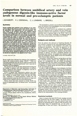
From www.bloodjournal.org by guest on February 6, 2015. For personal use only.
Studies of Human Cord Blood Dendritic Cells:
Evidence for Functional Immaturity
By David W.C. Hunt, Hans-lko Huppertz, Hui-Jun Jiang, and Ross E. Petty
We have isolated low-density, nonadherent, nonphagocytic,
HLA-DR+ve cells with the morphology of dendritic cells
(DCs) from the cord blood
of full-term newborninfants. Relative t o adult DCs, cord blood DC5 were poor stimulators of
the mixed leukocytereaction when either adult orcord
blood mononuclear cells (MNCs) or T lymphocytes were
used as responder cells. In contrast, cord blood T cells and
MNCs responded normally t o allogeneic adult DCs. Cord
blood DCs performed poorly as accessorycells for T-lymphocyte mitogenic responses at suboptimal concentrations of
concanavalin A (Con A) and phytohemagglutinin
A or at optimal concentrations of mitogen and low numbers of DCs.
Addition ofrecombinant interleukin-2(rlL-2) or recombinant
interferon-y (rlFN-y) t o cord blood DC-T-cell cultures containing a suboptimal concentrationof Con Apotentiated the
proliferative response. In contrast, rlL-2 and rlFN-y exerted
little effect on the Proliferative response of adult T cells CUItured with Con Aand DCs. Flow cytometric studies showed
that levels of intercellular adhesion molecule-l (CAM-1;
CD54) and major histocompatibility complex (MHC) class I
HLA-ABC and class II HLA-DR antigens on cord blood DCs
were significantly lower than those on adult blood DCs.
These findings suggest that therelative inefficiency of cord
blood DCs in the activation of T cells may be related t o
their low cell surface expression of MHC and cell adhesion
molecules. The demonstrated impairment of cord bloodDC
function could be of
importance in understanding the immunologicrelationshipbetween
the fetusandmotherand
could contribute to thesusceptibility of newborns t o infection.
0 1994 by The American Societyof Hematology.
C
laboratory personnel (25 to 51 years of age). In both groups, EDTA
or heparin were used as anticoagulants.
Isolation of DCs and T cells. Blood was mixed 1:l with RPM1
1640 medium (StemCell Technologies Inc, Vancouver, British Columbia, Canada) containing 25 mmolL HEPES, L-glutamine, 40
pmol/L nonessential amino acids, 40 pmolL Na pyruvate, 100 U/
mL penicillin, 100 pg/mL streptomycin, 2 pg/mL gentamycin, 40
ng/mL fungizone (GIBCO BRL, Burlington, Ontario, Canada), and
m o m 2-mercaptoethanol. The diluted bloodwas layered
5X
over Ficoll-Hypaque (Pharmacia, Baie D’Urf6, Quebec, Canada) and
centrifuged (30 minutes at 650g), and the mononuclear cell (MNC)
fraction at the interface was collected and washed twice with medium. To obtain T cells, MNCs were mixed (150) with sheep red
blood cells pretreated with Vibrio cholerae neuraminidase (GIBCO
BRL, 2 UlmL, 45 minutes, 37°C) for 10 minutes at 37°C. centrifuged,
and incubated on ice for a further 60 minutes.” Cells were gently
resuspended, layered over Ficoll-Hypaque, and centrifuged. Erythrocytes in the cell pellet were lysed with 0.14 m o m NH,CI to obtain
rosetting T cells (ER+ve). These cells were washed twice with
mediumand incubated overnight at 37”C, 5% CO2 in 10 mLof
medium containing 10% heat-inactivated fetal calf serum (FCS;
GIBCO BRL) in 10 X 1 0 0 mm plastic Petri dishes. Nonrosetting
(ER-ve) cells from the gradient interface were washed with medium
ONSIDERABLE evidence has been advanced that the
human neonatal immune system differs functionally
from that of the adult. Although often contradictory, the
majority of studies have indicated that cord blood T lymphocytes,’.’ B lymphocytes,’ and monocytes4 are deficient in a
variety of in vitro functional assays or express different levels of certain cell surface antigens than do the corresponding
cells of adult blood. The relative inability of neonatal T cells
to elaborate interferon-y (IFN-y)’.‘ is paralleled by the low
frequency of cells expressing the CD45RO (memory) isoform of the leukocyte common antigen.’ Because most cord
blood T lymphocytes bear the CD45RA isoform characteristic of naive T cells, it is probable that their activation requires
presentation of antigen by lymphoid dendritic cells (DCS).~
The existence of DCs in human cord blood was suggested
by earlier studies? and immunostimulatory DCs have been
propagated in vitro from CD34+ cells purified from human
cord blood.’0,”
We assessed DC function in the newborn by measuring the
accessory cell-dependent,’* T cell-proliferative response to
the mitogenic lectins concanavalin A (Con A) and phytohemagglutinin A (PHA) and the capacity of cord blood DCs
to stimulate the mixed leukocyte reaction (MLR). The functional and phenotypic attributes of cord bloodDCs were
compared with those of the corresponding cell population
isolated from adult peripheral blood. Our findings indicate
that, as accessory cells for T-cell responses, cord blood DCs
are functionally inferior to adult blood DCs andthat this
may be at least partly explained by their lower expression of
HLA-ABC, HLA-DR, and intercellular adhesion molecule-l
(ICAM-1, CD54).
MATERIALS AND METHODS
Blood samples. Small aliquots (3 to 5 mL) of cord blood were
obtained from the blood bank at British Columbia’s Children’s Hospital from samples routinely taken at delivery from healthy full-term
male and female newborns but not required for clinically indicated
laboratory studies. Samples were processed within 24 hours of birth.
Adult peripheral blood was donated by healthy male and female
Blood, Vol84, No 12 (December 15), 1994: pp 4333-4343
From the Department of Paediatrics and the Department of Microbiology and Immunology, University of British Columbia, Vancouver, British Columbia, Canada, andthe Children’s Hospital,
University of Wiirzburg, Wiirzburg, Germany.
Submitted March 1.5, 1994; accepted August 2, 1994.
Supported by grants from the George andFlorence Heighway
Foundation and theBritish Columbia Medical Research Foundation.
D. W.C.H. was a recipient of a British Columbia Science Council
G.R.E.A.T.award. H-I.H. was supported by the Deutsche Forschungsgemeinschaft.
Address reprint requests to Ross E. Petty, MD, PhD, Room 211,
The Children’s Variety Research Centre, 9.50 West 28th, Vancouver,
BC, Canada VSZ 4H4.
The publication costs of this article were defrayed in part by page
charge payment. This article must therefore be hereby marked
“advertisement” in accordance with 18 U.S.C. section 1734 solely to
indicate this fact.
0 1994 by The American Society of Hematology.
0006-4971/94/8412-0136$3.00/0
4333
From www.bloodjournal.org by guest on February 6, 2015. For personal use only.
4334
HUNT ET AL
and cultured separately overnight. Nonadherent cells were then carefully layered over 3-mL columns of hypertonic metrizamide (Sigma
Chemical CO, St Louis, MO;14.5 g plus 100 mL of RPMI 1640
containing 10% FCS) and centrifuged at 600g for 10 minutes at
room temperat~re.’~
T cells were recovered from the cell pellet,
whereas DCs were obtained at the gradient interface. As a source
of macrophages, plastic-adherent cells from the overnight culture
were obtained by gently scraping using a rubber policeman after
incubation on ice for 1 hour to facilitate detachment.
Characterization of cord blood DC. A combination of techniques was used to characterize cord blood DC, although their low
frequency precluded complete characterization of all preparations.
To assess phagocytic activity, cells were incubated in 24-well plates
in medium with fluorescent plastic beads (Fluoricon particles, Pandex, Mundelein, IL)at 3 7 T , 5% CO, overnight. Cells were then
washed and resuspended several times in the same well. The proportion of cells thattookup
beads was determined by examination
underthe fluorescence microscope. Indirect immunofluorescence
staining was performed on DC cytospin preparations usng a murine
antihuman HLA-DR monoclonal antibody (IgG,, clone L243; Becton Dickinson, Mississauga, Ontario, Canada) as the primary reagent
and fluorescein isothiocyanate (F1TC)-conjugated sheep antimouse
IgG (Fab’), (Sigma) as the second antibody. Cells were examined
under the fluorescence microscope.
Flow cvrornetry. T cells and DCs were analyzed for cell surface
expression of a variety of leukocyte markers by Ruorescence-activated cell sorting (FACS) analysis. Purified T cells ( 1 to 2 X
tube) were incubated for 1 hour on ice with directly labeled murine
anti-CD3 (IgG, , clone SK7)-phycoerythrin (PE), anti-CD I9 (IgG, ,
clone 4G7)-PE, anti-CD4 (IgG,, clone SK3)-FITC, or anti-CD8
(IgG, , clone SKI)-FITC monoclonal antibodies (Becton Dickinson)
in phosphate-buffered saline (PBS) containing 2% FCS and 0.03%
NaN,. PE- and FITC-labeled IgG, (clone MOPC-21) isotype control
monoclonal antibodies were purchased from Sigma. Cells were
washed twice with the assay buffer and fixed in 1% p-formaldehyde
in PBS. DCs ( I -5 X lo4celldtube) were stained with murine antihuman MHC Class I HLA-ABC (IgG2., clone B9.12.1,’5AMAC, Inc.,
Westbrooke, ME), antihuman MHC class 11-DR-specific (IgGb,
clone B8.12.2,I6 AMAC, Inc), or antihuman ICAM-I (IgG,. clone
15.2.” Boehringer Mannheim, Laval, Quebec, Canada) monoclonal
antibodies. Murine control monoclonal antibodies were MRC OX7
(IgG, antirat Thy- l,’* kindly supplied by Dr R.W. McMaster, Department of Medical Genetics, University of British Columbia), MRC
OX26 (IgG,,, antirat transfemn receptor,” Serotec Canada Ltd, Toronto, Ontario) and MOPC-141 (IgC,,, Sigma). After primary staining, cells were washed twice with PBS, incubated with FITC-labeled
antimouse IgG (Fah')* for 30 minutes, washed and fixed.Cells were
analyzed on an EPICS C flow cytometer (Coulter Diagnostics, Hialeah, FL) with a logarithmic fluorescence scale of three decades.
The flow cytometer was gated to exclude dead cells and debris.
Staining intensity is expressed as mean channel fluorescence, which
was derived using a linear scaling program (1 -256 channels). Specific fluorescence was calculated by the subtraction of themean
channel fluorescence obtained with the isotype-matched control from
that for the human leukocyte-specific monoclonal antibody.
In a separate set of experiments, metrizamide interface cells were
characterized with PE-labeled mouse monoclonal antibodies (Sigma)
to CD3 (IgG, . clone UCHT- l), CD14 (IgG?,, clone UCHM-l), and
CD19 (IgG, , clone SJ25-Cl). Expression of HLA-DR by these cells
was evaluated with theFITC-labeled monoclonal antibody 13 (IgGZ,,
clone 9-49, Coulter Immunology, Hialeah, K). PE- and FITC-labeled isotype-matched antibodies were obtained from Sigma. Cells
were stained as described above and analyzed on a Coulter XL flow
cytometer with a logarithmic scale of four decades.
1v/
Mitogen stimulation ossuys. T cells (2-4 X 10⁴/well)were incubated with irradiated (2,000 rad) autologous DCs in 96-well microtiter plates in RPMI 1640 medium containing 10% FCSat 0.2 mL/
well in triplicate or quadruplicate cultures. Mixing experiments were
performed by substituting allogeneic adult or cord DCs in the cultures. Control cultures contained T cells with or without mitogen,
or T cells and DCswithout mitogen. PHA and ConA were purchased
from Sigma. In some experiments, cultures were supplemented with
recombinant human IFN-y (rIFN-y; Boehringer Mannheim) or recombinant interleukin-2 (rIL-2; Amgen, Thousand Oaks, CA). Proliferative responses were assessed by the addition of 0.25 ~ C of’I
methyl-’H-thymidine (‘H-TdR; Du Pont Canada Inc, Mississauga,
Ontario) to each well for the final 24 hours of 96-hour cultures at
37°C 5% CO2. Cultures were collected onto glass fiber disks usmg
a PHD cell harvester (Cambridge Technologies, Watertown, MA)
and the incorporated radioactivity measured by liquid scintillation
spectroscopy using a Beckman LS6800 scintillation counter (Beckman Instruments Canada Inc, Toronto, Ontario). The kinetic profile
of the proliferative response of adult and cord blood T cells incubated
with Con A (2 pg/mL) with or without autologous DC was determined in an 8-day culture. Cells were harvested daily 6 hours after
the addition of 1 pCi of ‘H-TdR per well. In a separate experiment,
proliferative responses to Con A (0 to 8 pg/mL) by cord blood and
adult blood MNC preparations ( l X 10b/mL)were determined.
Bioussuy of IL-2. Levels of IL-2 in culture supernatants were
measured by assessing the proliferative response of the murine IL2-dependent CTLL-2 cell line” (American Type Culture Collect~on,
Rockville, MD)using a colorimetric assay.” Briefly, cells were
maintained in RPMl 1640, 5% FCS with rIL-2 (50 U/mL), washed
withmedium,andseeded
at 2 X 10⁴/well into microtiter plates
containing 5% FCSand culture supernatants (final dilution 1:4).
Known concentrations of rIL-2 were used to generate the standard
curve. After 24 hours of culture, 20 pL of a 5-mg/mL solution of
M l T [3-(4,5-dimethylthiazol-2-yl)-2,5-diphenyltetrazolium bromide; Sigma] was added for an additional 3 hours. One hundred
microliters of medium was removed, and 150 pL of acidified isopropanol was added to the wells and vigorously mixed to solubilize the
formazan crystals. Color intensity was measured at 590 nm using a
Titertek Multiskan (flow Laboratories Ltd, Mississauga, Ontario.
Canada). Supernatant 1L-2 concentrations were inierpolated from
the standard curve.
MLR. MLR assays were performed using cordblood or adult
bloodMNC (3.4 x Id/well) or purified T cells (2 X I@lwell)
cultured in RPMl 1640, 10% FCS. Responder cells were incubated
for 5 days with irradiated autologous or allogeneic DCs prepared
from cord or adult blood in quadruplicate flat-bottomed (MNCs) or
round-bottomed (T cells) wells of microtiter plates at 0.2 mL per
well. Proliferative responses were assessed by the addition of 0.25
pCi ’H-TdR for the final 24 hours of culture.
Statistical analysis. Where appropriate, mean values were comparedusing Student’s r-test. P values of <.05 were regarded as
significant. Data are reported as means t SEM.
RESULTS
Characterization of DCs and T cells. Fractionation of
cord bloodMNCs (n = 60) by T-cell rosetting,overnight
adherence, and a metrizamide density gradient yielded acell
population with the morphological appearance of DCs, representing an average of 0.5% (range, 0.1%to 1.5%)of the
Ficoll-HypaqueinterfaceMNCnumberwithanestimated
purity of 57% (range, 41% to 69%). Thesameisolation
procedure gave 1% (0.2% to 2.0%) DC-like cells from
MNCs preparedfrom the blood of 10 different adults (includ-
From www.bloodjournal.org by guest on February 6, 2015. For personal use only.
HUMAN CORD BLOOD DENDRITIC CELL FUNCTION
ing multiple repeats) with an estimated punty of 60% (50%
to 65%). Cell viability was greater than 95% by Trypan blue
dye exclusion. The majority of the cord blood low-density,
nonadherent cells obtained from the interface of metrizamide
gradients were irregularly shaped, sometimes with veils
around the cell, a barely visible nucleus, and projections
that slowly extended and retracted, features similar to those
observed for adult peripheral bloodDCs isolated via the
same technique. Essentially all plastic adherent cord blood
cells took up fluorescent beads, indicating that the overnight
culture had removed phagocytic cells. Sixty-five % 10%(n
= 6) of the cord blood nonadherent, metrizamide interface
cells were nonphagocytic. Immunofluorescent staining of
cord blood metrizamide interface cells with an anti-HLADR monoclonal antibody labeled 61.3% f 3.7%(n = 3)
of these cells, with the majority of these cells having the
morphological appearance of DCs.
3H-TdR incorporation by purified cord blood or adult T
cells with or without Con A was less than 1,OOO cpm. Addition of rIL-2 (200 U/mL) or rIFN-y (500 U/&) to cultures
containing T cells alone or T cells and Con A (2 pg/mL)
did not increase 3H-TdR incorporation significantly above
these background levels. However, addition of purified autologous DCs to T cells in the presence of Con A stimulated
incorporation of the 3H label in both adult and cord blood
cultures, although the response in the adult cultures was
characteristically much greater (see below). Plastic-adherent
cells prepared from adult MNCs functioned poorly as accessory cells for T-cell responses to Con A (2 pg/mL), supporting proliferative responses 8% to 20% of that obtained using
the same concentration of DC (data not shown).
FACS analysis. The majority (80% to 90%) of the
ER+ve cells isolated from adult and cord blood for use in
this study were CD3' and contained fewer than 5% cells
that expressed the B cell-restricted marker CD19. Cell surface CD3 labeling intensity (mean channel linear fluorescence) was comparable for adult blood (mean 45.7 5 1.9, n
= 4) and cord blood (mean 50.9 2 5.9, n = 5) T-cell preparations. Adult and cord blood T-cell preparations were composed of CD4+ and CD8+ cells in a ratio of approximately
151.
Flow cytometric studies demonstrated that metrizamide
interface cells prepared from cord blood (n = 6) contained
cells that expressed CD3 (3.1% 2 1.5%), CD14 (45.5% -+
5.9%), CD19 (2.7% _f 1.4%), or HLA-DR (67.6% % 6.0%).
The same low-density fraction from adult blood (n = 3) was
comprised of cells that expressed CD3 (6.4% 2 4.0%), CD14
(45.4% ? 5.9%), CD19 (4.5% 2 4.4%), or HLA-DR (74.8%
% 8.8%).
Flow cytometric analysis of large metrizamide interface
cells (70% to 85% of total cells) for surface expression of
HLA-ABC, HLA-DR, and ICA"1 indicated that these antigens were expressed with a significantly greater frequency
and higher density on cells prepared from adult peripheral
blood than on the corresponding cells isolated from cord
blood (Table 1). FACS analyses of these cells isolated at
different times from the same adults gave highly reproducible results with respect to both percentage of positivity and
4335
Table 1. Results of FACS Analyses for the Nonadherent, ER-ve,
Metrizarnide InterfaceCells (DC)isolated From
Cord Blood and Adult Blood
Cell
Surface
Antigen
HLA-ABC
HLA-DR
CAM-1
DC Source
n
Adult blood
Cord blood
Adult blood
Cord blood
Adult blood
Cord blood
93.3
6
12
7
17
86.8
5
6
96 Positive
Mean Channel
Fluorescence
i 125.9
1.7'
84.4 t 3.1
89.1 ? 2 . l t
70.2 2 3.3
5 2.lt
28.5 5 12.6
2 9.7t
94.7 i 5.0
101.9 5 10.2t
56.2 ? 3.7
54.9 ? 6 . l t
18.8 i 8.6
Dead and small cells have been gated out, and results obtained for
cells with large forward scatter are given. Mean i SEM percentage
of positive cells and linear fluorescence values were obtained by subtraction of the result for theisotype-matched control monoclonal antibody. Mean fluorescence values for control antibodies ranged between channels 40 and 60. n = individuals analyzed.
* f < .05, tf < ,005 for adult blood/cord blood DC comparisons.
mean channel fluorescence intensity values. Characteristic
monoclonal antibody staining patterns of adult peripheral
blood and cord blood nonadherent, ER-ve, metrizamide gradient interface cells (DCs) are presented in Fig 1.
M N C proliferative response to C o n A . The culture of
cord and adult MNC preparations with increasing amounts
of Con A (0.5 to 8 pg/mL) demonstrated that, compared
with the corresponding cells prepared from adult blood, cord
blood MNCs responded poorly to concentrations of Con A
of 5 2 pg/mL (Fig 2). Whencord blood MNCs were cultured
with Con A at 4 pg/mL, 'H-incorporation was equivalent to
that of adult bloodMNCs;withCon
A at 8 &mL, 'Hincorporation by cord blood MNCs was significantly greater
than that of adult MNCs.
Response of adult and cord blood T cells to mitogen.
Distinct differences in the patterns of proliferation were observedwhen T cells purifiedfrom adult and cord blood
were cultured with a constant number of DCs and varying
concentrations of Con A or PHA (Fig 3). Thus, cord blood
T cells proliferated weakly in response to low concentrations
of either mitogen but gave proliferative responses that were
equivalent to that observed for adult T cells when cultured
with Con A at 10 pg/mL or with PHA at 2 1 0 pg/mL. When
either a high (10 pg/rnL) or a low concentration of mitogen
(Con A at 2 pg/mL; PHA at l pg/mL) was added to T
cells and titrated DCs, distinct differences in proliferative
responses were again observed for adult and cord blood DCT cell mixtures (Fig 4). Relative to the corresponding cell
cultures, cord blood T cells proliferated weakly with the low
concentration of Con A and PHA, even atthe highest number
of autologous DCs added. With the higher concentration of
mitogen (10 pg/mL), cord and adult T cell-proliferative
responses were comparable at DC numbers of 2 5 X 10' per
well. At lower DC concentrations, cord blood DCs were
much less effective than were adult DCs in providing accessory activity for T cell-proliferative responses, even at the
higher concentration of Con A or PHA. Proliferative responses to Con A in adult DC-T cell cultures were strongly
From www.bloodjournal.org by guest on February 6, 2015. For personal use only.
HUNT ET AL
4336
Cord Blood DC
Adult Blood DC
I
1 CAM-1
L
I
r
1oo
IO'
1o2
1o3
1oo
IO'
Log Fluorescence Intensity
1o2
1o3
Fig 1. Characteristic FACS
profiles of metrizamideinterface
cells isolated from adult blood
and cord blood for their expression of HLA-ABC,HIA-DR. and
CAM-1 molecules.Background
staining obtained with &typematched monoclonal antibodies
is indicated by broken lines.
1o5
Fig 2. Prolierative response of four adult blood
(0)and seven cord blood (0)
MNC preparations
cultured with a concentration gradient of Con A.
'H-incorporation in the absence of ConA was 128
k 19 cpm for adult and 285 k 58 cpm for cord blood
MNC. * * P < ,025. * P < .05.
10'
Con A (uglml)
From www.bloodjournal.org by guest on February 6, 2015. For personal use only.
HUMAN CORDBLOODDENDRITICCELLFUNCTION
4337
Fig 3. Proliferative response
of adult blood (0)and cord blood
( 0 , O ) T cells cuttured with 1 x
10’ individual-matched DC and
concentration gradients of Con
A or PHA. Error bars have been
omitted for dam of presentation. SEMI were less than 10%
of mean cpm values.
Mitogen added(uglml)
inhibited by the addition of an anti-HLA-DR monoclonal
antibody (data not shown).
The kinetic profiles of adult and cord blood T cell-proliferative responses to Con A (2 pg/mL) in the presence of
autologous DCs over an %day period were similar, increasing to maximum levels of 3H-incorporationafter 3 to 4 days
and then declining to approximately 25% of maximum values in the adult cultures and to background levels in the
5
PHA (1 PHA
ug/ml)
(10 uglml)
Fig 4. Proliferative response
of adutt blood (01and cord blood
(01T celh cutturedwith titrated,
0.2
1
2
10 20 0.2
1
DC added (x lom3)
2
10 20
individual-matched DC at two
different concentrations of Con
A and PHA. One of two representative experiments is shown except for expariments with Con A
at 2 pmlmL, which was repeated 19 timeswith highly dmilar results.
From www.bloodjournal.org by guest on February 6, 2015. For personal use only.
HUNT ET AL
0.2
1
2
DC added (x 10')
cord blood cultures after 8 days. Maximum observed 3Hincorporation by 4 days was 93,400 2 11,754 cpm in the
adult and 9,957 2 4,354 cpm in the cord blood cell cultures.
Supernatants from adult T cell-DC cultures containing Con
A (2 &mL) generated readily measurable levels of IL-2
within 24 hours of the start of the culture, increasing to
maximum (1 3 t 7.3 U/mL, three experiments) levels after
3 days. In contrast, the corresponding cord blood T cellDC Supernatants did not contain levels of cytokines ,that
could support the proliferation of the CTLL-2 indicator cell
line.
Cell-mixing experiments. When adult DCs were substituted for cord blood DCs, cord blood T cell-proliferative
responses to Con A were comparable with those of adult T
cells cultured with autologous DCs (Fig 5). The cell densities
used in these assays did not generate significant allogeneic
T-cell responses in the absence of mitogen. Substitution of
cord blood DCs for adult DCs diminished the adult T-cell
response to Con A to the level observed in the cord blood
DC-T cell cultures.
Effects of rIFN-y and rIL-2. Addition of rIFN- y to cord
blood DC-T cell cultures containing Con A (2 pg/mL) increased 3H-TdR incorporation up to threefold above that of
cultures without added rIFN-y (Fig 6). r m - y had little
effect on the proliferative response of the corresponding
adult cell cultures. Addition of rIFN-y (500 U/mL) to cultures containing up to 4 X lo4 DCs and ConA (2 ,ug/
mL) significantly increased the proliferative response of cord
blood T cells, although 3H-incorporationwas still lower than
in the Corresponding adult DC-T cell cultures (Fig 7). rIFNy did not significantly alter the proliferative response of the
corresponding adult cell cultures througha range of DC
10
Fig 5. Proliferativeresponse of cordblood T
cells culturedwith Con A (2 pg/mLJ and either autologous cord blood DC(0)
or allogeneic adult DC
(0)
and adult T cells cultured with either autologous DC (@I or allogeneic cord blood DC (H) and
the same concentration of Con
A. 3H-incorporation
in the absence of Con A was less than 1,OOO cpm
for all cell culture combinations. Two additional
experiments gave similar results.
concentrations. Addition of rIL-2 up to 1,OOO U/mL stimulated a minor (less than 15%) increase in 3H-incorporation
by adult T cell-DC cultures containing Con A, whereas the
same concentration of rIL-2 enhanced the cord blood cell
response up to twofold (data not shown). When both rIL-2
and rIFN-y were added to the adult and cord blood cell
culture systems, no additional augmentation of proliferation
occurred (data not shown).
MLR studies. The proliferative response of adult and
cord blood MNCs to allogeneic adult DCs was significantly
greater than that to allogeneic cord blood DCs (Fig 8). The
response of purified adult T cells to allogeneic adult DCs
was also much greater than to allogeneic cord blood DCs
(Fig 9). 3H-incorporationby adult T cells cultured with allogeneic adult DCs was still greater than fivefold above background levels at DC:T cell ratios of 1500 (data not shown).
When the converse experiment was performed, cord blood
T cells responded strongly to allogeneic adult DCs but
weakly to allogeneic cord blood DCs. The proliferative response of adult T cells to allogeneic adult DCs was 6 to 20
times greater than that against the same concentration of
plastic-adherent cells prepared from the same individual
(data not shown).
DISCUSSION
DCs occupy a crucial position in the initiation of primary
immune reponses and are characteristically potent accessory
cells for T-cell responses to mitogenic lectins and as stimulators of the MLR?' The study of human DCs is confounded
by their low yield from all tissues and the unavailability of
lineage-specific monoclonal antibodies for their identification. Using a series of techniques, we isolated from cord
From www.bloodjournal.org by guest on February 6, 2015. For personal use only.
HUMAN CORD BLOOD DENDRITIC CELL FUNCTION
4339
t
T
cn
"
20
-
15
-
10 -
v,
T
~~
43
r
E
5-
=h
cn
a
z
Fig 6. Promerativa responseof adult blood I*)
and cord blood (0)l cells cultured with l x IO'
individual-matched DC and Con A (2 fig/mLl and
a concentrationgradient of rlFN-y. Two addkional
experiments gavesimilar results.
T
c3
0'
I
I
I
0 500 1000
I
l
2000
1
I
3000
I
I
4000
I
I
5000
rlFN-y (U/ml)
30
25
20
15
10
5
0
0.2
0.5
1
2
5
10
DC added (x lo3)
20
4c
Fig 7. Prolierative response of cord blood T
cells cultured with Con A 12 pg/mL) and titrated
autologous DC without (01or with rlFN-y ( 5 0 0 U/
ml, Q). R e s u l t s for adult T cells cultured with titrated autologous DC and Con A without (0)or
with rlFN-y (.I are ah0 presented. An additional
experiment gave similar results.
From www.bloodjournal.org by guest on February 6, 2015. For personal use only.
HUNT ET
4340
1
T
*
Cord
Blood MNC
molecules, including ICAM- 1 :*
BB llB7 (CD80),2hand leukocyte function-associated antigen-3 (LFA-3)." These molecules serve to initiate and stabilize DC interaction with T
cells through specific counter-ligands expressed on T cells.
The density of MHC class I1 molecules on APCs correlates
with their ability to present antigen to T-cell clones," although expression of high levels of MHC gene products does
not necessarily confer immunostimulatory activity." The observation that cord blood DCs are inefficient accessory cells
for T-cell replication parallels their relatively low expression
of HLA-ABC and HLA-DR molecules. Human cord blood
monocytes were shown to express significantly less HLADR than adult bloodmonocytes." Splenic macrophage la
expression inneonatal mice was considerably lower than
that of adult mice and correlated with an impaired capacity
to present antigen.zgHowever, a recent study suggested that
the antigen-presenting function of human cord blood MNCs
is comparable with that of adult blood MNCs.'" Relative to
adult blood DCs, cord blood DCs also expressed significantly
lower levels of ICAM-l, a molecule involved with the stabilization of intercellular interactions through binding to LFA1 ." ICAM-I is upregulated on a variety of activated leuko-
51
1-
AL
l
Adult
Blood MNC
Responder
Proliferative res~onsesbv 3.4 x lo5 cord blood (n = 7) and
adult blood (n = 61 MNC cultured with 2 x lo' allogeneic cord blood
(n = 6, M) in an MLR.
DC (n = 8, 0 ) orallogeneicadultbloodDC
Mean *H-incorporation for wells containing MNC alone was subtracted fromthe mean result obtained for cultures containing allogeneic DC and correspondedto 349 f 65 for cord bloodMNC and 1,018
f 255 for adult blood MNC. * P < .01,+P < .05.
blood a low-density, HLA-DRtve, ICAM-l'", ER-ve, nonphagocytic, nonadherent fraction containing cells with the
morphological appearance of DCs. Estimates of DC purity
for cord and adult blood preparations were within the range
reported by others using highly similar cell isolation protocols.2'.22However, categorizing DCs by purely morphological criteria may be somewhat misleading because other leukocytes may display a dendritic or veiled appearance under
certain condition^?^*^^ We found that cord blood DCs, relative to DCs isolated from adult blood using the same methodology, were inefficient accessory cells for T-cell mitogenic
responses and as MLR stimulators. The immunostimulatory
strength of adult DCs was confirmed by the observation that
these cells supported T cell-proliferative responses many
times greater than those generated when plastic adherent
cells were employed, as described p r e v i ~ u s l y . ~ ~
The potency ofDC as antigen-presenting cells (APCs)
correlates with their constitutive expression of high levels
of cell surface MHC class I and I1 antigens and cell adhesion
4 -
T
3-
2-
Cord
Blood
Adult
Blood
Responder T Cell
Fig 9. Proliferative responm by 2 x lo6 cord blood and adult T
cells culturedwith either 1 x 10. autologous DC (0).allogsneic cord
blood DC (cross-hatched),or allogeneic adult DC(I.
in the MU. The
same panelof DC preparations was tested against
the adult andthe
cord blood T cells.SH-incorp~ation
in the absence of added DC was
84 & 20 cpm for cord blood Tcelh and 110 k 47 cpm for adultT cells.
A second experiment gave similar results.
From www.bloodjournal.org by guest on February 6, 2015. For personal use only.
HUMAN CORDBLOODDENDRITICCELL
FUNCTION
~ y t e s , ~although
’
it is constitutively expressed on adult blood
DCs.” Thus, in contrast to adult DCs, neonatal DCs do not
display the dense array of some of the cell surface molecules
required for interaction with T-lymphocytes.
The hyporesponsiveness of cord blood T cells to low concentrations of Con A or PHA as described in this study could
represent an intrinsic defect in a cellular activation pathway
through the T-cell receptor (TCR). Relative to those generated by adult blood MNCs, low proliferative responses to
soluble anti-CD3 monoclonal antibody by cord blood MNC
and T cells have been observed.” This deficiency was not
reversible using adult macrophages as accessory cells.33
However, adult and neonatal T cells proliferated to the same
degree when cultured with immobilized anti-CD3 monoclonal antibody5 in the absence of accessory cells. Upon
activation, cord blood T cells release IL-2, whereas adult T
cells also synthesize IFN-y and IL-4’ and transcribe mRNAs
for IL-3, IL-5, IL-6, granulocyte-macrophage colony-stimulating factor (GM-CSF). We showed that cord blood T cells
proliferated as strongly as adult T cells when cultured with
optimal concentrations of mitogen at higher densities of autologous DCs or with a suboptimal concentration of Con A
and adult DCs. Correspondingly, cord blood MNCs responded poorly to low concentrations of Con A but proliferated as well as or even more strongly than adult MNCs at
higher concentrations of the mitogen. In addition, cord blood
T cells proliferated strongly in an MLR when cultured with
allogeneic adult DCsbutnot allogeneic cord bloodDCs.
The lectins PHA and ConA act as polyclonal activators of Tlymphocytes and bind to multiple cell surface glycoproteins
including the TCWCD3 ~ o m p l e x . ”The
~ ~ ~density of CD3
on cord blood T cells was equivalent to that on adult T cells,
a finding that differs from an earlier study.’ Thus, the weak
proliferative response of cord blood T cell-DC cultures to
low concentrations of mitogen, as described in this study,
was not related to an intrinsic deficiency of the cord blood
T cell. In the response to a low concentration of ConA,
cord blood T cells incorporated low amounts of the ’H label,
produced no detectable IL-2, but were responsive to exogenous IL-2. This suggests that cord blood DCs do,not provide
the appropriate cellular signals to maximally activate T cells.
At higher concentrations of mitogen, cordblood T cellproliferative responses were comparable with those of adult
T cells with the highest concentrations of DCs. The explanation for this result is unclear, but higher mitogen concentrations may enhance cellular interactions through linkage of
T-cell and DC lectin-binding domains.
In a recently published study, cord blood DCs, isolated
with a method similar to the one described herein, were
evaluated for their ability to elicit primary T-cell responses
against the major outer membrane protein (MOMP) of Chlamydia trachomatis.36Antigen-specific proliferation was detected in fewer than half of the cord blood T-cell cultures
containing
IFN-y was detectable in the majority of
the antigen-pulsed cord blood cell cultures but at relatively
low levels. In contrast, essentially all experiments testing
MOMP-pulsed DCs from nonsensitized, naive adults generated significant T cell-proliferative responses and high lev-
4341
els of supernatant IFN-y. The capacity of cord blood DCs
to promote primary T cell responses and elicit formation of
IFN-y will require further study. In the present investigation,
addition of rIFN-y to cord blood but not toadult blood DCT cell cultures containing a suboptimal concentration of Con
A significantly enhanced cord blood T-lymphccyte-proliferative responses. Although we have no evidence that IFN-y
acted directly on cord blood T cells, an earlier study showed
that IFN-y potentiated the proliferation of mitogen-activated
cord blood T cells and dramatically augmented their expression of HLA-DR.37Current evidence22indicates that IFN-y
does not modify expression of MHC class I1 molecules by
DCs, and one study found that IFN-y diminished murine
splenic DC f~nction.~’
Because CD14’ cells represent the
major contaminant of adult and cord blood DC preparations,
it is conceivable that rIFN-y may have influenced cord blood
T-cell responses through an interaction with macrophages.
Although human blood DCs may weakly
express CD14,”
use of a more stringent DC purification scheme, as recently
described,24339
would limit the influence of other cell types
on T-lymphocyte responses.
Differential effects of various cytokines on the immunostimulatory activity of DCs have been ~bserved.~~.~’.~’
IL10, in contrast to its potent inhibitory effect on macrophage
activity, did not alter the ability of DCs to support proliferation and IL-2 secretion by antigen-specific murine T-cell
clones but did impair IFN-y release by these T celk4’ The
cytokine profile of differentiating T-lymphocytes maybe
dictated by the APCs during interaction with the T ceIL4’
IFN-y production could be induced in neonatal T cells cultured with PHA and adult but not cord blood monocyte^.^ It
has been postulated that an immunoregulatory circuit may
exist in the fetus in which IL-10 has a central role in the
modulation of T-cell production of inflammatory cytokines,
including IFN-Y.~’The delineation of the cytokine network
that controls fetal DC formation could provide insights into
immunological mechanisms that permit tolerance of the fetus
and also identify avenues for immunomodulation in episodes
of neonatal infection. The identification of GM-CSF and
tumor necrosis factor as cytokines that synergistically promote the in vitro production of DCs from early stem cells43
suggests that these factors may also be important in fetal
DC maturation.
The biological and clinical significance of the findings
reported herein are uncertain. Survival of the fetus in a semiallogeneic environment maybe facilitated by a relatively
impotent fetal immune system. On the other hand, because
of the limited ability of the newborn DCs to support Tcell responses, the neonate may be especially vulnerable to
pathogens. Thus, the functional immaturity of the neonatal
DC may becentral to the susceptibility of newborns to infection with intracellular bacteria and viruses.
ACKNOWLEDGMENT
We thank Marinda Fung for technical assistance; Angela Tsang,
Angela Yip, Diane Berry, and Beth Hay of the Department of Immunology, B.C. Children’s Hospital for assistance with flow cytometry;
Carol Stanley and Dr Louis Wadsworth and staff of the B.C. Chil-
From www.bloodjournal.org by guest on February 6, 2015. For personal use only.
HUNT ET AL
4342
dren’s HospitalBloodBank for assistance in the procurement of
cord blood samples; and Drs Julia Levy and Michael Steward for
critical reading of the manuscript.
REFERENCES
1. Wilson CB: The ontogeny of T lymphocyte maturation and
function. J Pediatr 118:S4, 1991
2. Harris DT, Schumacher MJ, Locascio 3, Besencon FJ, Olson
GB, DeLuca D, Shenker L, Bard 1, BoyseEA: Phenotypic and
functional immaturity ofhumanumbilical cord blood T lymphocytes. Proc Natl Acad Sci USA 89:10006, 1992
3. Tucci A, Mouzaki A, James H, Bonnefoy J-Y, Zubler RH: Are
cord blood B cells functionally mature? Clin Exp Immunol 84:389,
1991
4. Taylor S, Bryson YJ: Impaired production of y-interferon by
newborn cells in vitro is due to a functionally immature macrophage.
J Immunol 134:1493, 1985
5 . Ehlers S, Smith KA: Differentiation of T cell lymphokine gene
expression: The in vitro acquisition of T cell memory. J Exp Med
173:25, 1991
6. Lewis DB, Larsen A, Wilson CB: Reduced interferon-gamma
mRNA levels in human neonates. Evidence for an intrinsic T cell
deficiency independent of other genes involved in T cell activation.
J ExpMed l63:1018, 1986
7. Lewis DB, Yu CC, Meyer J, English BK,Kahn SJ, Wilson
CB: Cellular and molecular mechanisms for reduced interleukin 4
and interferon-y production by neonatal T cells. J Clin Invest 87: 194,
1991
8. Inaba K, Steinman RM: Resting and sensitized T lymphocytes
exhibit distinct stimulatory (antigen-presenting cell) requirements
for growth and lymphokine release. J Exp Med 169: 1717, 1984
9. Gothelf Y, Sharon N, Gazit E: A subset of human cord blood
mononuclear cells is similar to Langerhans cells of the skin: A study
withpeanut agglutinin and monoclonal antibodies. Hum Immunol
15:164, 1986
10. Caux C, Dezutter-Dambuyant C, Schmidt D, Banchereau J:
GM-CSF and TNF-a cooperate in the generation of dendritic Langerhans cells. Nature 360:258, 1992
l I . Santiago-Schwartz F, Belilos E, Diamond B, Carsons SE:
TNF in combination with GM-CSF enhances the differentiation of
neonatal cord blood stem cells into dendritic cells and macrophages.
J Leuk Biol 52:275, 1992
12. RockKL: The role of la molecules inthe activation of T
lymphocytes. I. The activation of an IL1-dependent IL2-producing
T cell hybridoma by Con A requires an interaction, which is not H2-restricted, with an la-bearing accessory cell. J Immunol 129: 1360,
1992
13. Van Voorhis WC, Hair LS. Steinman RM, Kaplan G: Human
dendritic cells. Enrichment and characterization from peripheral
blood. J Exp Med 155:1172, 1982
14. Knight SC, Mertin J, Stackpoole A, Clarke J: Induction of
immune responses in vivo with small numbers of veiled (dendritic
cells). Proc Natl Acad Sci USA 80:6032, 1983
15. Malissen B, Rebai N, Liabeuf A, Mawas C: Human cytotoxic
T cell structures associated with expression of cytolysis 1. Analysis at
the clonal cell level of the cytolysis-inhibiting effect of 7 monoclonal
antibodies. Eur J Immunol 12:739, 1982
16. Rebai N, Malissen B, Pierres M, Accolla RS, Corte G, Mawas
C: DistinctHLA-DR epitopes and distinct families ofHLA-DR
molecules defined by 15 monoclonal antibodies (mAb) either antiDR or allo-anti-Ia’ cross-reacting with human DR molecule I. Crossinhibition studies of mAb cell surface fixation and differential binding of mAb to detergent-solubilized HLA molecules immobilized to
a solid phase by a first mAb. Enr J Immunol 13:106, 1983
17.Dransfield I, Cabanas C, Barrett J, Hogg N: Interaction of
leukocyte integrins with ligand is necessarybutnotsufficient
for
function. J Cell Biol 1 16:1527, 1992
18. Mason DY, Williams AF: The kinetics of antibody binding
to membrane antigens in solution and at the cell surface. Biochem
J 187:1,1980
19. Jefferies WA, Brandon MR, Hunt SV, Williams AF, Gatter
KC, Mason DY: Transferrin receptor on endothelium of brain capillaries. Nature 312:162, 1984
20. Baker PE, Gillis S . Smith KA: Monoclonal cytolytic T-cell
lines. J ExpMed 149:273, 1979
21. Mosmann TR: Rapid colorimetric assay for cellular growth
and survival: Application to proliferation and cytotoxicity assays. J
Immunol Methods 6555, 1983
22. Steinman RM: The dendritic cell system and its role in immunogenicity. AnnRev Immunol 9:271, 1991
23. Knight SC, Farrant J, Bryant A, Edwards AJ, Burman S,
Lever A, Clarke J, Webster ADB: Non-adherent, low-density cells
from human peripheral blood contain dendritic cells and monocytes,
both with veiled morphology. Immunology 57595, 1986
24. Thomas R, Davis LS, Lipsky PE: Isolation and characterization of human peripheral blood dendritic cells. .l Irnmunol I50:821.
1993
25. Van Voorhis WC, Valinsky J, Hoffman E, Luban I, Hair LS,
Steinman RM: Relative efficacy of human monocytes and dendritic
cells for T cell replication. J Exp Med 158: 174, 1983
26. Young JW, Koulova L, Soergel SA, Clark EA, Steinman RM,
Dupont B: The B7/BB I antigen provides one of several costimulatory signals for the activation of CD4+ T lymphocytes by human
blood dendritic cells in vitro. J Clin Invest 90:229. 1992
27. Matis LA, Glimcher LH, Paul WE, Schwartz RH: Magnitude
of response of histocompatibility-restricted T-cell clones is a function of the product of the concentrations of antigen and Ia molecules.
Proc Natl Acad Sci USA
80:6019, 1983
C,
28. Stiehm ER, Sztein MB, Steeg PS, MannD,Newland
Blaese M, Oppenheim JJ: Deficient DR antigen expression on human
cord blood monocytes: Reversal with lymphokines. Clin Immunol
Immunopathol 30:430, 1984
29. Lu CY, Calami EG, UnanueER: A defect in the antigenpresenting function of macrophages from neonatal mice. Nature
282:327, 1979
30. Clerici M, DePalma L, Roilides E, Baker R, Shearer GM:
Analysis of T helper and antigen-presenting cell functions in cord
blood and peripheral blood leukocytes from healthy children of different ages. J Clin Invest 91:2829, 1993
3 I . Marlin SD, Springer TA: Purified intercellular adhesion molecule- 1 (ICAM- \ ) is a ligand for lymphocyte function-associated antigen 1 (LFA-I). Cell 51:813, 1987
32. Springer TA: Adhesion receptors of the immune system. Nature 346:425, 1990
33. Bertotto A, Gerli R, Lanfrancone L, Crupi S, Arcangeli C.
Cernetti C, Spinozzi F, Rambotti R: Activation of cord T lymphocytes. 11. Cellular and molecular analysis of the defective response
induced by anti-CD3 monoclonal antibody. Cell Immunol 127247,
1990
34. Ohashi PS, Mak T W , van den Elsen P, Yanagi Y, Yoshikai
Y, Calman AF, Terhorst C, Stobo JD, Weiss A: Reconstitution of
an active surface T3/TT-cellantigen receptor by DNA transfer. Nature
3 16:606, 1985
35. Valge VE, Wong JGP, Datlof BM, Sinskey AJ. Rao A: Protein kinase C is required for responses to T cell receptor ligands but
not to interleukin-2 in T cells. Cell 55:101, 1988
36. Stagg AJ, Elsley WAJ, Pickett MA, Ward ME, Knight SC:
From www.bloodjournal.org by guest on February 6, 2015. For personal use only.
HUMAN CORDBLOODDENDRITICCELL
FUNCTION
Primary human T-cell responses to the major outer membrane protein of Chlamydia trachmatis. Immunology 79:1, 1993
37. Miyawaki T, Seki H, Taga K, Taniguchi N: Interferon-y can
augment expression ability of HLA-DR antigens on pokeweed mitogen-stimulated human T lymphocytes. Cell Immunol 89:300,
1984
38. Koide SL, Inaba K, Steinman RM: Interleukin 1 enhances Tdependent immune responses by amplifying the function of dendritic
cells. J Exp Med 165515, 1987
39. O’Doherty U, Steinman RM, Peng M, Cameron PU, Gezelter
S, Kopeloff I, Swiggard WS,Pope M, Bhardwaj N Dendritic cells
freshly isolated from human blood express CD4 and mature into
typical immunostimulatory dendritic cells after culture in monocyteconditioned medium. J Exp Med 178:1067, 1993
40. Koch F, Heufler C, Kampgen E, Schneeweiss D, Bock G,
4343
Schuler G: Tumor necrosis factor a maintains the viability of murine
epidermal Langerhans cells in culture, but in contrast to granulocyte/
macrophage colony-stimulating factor, without inducing their maturation. J Exp Med 171:159, 1990
41. Macatonia SE, Doherty TM, Knight SC, O’Garra A: Differential effect of IL-10 on dendritic cell-induced T cell proliferation and
IFN-y production. J Immunol 1503755, 1993
42. Wegmann TG, Lin H, Guilbert L, Mosmann TR: Bidirectional
cytokine interactions in the maternal-fetal relationship: Is successful
pregnancy a TH2phenomenon? Immunol Today 14:353, 1993
43. Santiago-Schwartz F, Divaris N. Kay C, Carsons SE: Mechanisms of tumor necrosis factor-granulocyte-macrophage colonystimulating factor-induced dendritic cell development. Blood
82:3019, 1993
From www.bloodjournal.org by guest on February 6, 2015. For personal use only.
1994 84: 4333-4343
Studies of human cord blood dendritic cells: evidence for functional
immaturity
DW Hunt, HI Huppertz, HJ Jiang and RE Petty
Updated information and services can be found at:
http://www.bloodjournal.org/content/84/12/4333.full.html
Articles on similar topics can be found in the following Blood collections
Information about reproducing this article in parts or in its entirety may be found online at:
http://www.bloodjournal.org/site/misc/rights.xhtml#repub_requests
Information about ordering reprints may be found online at:
http://www.bloodjournal.org/site/misc/rights.xhtml#reprints
Information about subscriptions and ASH membership may be found online at:
http://www.bloodjournal.org/site/subscriptions/index.xhtml
Blood (print ISSN 0006-4971, online ISSN 1528-0020), is published weekly by the American
Society of Hematology, 2021 L St, NW, Suite 900, Washington DC 20036.
Copyright 2011 by The American Society of Hematology; all rights reserved.
© Copyright 2026