
Origin of Human Mast Cells: Development From
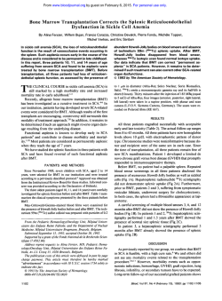
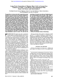
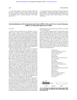
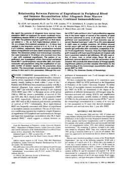
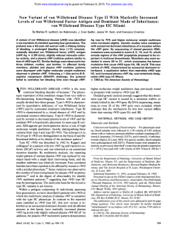
Origin of Human Mast Cells: Development From Transplanted
Hematopoietic Stem Cells After Allogeneic Bone Marrow Transplantation
By M. Fodinger, G. Fritsch, K. Winkler, W. Emminger, G. Mitterbauer, H. Gadner, P. Valent, and C. Mannhalter
Although mast cells are hematopoietic cells, little is known
about the origin of their precursors in vivo. In this study,
the origin(donor Y recipient genotype) ofhuman mastcells
(MCs) was analyzed in a patient who underwentallogeneic
bone marrow transplantation (BMT). The patient presented
with secondary acute myeloid leukemia (French-AmericanBritish classification, M21 arising from refractory anemia
with excess of blast cells and bone marrow (BM) mastocytosis. Transplantation was performed in chemotherapyinduced complete remission. On days 88, 126, 198, and 494
after BMT, mast cells were enriched t o homogeneity from
bone marrow mononuclear cells (BM MNCs) by cell sorting
for CD117'/CD34- cells. Purified mast cell populations were
CD117(c-kit)+ (>95%),CD34(<l%), CD3- (<l%), CD14(<l%), and virtuallyfree of contaminating cells as assessed
by Giemsa staining. The genotype of MCs was analyzed
after amplification by polymerase chain reaction (PCR)
of a variable number tandem repeat (VNTR) region within
intron 40 of the vonWillebrand factor(vWF) gene.Unexpectedly, on days 88 and 126 after BMT, sorted MCs displayed
recipient genotype as shown by vWF.VNTR-PCR. However,
on days 198 and 494, PCR analysis showed a switch t o donor
genotype in isolated mast cells. Peripheral blood (PS) and
BM MNC as well as highly enriched (sorted) CD3+ T cells
(PB, BM), CD4+ helper T cells (PB), CD8' T cells (PB), CD19'
B cells (PB), CD14+ monocytes (PB, BM), and CD34' precursor cells (BM) showed donor genotype throughout the observation period. Together, these results provide evidence
that human MCs developed in vivo fromtransplanted hematopoietic stem cells. Engraftment and in vivo differentiation
of MCs from early hematopoietic progenitor cells may be a
prolonged process.
0 1994 by The American Society of Hematology.
M
transplantation (BMT) in complete remission (CR). For discrimination between donor and recipient genotype of MCs
we used amplification by polymerase chain reaction (PCR)
of a highly variable region (variable number tandem repeat,
VNTR) within intron 40 of the von Willebrand factor(vWF)
gene.
AST CELLS (MCs) represent a distinct cell lineage
within the hematopoietic cell ~ y s t e m . "These
~
cells
have characteristic secretory granules and produce a number
ofvasoactive andimmunomodulatingsubstances
suchas
histamine,heparin, ortumor necrosis factor
Mast cells
reside in various organs including lung, skin, and thegastroheintestinal
However, although MCs
belong to the
matopoietic cell system, the number of human MCs in normal bone marrow (BM) usually is very low.
The origin of MCs from hematopoietic progenitor cells
isagenerallyaccepted
hyp~thesis.'.''"~Differentiationof
humanMCscanbe
inducedinvitro
from c-kit', CD34'
colony-forming cell^.'^^'^ However, whether human MCs are
replenished from BM-derived or circulating stemcells
throughout life remains unknown at present. Alternatively,
MCs may develop fromlocal progenitor cell pools in various
tissues.
To study in vivo originof human MCs from BMprecursor
cells, we analyzed the MC genotype (donor v recipient) in
a patient with acute myeloid leukemia (AML) (after myelodysplastic syndrome [MDS]) who underwent allogeneic BM
From the Clinical Institute of Medical and Chemical Lab Medicine, Department of Molecular Biology: and the I. Medical Department, Division of Hematology and Hemostaseology, Universiv of
Vienna, and St Anna Childrens ' Hospital, Vienna, Austria.
Submitted March 15, 1994; accepted June 30, 1994.
Supported by Fonds zur Forderung der Wissenschaftlichen
Forschung in Osterreich and Grant No. P-9359.
Address reprint requests to Christine Mannhalter, PhD, Clinical
Institute of Medical and Chemical Lab Medicine, Department of
Molecular Biology, The University of Vienna, Wahringer Giirtel 1820, A-1090 Vienna, Austria.
The publication costsof this article were defrayedin part by page
chargepayment. This article must therefore be hereby marked
"advertisement" in accordance with 18 U.S.C. section 1734 solely to
indicate this fact.
0 1994 by The American Society of Hematology.
0006-4971/94/8409-0016$3.00/0
2954
MATERIALS AND METHODS
Patient's description. In December 1991, a IO-year-old girl presented with anemia and thrombocytopenia. The hemoglobin concentration was 5.7 g/dL; the red blood cell count, 1.67 TiL; the white
blood cell count, 8.4 G/L; and the platelet count, 20 GiL. BM examination showed myelodysplasia with 16% blast cells and BM mastocytosis (Fig 1). The diagnosis of a MDS (French-American-British
[FAB] subgroup: refractory anemia with an excess of blast cells
[RAEB]) was established. After transition to AML [FAB: M2, 32%
blast cells, karyotype: t(8; l)(q22,q21),de1(5)(q13,q23)] the patient
was treated with chemotherapy (AML-BFM 83 protocol)Ib and CR
was achieved. However, BM mastocytosis persisted. In September
1992, the patient was still in CR and received allogeneic BMT (1.1
X IOx BM mononuclear cells [MNCsIkg body weight) from her
HLA-identical sister. The conditioning regimen consisted of 12 mg/
kg busulfan, 40 mgkg etoposide, and 120 m a g cyclophosphamide.
Methotrexate and cyclosporin-A were administered as graft-versushost disease (GVHD) prophylaxis. Because of late engraftment and
acute GVHD, grade I1 (day 26 after BMT), the patient received
prednisolon and anti-interleukin-2 (anti-IL-2; 0.15 to 0.3 mg/kg/d),
high-dose lgs and recombinant human IL-3. On day 121, complete
hematopoietic recovery was noted and successful engraftment of all
cell lineages (except MCs) was evidenced by vWF.VNTR-PCR.
During the whole observation period, mastocytosis persisted. Five
hundred days after BMT, all cell lineages displayed donor genotype
and the patient was still in CR.
Monoclonal antibodies (MoAbs). The following MoAbs were
used to analyze and purify MNCsubpopulations. The phycoerythrinor fluorescein isothiocyanate-conjugated MoAbs UCHTl (CD3),
MT310 (CD4).DK25(CD8). TUK2 (CD14), and HD37 (CD19)
were purchased from Dakopatts (Glostrup, Denmark) and MoAb
8G12 (CD34) was purchased from Becton Dickinson (Sunnyvale,
CA). The anti-c-kit MoAb YB5.B8 (CD1 17)" was kindly provided
by Leonie Ashman (University of Adelaide, Australia).
Flow-cytometric analysisand cell sorting. Peripheral blood (PB)
Blood, Vol 84. No 9 (November l ) , 1994: pp 2954-2959
ORIGIN
OF HUMANMAST CELLS
2955
Fig l. BM MCs on day 88
after BMT Giemsa staining.
and BM MNCs were separatedby density centrifugation (Nycoprep;
Nycomed, Oslo, Norway). MNCs were prepared on days 78 (PB),
88 (BM),126(BM),
133 (PB), 198(BM),and
494 (BM) after
BMT and incubated with the appropriately diluted MoAbs. Flowcytometric analysis and cell sorting were performed on a FACStar
Plus(BectonDickinson,MountainView,CA)
as previously described.” MNC wereenriched for CD3+T cells, CD4+T cells, CD8’
T cells, CD19’ B cells, CD34+ precursor cells, and CD117+ MCs.
In case of sorting for MCs, double staining with MoAbs YBS.B8
( 0 1 1 7 , c-kit) and 8G12 (CD34) was used to enrich for CD117’/
CD34- cells (exclusion of c-kit+ precursor cell subsets). The purity
of each sort was assessed by reanalysis and usually ranged between
96% and 99.5%. The presence of MCs in the c-kit+ cell fractions
was confirmed by Giemsa staining of cytospin preparations as well
as by measurement of histamine in cell lysates. Histaminewas measured by a radioimmunoassay (Immunotech, Marseille, France) as
described.’~‘~
PCR analysis of a VNTR region within the vWF gene in highly
enriched leukocyte subsets. The vWF gene contains a polymorphic region of ATCT repeats (VNTR) within intron 40. This region was shown to vary in length between different individuals.”
Analysis of the vWF gene has been used for carrier detection and
prenatal diagnosis in vWF disease”.” and more recently for the
investigation of engrafted hematopoietic cells after allogeneic BM
transplantation.” In this study, the vWF.VNTR-PCR was used to
analyze the genotype (donor v recipient) of individual cell lineages in a patient after allogeneic BMT. The PCR technique was
applied on highly enriched CD1 17+/CD34- BM MCs (days 88,
126, 198, and 494 after BMT), CD34+ BM precursor cells (days
72 and 494). CD3’ BM T cells (days 126 and 198), CD14’ BM
cells (day 126). isolated BM MNCs (day 494), isolated BM polymorphonuclear cells (PMNCs) (days 198 and 494). CD3+ PB T
cells (day 78), CD4’ PB T cells (day 133), CD8+ PB T cells (day
133), CD4+/CD8+ PB T cells (day 133), CD19+ PB B cells (day
78), isolated PB MNCs (day 133), isolated PB PMNCs (day 133)
as well as on citrated whole blood (days S9 and 64). After sorting,
cell fractions were washed once in phosphate-buffered saline,
resuspended to a final concentration of 500 cells per pL and frozen
at -20°C. Cellular DNA (PB, MNCs, PMNCs, and sorted cell
fractions) was obtained by thawing and boiling for 10 minutes,
followed by centrifugation at 12,OOOg (for 10minutes). The supernatants were collected and stored at -20°C. The PCR was performed as described by Peake et al.”The PCR amplification products were analyzed by electrophoresis on vertical 8%
polyacrylamide (PAA) gels in 1 X TBE buffer (90 mmoliL TRIS,
90 mmoVL boric acid, 1.25 mmol/L EDTA) and stained with
ethidium bromide.
The sensitivity of the vWF.VNTR-PCR was determined by dilution experiments using DNA samples from two healthy individuals
whose VNTR polymorphism could be distinguished by PCR. Isolated PBMNCsof one inividual weremixedwith l%, 2%, S%,
lo%, 20%, 30%,40%, SO%, 60%, 70%, 80%, 90%,95%. 98%, and
99% MNCs from the other individual. Each aliquotof IO’ cells was
as
boiledandtheSupernatantswereusedforPCRamplification
described above.
The PAA gel electrophoresisof the PCR products showed amplification fragments of both genotypes when at least 10% MNCs of
one and 90% MNCs of the other individual were present. Below
10%. a reliable detection of the smaller MNC population was not
possible.
RESULTS
Isolation and purity of leukocyte populations. The purity
of the FACS-sorted cell fractions was analyzed by morphol-
ogy, flowcytometry, andbymeasuring cellularhistamine
levels. Reanalysis of sorted (c-kit+/CD34-) MCs
with
YB5.B8 MoAb revealed a purity of >95% (Fig 2). h addition, the isolated MCs were more than 95% pure as assessed
by Giemsa staining. A significant contamination of purified
MCswith CD3+ T cells (<l%), CD34’precursor cells
(< 1 %) or CD14+ monocytes (<1%) could be excluded by
flow-cytometric examination. Selective enrichment of MCs
was also analyzedbymeasurement of cellularhistamine.
2956
FODINGER ET AL
10:F1401038
I
~
B
R1
CD34
1
FLl-H\FLi-Hei&t
--->
10rF1401040
10:F1401041
c
I
r\4
D
C
R2
The calculated amounts of cellular histamine in the pure MC
populations ranged between 0.6 and 25 pg per cell, whereas
in other cell fractions (CD3' T cells, CD19+ B cells, CD14+
monocytes, and CD34+ precursor cells) the histamine levels
were below the detection limit. Approximately 1 to 2 X lo4
BM MCs were recovered from MNC specimens (percentage
of MCs in primary MNC samples: 0.1% to 0.3%). The viability of the cells was more than 70% as assessed by trypan
blue exclusion. In each MC preparation, 5.000 cells were
used for PCR analysis and 5,000 cells for cytospin preparation. Remaining cells were used for control experiments.
The punty of the CD34+ BM precursor cells was 94% to
98%; of CD3+ T cells, 98% to 99%; of CD4+ T cells, 99%;
of CD8+ T cells, 98%; of CD19' B cells, 98%; of CD14'
PB monocytes, 95% to 98%.
Analysis of vWF.VhTR on whole blood cells before BMT.
2957
ORIGIN OF HUMAN MAST CELLS
A
DISCUSSION
l 2 3 4 5 6 7 8 9 1 0
90 b
B
Fig 3. Comparison of BM MCs genotype with various cell fractions
as assessed by PAA gel electrophoresis of PCR amplification products. (A) Recipient genotype of BM MCs on day 88 after BMT is
shown. PCR was performed with donor's PB (lane 2). recipient's PB
before BMT (lane 3). recipient's PB on day 59 after BMT (lane 4). CD3'
T cells (day 78, lane 5). CD19' B cells (day 78, lane 6), CD14' monocytes (day 126, lane 7 ) . CD34' progenitor cells (day 72, lane 8). and
CD117+/c-kit' MCs (day 88, lane 9). Markers (lanes 1, 10): Msp Idigested plasmid pBR322 (Clontech, Palo Alto, CA). (B) Donor genotype of MCs on day 198 after BMT is shown: donor's PB (lane 2).
recipient's P6 before BMT (lane 3), recipient's PB after BM (day 198,
lane 41, BM MNC (day 198, lane 5). BM PMNC (day 198, lane 6). and
BM MCs (day 198,lane7). Markers (lanes 1,8): MspI-digested plasmid
pBR322.
In a first set of experiments, we amplified DNA extracted
fromthe recipient's andthe donor's wholebloodbefore
BMT. Gel electrophoresis of the PCR products showed two
different banding patterns in the two individuals (Fig 3)
representing the donor's and recipient's genotype.
At1clIwi.v of VWF.VNTR on highly cwrichd RM MCs. To
determine the genotype and origin ofBMMCs
and other
leukocyte subsets on various days after BMT, fractionated
and unfractionated PB and BM cells of thepatient were
analyzed by vWF.VNTRPCR.Highly enriched BM MCs
were analyzed on days 88, 126. 198. and 494 (Table I ) . On
days 88 and 126, MCs displayed recipient genotype in PCR
analyses (Fig 3A). However, a consecutive switch in the
genotype was observed. and on days 198and 494, MCs
exhibited donor genotype (Fig 3B).
At1crIwi.v of vWF. VNTR it1 P R crncl BM leukocyte suhsets.
In contrast withpurifiedMCs,all
other leukocyte subsets
showed donor origin in vWF.VNTR PCR experiments during theobservation period. On days S9 and64, analysis of the
patient's whole blood displayed donor genotype, suggesting
successful engraftment. On days 78 and 133. PB MNC and
PMNC as well a s purified (sorted)T cells, B cells. and
monocytes were analyzed. VNTR-PCR showed donor origin
of all circulating leukocyte subsets tested (data not shown).
BM cells and subfractions were analyzed on days 72, 88,
126,198,and 494 after BMT. PCR analysis ofDNA extracted from pure BM precursor cells (CD34'k-kit' cells)
on days 72 and 494 showed donor genotype. BM-derived
(CD14') monocytes and (CD3 + ) T cells were analyzed on
days 126 and 198 and displayed donor genotype. PCR data
onBM cells are summarized in Table I .
Although it iswell established thatMCsbelongto
the
hematopoietic cell system,'.I3little is knownabout origin and
differentiation pathways of cells that become committed to
and differentiate into mature MCs. Studies using clonal cell
assays and MC growth factor (MGF) (c-kit ligand) have
shown origin of human MCs from multipotent colony-forming precursor cells in vitro.".I5
However, confirmation for the situation in vivo has not
been provided so far. In the present study, origin of human
MCs from transplanted stem cells in vivo was investigated
by vWF.VNTR-PCR. MCs were purified from BMbyuse
of anti-c-kit MoAb YBS.B8 and analyzed on various days
after BMT. Unexpectedly, on days 88 and 126 after BMT,
PCR analysis showed recipient origin of MCs. However, on
days 198 and 494 after BMT, a switch to donor genotype
was evidenced.
Like all other hematopoietic cells, MCs develop from immature, uncommitted hematopoietic progenitor cells. In the
case of MCs, it is unknown whether the BM remains the
constant source of precursor cells throughout life or whether
these cells develop from a local pool of precursor cells in
extramedullary tissues. Our data support the concept that
MCs are constantly replenished from BM precursor cells,
although one has to take into account that in contrast with the
situation in normal tissue, MCs were obtained and analyzed
shortly after BMT (a highly abnormal situation). Moreover,
these MCs could onlybe analyzed in BM specimens, but
not in other organs or tissues, and although the BMof the
donor did not contain MCs, the donor MCs developed in a
patient who had persistent BM mastocytosis. Nevertheless,
our data provide the first evidence that BM stem cells give
risetohumanMCs
in vivo.However, further studies are
required to elucidate whether this canbe translated to all
MC systems in humans. It is noteworthy in this regard, that
in MC-deficient WIW' mice, transplanted hematopoietic precursor cells (obtained from normal littermates) gave rise to
MCs in various extramedullary organs.'3.23
In contrast with other hematopoietic cells, MCsneed a
longer time to develop from their precursor cells and (as
mature cells) exhibit prolonged survival. Development and
differentiation of human MCs in vitro (with c-kit ligand as
Table 1. Genotype of Sorted BMMNC Fractions as Assessed by
vWF.VNTR-PCR
Days
After
BMT
72
88
126
198
494
CD34'l
CD117'
Precursors
CD14'
CD117'1
CD34 MC
Donor
-
-
Recipient
Donor
Recipient
Donor
Donor
-
CD3'
Lymphocytes
Granulocytes
Monocytes
-
-
-
Donor
Donor
Donor
Donor
-
-
IPMNCI
-
BM MNC fractions were obtained by cell sorting with MoAbs. The
purity ofthe isolated cells ranged between 97% and 99.5%. The genotype(donor v recipient) of the MNC fractions was analyzed by
vWF.VNTR-PCR as described in Materials and Methods.
2958
FODINGER ET AL
growthfactor)takesabout
4 to 10 ~ e e k s . ’ ~ * In
’ this
~ , ~ ~ , ~ ~ 10. Gordon RG,Burd PR, Galli SJ: Mast cells as a source of
multifunctional cytokines. Immunol Today 11:458, 1990
study, the genotype ofMCs had not switched from recipient
I 1. Gordon JR, Galli SJ: Mast cells as a source of both preformed
todonortypebeforeday
180 after BMT, whereas donor
and immunologically inducable TNF-alphdcachectin. Nature
origin of othercelltypeswasidentified
earlier (day 50).
346:274, 1990
These observations suggest that in vivo differentiation of
12. Kmhenbaum AS, Goff JP, Kessler SJ, Mican JM, Zsebo M.
MCs from their precursor cells is a prolonged process as
Metcalfe DD: Effect of L-3 and stem cell factor on the appearence
compared withgranulocyte, monocyte, or lymphocyte differof human basophils and mast cells from CD34’ pluripotent progenientiation. Alternatively, the prolonged presence of recipient
tor cells. J Immunol 148:772, 1992
MCs could have inhibited rapid growth
of donor MCs (nega13. Kitamura Y, Yokoyama M, Matsuda H, Ohno T: Spleen coltive inhibition).
ony-forming cell as common precursor for tissue mast cells and
granulocytes. Nature 291:159, 1981
A number of studies have suggested
an association be14. Agis H, Willheim M, Sperr WR, Wilfing A, Kromer E,
tween BM mastocytosis andmyelodysplastic or myeloprolifKabrna E, Spanbiichl E, Strobl H, Geissler K, Spittler A, Boltzerativesyndromes.26-3’In our patient, the initial diagnosis
Nitulescu G, Majdic 0, Lechner K, Valent P: Monocytes do not
was RAEB associated with BM mastocytosis. Interestingly,
make mast cells when cultered in the presence of SCF. J Immunol
BM mastocytosis persisted throughout the observation
pe151:4221, 1993
riod, even when donor stem cells were the apparent source
15. Kirshenbaum AS, Kessler SW, Goff JP, Metcalfe DD: Demof MCs. Possibly, MC proliferationin BM tissuewas a
onstration of the origin of human mast cells from CD34’bone marreactive process. The hypothesis of a reactive MC process
row progenitor cells. J Immunol 146:1410, 1991
would be in line with the mature morphology of MCs in BM
16. Sartori PC, Taylor MH, Stevens MC, Darbyshire PJ, Mann
specimens and with the observation that the MC genotype
JR: Treatment of childhood acute myeloid leukaemia using the B F ”
83 protocol. Med Pediatr Oncol 2123, 1993
switched from recipient to donor.
17. Mayrhofer G, Gadd SJ, Spargo LDJ, Ashman LK: Specificity
To confirm the hypothesis of
a reactive process and to
of a mouse monoclonal antibody raised against acute myeloid cells
excludethe recurrence of clonal disease of donor origin
for mast cells in human mucosal and connective tissues. Immunol
after BMT, which has been described for various types of
Cell Biol 65:241, 1987
leukemias,32s33 genetic studiesare currently under way.
18. Fritsch G, Buchinger P, Printz D, Fink FM, Mann G, Peters
For most hematopoietic cell lineages donor origin after
C, Wagner T, Adler A, Gadner H: Rapid discrimination of early
BMT has been d e s ~ r i b e d .Origin
~ ~ , ~ of
~ MCs from early preCD34+ myeloid progenitors using CD45-RA analysis. Blood
cursor cells has long been a matter of intensive discussions.
81:2301, 1993
Our data provide the first evidence that human MCs derive
19. Valent P, Spanblochl E, Sperr W, Sillaber C, Agis H, Strobl
in vivo from the earliest (transplantable) hematopoietic proH, Zsebo KM, Geissler K, Bettelheim P, Lechner K: Induction of
differentiation of human mast cells from bone marrow andperipheral
genitor cells. The development of human MCs in vivo from
blood mononuclear cells by recombinant human stem cell factor
their precursor cells seems to be a prolonged process.
(SCF)/kit ligand (KL) in long term culture. Blood 80:2237, 1992
20. Peake IR, Bowen D, Bignell P, Lidell MB, Sadler JE, Standen
ACKNOWLEDGMENT
G, Bloom AL: Family studies and prenatal diagnosis in severe von
We thank P. Buchinger, D. Printz, H. Semper, B. Keck, 0. Haas,
Willebrands disease by polymerase chain reaction amplification of
M. Veitl, and H.C. Bankl for excellent technical assistance.
a variable number of tandem repeat region of the von Willebrand
factor gene. Blood 76:555, 1990
REFERENCES
21. Mannhalter C, Kyrle PA, Brenner B, Lechner K: Rapid neonatal diagnosis of type IIB von Willebrand disease using the poly1. Galli SJ: New concepts about the mast cells. N Engl J Med
328:257, 1993
merase chain reaction. Blood 77:2539, 1991
22. Gaiger A, Mannhalter C, Hinterberger W, Haas 0, Marosi C,
2. Schwartz LB, Huff TF: Mast cells, in Crystal RG, West JB
Kier P, Eichinger S, Funovic M, Lechner K: Detection of en(eds): The Lung. New York, NY, Raven, 1991, pp 601-16
graftment and mixed chimerism following bone marrow transplanta3. Valent P, Ashman LK, Hinterberger W, Eckersbexger F, Majtion using PCR amplification of a highly variable region-variable
dic 0, Lechner K, Bettelheim P: Mast cell typing: Demonstration
number of tandem repeats (VNTR) in the von Willebrand gene. Ann
of a distinct hematopoietic cell type and evidence for immunophenoHematol 63:227, 1991
typic relationship to mononuclear phagocytes. Blood 73:1778, 1989
23. Qiu F, Ray P, Brown K, Barker PE, Jhanwar S, Ruddle FH,
4. Valent P, Bettelheim P Cell surface structures on human basoBesmer P: Primary structure of c-kit: Relationship with the CSF-l/
phils and mast cells: Biochemical and functional characterization.
PDGF receptor kinase family. EMBO J 7:1003, 1988
Adv Immunol 52:333, 1992
24. Mitsui H, Furitsu T, Dvorak AM, bani AMA, Schwartz LB,
5. Galli SJ: Biology of disease. New insights into “The riddle of
Inagaki N, Takei M, Ishizaka K, Zsebo KM, Gillis S , Ishizaka T:
the mast cell”: Microenviromental regulation of mast cell developDevelopment of human mast cells from umbilical cord blood cells
ment and phenotypic hterogeneity. Lab Invest 62:5, 1990
by recombinant human and murine stem cell factor c-kit ligand. Proc
6. Valent P, Sillaber C, Bettelheim P: The growth and differentiaNatl Acad Sci USA 90:735, 1993
tion of mast cells. Prog Growth Factor Res 3:27, 1991
25. Irani AM, Nilsson G, Miettinen U, Craig S S , Ashman LK,
7. Denburg JA: Basophil and mast cell lineages in vitro and in
Ishizaka T, Zsebo KM, Schwartz LB: Recombinant human stem cell
vivo. Blood 792346, 1992
factor stimulates differentiation of human mast cells from dispersed
8. Johnston SL, Holgate ST: Cellular and chemical mediatorsfetal liver cells. Blood 80:3009, 1992
Their roles in allergic diseases. Cum Opin Immunol 2:513, 1990
26. Horny HP, Ruck M, Wehrmann M, Kaiserling E: Blood find9. Serafin WE, Austen KF: Mediators of immediate hypersensiings in generalized mastocytosis: Evidence of frequent simultaneous
tivity reactions. N Engl J Med 317:30, 1987
ORIGIN OF HUMAN MAST CELLS
occurrence of myeloproliferative disorders. Br J Haematol 76:186,
1990
27. Lewis JP, Welborn JL, Meyers FJ, LevyNB, Roschak T:
Mast cell disease followed by leukemia with clonal evolution. Leuk
Res 11:769,1987
,
RG: Sig28. Travis WD, Li CY, Yam LT, Bergstralh €3Swee
nificance of systemic mast cell disease with associated hematologic
disorders. Cancer 62:965, 1988
T, Kandels S, Radaszkiewicz T, UllrichA,
29. vonRuden
Wagner EF: Development of a lethal mast cell disease in mice
reconstituted with bone marrow cells expressing the v-erbB oncogene. Blood 79:3145, 1992
30. Webb TA, Li CY, Yam LT: Systemic mast cell disease: A
clinical and hematopathologic study of 26 cases. Cancer 49:927,
1982
2959
31. Prokicimer M, Polliack A: Increased bone marrow mast cells
in preleukemic syndromes, acute leukemia, and lymphoproliferative
disorders. Am J Clin Pathol 75:34, 1981
32. Boyd CN, Ramberg RC, Thomas ED: The incidence of recurrence of leukemia in donor cells after allogeneic bone marrow transplantation. Leuk Res 62333, 1982
33. Smith JL, Heerema NA, Provisor A: Leukaemic transformation of engrafted bone marrow cells. Br J Haematol 60:415, 1985
34. Ginsburg D, Antin JH, Smith BR, Orkin SH, Rappeport JM:
Origin of cell populations after bone marrow transplantation. Analysis using DNA sequence polymorphisms. J Clin Invest 75596, 1985
35. Ault KA, Antin JH, Ginsburg D, Orkin SH, Rappeport JM,
Keohan ML, Martin P, Smith BR: Phenotype of recovering lymphoid
cell populations after marrow transplantation. J Exp Med 161:1483,
1985


© Copyright 2026