
Recent advances in central acute vestibular syndrome of a vascular
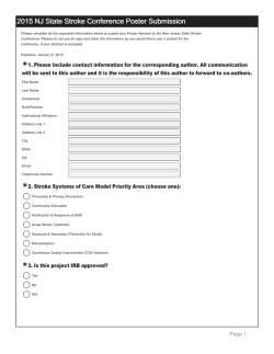
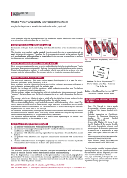
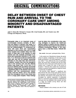
Journal of the Neurological Sciences 321 (2012) 17–22
Contents lists available at SciVerse ScienceDirect
Journal of the Neurological Sciences
journal homepage: www.elsevier.com/locate/jns
Review article
Recent advances in central acute vestibular syndrome of a vascular cause
Hyun-Ah Kim, Hyung Lee ⁎
Department of Neurology, Keimyung University School of Medicine, Daegu, South Korea
Brain Research Institute, Keimyung University School of Medicine, Daegu, South Korea
a r t i c l e
i n f o
Article history:
Received 25 April 2012
Received in revised form 20 July 2012
Accepted 23 July 2012
Available online 17 August 2012
Keywords:
Isolated vertigo
Acute vestibular syndrome
Cerebellar stroke
Vestibular neuritis
a b s t r a c t
Acute vestibular syndrome (AVS) is characterized by acute onset of spontaneous prolonged vertigo (lasting
days), spontaneous nystagmus, postural instability, and autonomic symptoms. Peripheral AVS commonly
presents as vestibular neuritis, but may also include other disorders such as Meniere's disease. Vertigo in central AVS due to vertebrobasilar ischemic stroke is usually accompanied by other neurological dysfunction.
However it can occur in isolation and mimicking peripheral AVS, particularly with cerebellar strokes. Recent
large prospective studies have demonstrated that approximately 11% of patients with isolated cerebellar infarction presented with isolated vertigo mimicking peripheral AVS, and the bedside head impulse test is the
most useful tool for differentiating central from peripheral AVS. Herein we review the keys to the diagnosis of
central AVS of a vascular cause presenting with isolated vertigo or audiovestibular loss.
© 2012 Elsevier B.V. All rights reserved.
Contents
1.
2.
3.
Introduction . . . . . . . . . . . . . . . . . . . . . . . . . . . . . . . . . . . . . . . . . . . . . . . . . . . . . . .
Possible anatomical structures responsible for central AVS of a vascular cause . . . . . . . . . . . . . . . . . . . . . . . . .
Classification of central AVS . . . . . . . . . . . . . . . . . . . . . . . . . . . . . . . . . . . . . . . . . . . . . . . .
3.1.
Cerebellar ischemic stroke . . . . . . . . . . . . . . . . . . . . . . . . . . . . . . . . . . . . . . . . . . . . . .
3.1.1.
Frequency, pattern of involved vascular territory, and associated vestibular dysfunction . . . . . . . . . . . .
3.1.2.
Clinical implication of central AVS due to cerebellar ischemic stroke from the standpoint of mechanism of stroke
3.2.
Central AVS associated with brainstem ischemia . . . . . . . . . . . . . . . . . . . . . . . . . . . . . . . . . . .
4.
Which of the neurological examinations at the bed side is most useful for differentiating central AVS from more benign disorders
involving the inner ear? . . . . . . . . . . . . . . . . . . . . . . . . . . . . . . . . . . . . . . . . . . . . . . . . . . . . . .
5.
Why infarction in the territory of the anterior inferior cerebellar artery (AICA) seldom serves as a common cause of central AVS of a
vascular cause presenting with isolated vertigo? . . . . . . . . . . . . . . . . . . . . . . . . . . . . . . . . . . . . . . . . . .
6.
Prolonged vertigo and hearing loss as the presenting symptoms of VBIS may be misdiagnosed as Meniere's disease . . . . . . .
7.
When does the patient with isolated vertigo need an urgent brain scan? . . . . . . . . . . . . . . . . . . . . . . . . . . .
Conflict of interest . . . . . . . . . . . . . . . . . . . . . . . . . . . . . . . . . . . . . . . . . . . . . . . . . . . . . . .
Acknowledgments . . . . . . . . . . . . . . . . . . . . . . . . . . . . . . . . . . . . . . . . . . . . . . . . . . . . . . .
References . . . . . . . . . . . . . . . . . . . . . . . . . . . . . . . . . . . . . . . . . . . . . . . . . . . . . . . . . . .
1. Introduction
Acute vestibular syndrome (AVS) is characterized by acute onset
of spontaneous prolonged vertigo (lasting days), spontaneous nystagmus, postural instability, and autonomic symptoms [1,2]. AVS can be
⁎ Corresponding author at: Department of Neurology, Keimyung University School
of Medicine, 194 Dongsan dong, Daegu 700‐712, South Korea. Tel.: + 82 53 250 7835;
fax: + 82 53 250 7840.
E-mail address: [email protected] (H. Lee).
0022-510X/$ – see front matter © 2012 Elsevier B.V. All rights reserved.
http://dx.doi.org/10.1016/j.jns.2012.07.055
.
.
.
.
.
.
.
.
.
.
.
.
.
.
.
.
.
.
.
.
.
17
18
18
18
18
19
19
. . . . . . .
19
.
.
.
.
.
.
20
21
21
21
21
21
.
.
.
.
.
.
.
.
.
.
.
.
.
.
.
.
.
.
.
.
.
.
.
.
.
.
.
.
.
.
.
.
.
.
.
.
.
.
.
.
.
.
.
.
.
.
.
.
.
.
.
.
.
.
.
.
.
.
.
.
.
.
.
.
divided into peripheral (i.e., inner and vestibular nerve) and central
(i.e., brainstem and cerebellum) causes. The peripheral causes of
AVS included acute vestibular neuritis (VN), Meniere's disease, and
migraine. Vertebrobasilar ischemic stroke (VBIS) can also cause isolated prolonged vertigo mimicking peripheral AVS [3–5]. Recent studies have shown that cerebellar infarction simulating peripheral AVS is
more common than previously thought and the bedside head impulse
test (HIT) is the most useful tool for differentiating central AVS from
other more benign disorders involving the inner ear [4,5]. Clinically,
it is important to differentiate central AVS of a vascular cause from
18
H.-A. Kim, H. Lee / Journal of the Neurological Sciences 321 (2012) 17–22
peripheral AVS because therapeutic strategy and prognosis are different in the two conditions. Early recognition of the central AVS of a vascular cause may allow specific management. This review aims to
highlight recent advances in central AVS of a vascular cause presenting
with isolated vertigo or audiovestibular loss and to address their clinical
significance.
2. Possible anatomical structures responsible for central AVS of a
vascular cause
Vertigo is resulted from imbalance of tonic discharge of the vestibular systems arising from the inner ears on both sides. The origin of vertigo may be peripheral or central. When the vertigo occurs as a
symptom of VBIS, it is usually associated with other neurological symptoms or signs [6]. Three possible structures responsible for central AVS
of a vascular cause are the nodulus, root entry zone of the eighth
nerve in the pontomedullary junction, and vestibular nucleus (Fig. 1).
Theoretically, a small infarct localized to these structures can cause vertigo with no accompanying other neurological symptoms or signs since
all of these structures receive afferent vestibular inputs from the inner
ear (Fig. 2). Because none of these structures are known to be more sensitive to ischemia than other surrounding structures, the incidence of
central isolated vertigo associated with ischemic stroke is low. Rarely,
lesions involving the flocculus lobe or dorsal insular cortex can also
cause isolated vertigo (Fig. 2) [7–9]. Vertigo due to a lesion involving
the dorsal insular cortex is usually not associated with nystagmus and
a flocculus lesion is commonly associated with other central signs
with gaze-evoked nystagmus and asymmetrical oculomotor dysfunction [7,9]. Therefore, in such cases, clinicians may conclude that vertigo
is caused by damage to the central vestibular structure.
3. Classification of central AVS
3.1. Cerebellar ischemic stroke
3.1.1. Frequency, pattern of involved vascular territory, and associated
vestibular dysfunction
Vertigo is one of the commonest symptoms in patients with cerebellar stroke syndrome. Among cerebellar stroke syndrome, cerebellar ischemic stroke probably ranks first as central AVS of a vascular cause.
A small retrospective study showed that as many as 25% of patients
with vascular risk factors who presented to an emergency medical setting with isolated severe vertigo, nystagmus, and postural instability
have a cerebellar infarction in the territory of the medial branch of the
PICA (mPICA) [10]. A recent large prospective study on clinical findings
of 240 patients with isolated cerebellar infarction also demonstrated
similar results. In this study, about 11% (25/240) with isolated
cerebellar infarction had isolated vertigo only and most (24/25: 96%)
patients with isolated vertigo had an infarct in the territory of the
mPICA including the nodulus [5]. Another more recent study using
diffusion-weighted imaging found that 75% of patients with at least
one vascular risk factor who presented with acute isolated vertigo had
acute stroke, mostly involving the caudal cerebellum in the mPICA territory [4]. In PICA territory cerebellar infarction, the key structure responsible for vertigo is the nodulus. The nodulus is strongly connected
to the ipsilateral vestibular nucleus and receives direct projections
from the labyrinth [11,12]. Functionally, nodulovestibular Purkinje fibers have an inhibitory effect on the ipsilateral vestibular nucleus
[11,12]. A predominant involvement of mPICA territory cerebellar infarction associated with central AVS may be explained in several
ways. First, the mPICA usually supplies the nodulus, a part of the
vestibulocerebellum [13]. Thus, infarction in the territory of the medial
PICA can cause severe vertigo. Second, dysmetria, a major finding of the
cerebellar lesion, may be minimal or absent after a cerebellar infarction
in the territory of the mPICA if the size of an infarct is not large [5,14,15].
Third, gaze-evoked asymmetrical nystagmus, which commonly occurred in central vestibulopathy of cerebellar origin, is sometimes absent in the PICA territory cerebellar lesion [5,14–18]. Finally, hearing
loss that is generally considered a peripheral sign commonly accompanies anterior inferior cerebellar artery (AICA) stroke, not PICA stroke
[19]. Since the superior cerebellum supplied by the superior cerebellar
artery (SCA) does not have significant vestibular connections, cerebellar
infarction in the SCA territory rarely causes vertigo [20,21]. The
vestibulo-ocular portion of the cerebellum is located primarily in the
flocculonodular lobes, which are supplied by branches of the AICA and
PICA. The low incidence of vertigo in SCA distribution may be a useful
clinical distinction from PICA or AICA cerebellar infarction in patients
with acute vertigo and limb ataxia [20,21].
In PICA territory cerebellar infarction, the direction of nystagmus
and degree of postural instability were variable. The prominent cerebellar signs, particularly severe axial instability and direction changing gaze-evoked nystagmus (occurring in 71% and 54%, respectively,
in the aforementioned series [5]), can help in the differential, but
these findings are less reliable.
Similarly, perverted head shaking (mostly downbeating) and positional downbeating nystagmus as important signs of central vestibular
dysfunctions are found in only half of the cases with cerebellar infarction [22]. In contrast, the vestibular dysfunction in some patients with
PICA territory cerebellar infarction is similar to that in those with VN.
For example, spontaneous unidirectional, ipsilesional nystagmus and
mild postural instability with standing or walking independently could
be seen in cases with PICA territory cerebellar infarction (occurring in
17% and 29%, respectively, in the aforementioned series [5]). The mechanism of spontaneous unidirectional, ipsilesional nystagmus may have
Fig. 1. Structures related to the central isolated vertigo syndrome. F = flocculus, 8 = vestibule-cochlear nerve,
fascicle), = vestibular nucleus, * = nodulus, ** = oculomotor vermis.
= intraparenchymal portion of vestibular nerve (i.e., vestibular
H.-A. Kim, H. Lee / Journal of the Neurological Sciences 321 (2012) 17–22
19
Fig. 2. Focal infarction selectively involving the structures responsible for isolated vertigo. A. Isolated nodulus infarction. B. AICA territory infarct involving the lateral caudal pons
extending from the root entry zone (*) of the eighth nerve to the most proximal portion of the vestibular fascicle. C. Isolated vestibular nucleus infarction. D. Focal infarction selectively
involving the dorsal insula. D. The flocculus lobe is selectively infracted.
involved an increased tonic activity of ipsilesional medial and superior
vestibular nucleus neurons, caused by disconnection of inhibitory
nodulovestibular Purkinje fibers from neurons in the vestibular nuclei
[16–18]. The nystagmus in PICA territory cerebellar infarction involving
the nodulus may be contrasted with that of a unilateral VN, in which
the tonic resting firing rate of semicircular canal afferents is decreased
and contralesional nystagmus is due to unopposed activity of the intact
vestibular nucleus. Overall, although the severity of imbalance and the
appearance of nystagmus in PICA territory cerebellar infarction can help
in differentiating central AVS from VN, these findings are less reliable
for differentiating two conditions. PICA territory cerebellar infarction
should be considered in the differential diagnosis of central AVS, even if
the nystagmus and imbalance are more typical of VN.
3.1.2. Clinical implication of central AVS due to cerebellar ischemic stroke
from the standpoint of mechanism of stroke
Isolated vertigo associated with cerebellar infarction (mainly in
the distribution of the PICA) may give clinicians two important messages. First, although small PICA territory cerebellar infarction causing
vertigo generally has a benign prognosis, isolated PICA territory cerebellar infarction usually results from emboli originating from the
heart or great vessels [23], and recurrent emboli should need to be
treated. Second, patients with cerebellar infarction may have isolated
vertigo with no other cerebellar signs, even though there is a relatively large size of a lesion on brain MRI because the threshold of damage
to the central nervous system for producing neurological symptoms
or signs appears to be different in individuals and other areas of the
cerebellum may be able to compensate for the mPICA territory damage. These situations have been previously reported in the literature
[5,24]. Cerebellar infarction may develop a mass effect in 10% to 25%
of cases and PICA territory infarcts are more likely to produce a
mass effect than SCA territory infarcts [25,26]. Large PICA territory
cerebellar infarction can cause brainstem compression, hydrocephalus, cardiorespiratory complications, coma, and death [27–29]. Thus,
in view of the different therapeutic strategies and potentially grave
prognosis of the strokes involving the vertebrobasilar artery territory,
it is of great importance to differentiate central AVS of a vascular
cause from more benign disorders involving the inner ear.
3.2. Central AVS associated with brainstem ischemia
As noted above, the central vestibular system located in the brainstem is not more vulnerable to ischemia than other surrounding structures. Thus, mono-symptomatic attacks of vertigo and nystagmus
without any other brainstem symptoms and signs would be unusual
in brainstem ischemia. Selective damage to the vestibular nucleus and
root entry zone of the eighth nerve in the pontomedullary junction
can cause isolated vertigo [30–34]. Because the root entry zone of the
eighth cranial nerve has a rich network of anastomotic vessels arising
from the lateral medullary artery, anterior inferior cerebellar artery, inferior lateral pontine artery, and arteries supplying adjacent dura matter and petrous bone [35,36], the possibility of focal infarction in that
area is extremely low in a clinical practice. There were case reports of
central AVS due to a demyelinating lesion localized to the root entry
zone of the eighth nerve [30,31], but isolated vertigo due to focal infarction in the root entry zone of the vestibular nerve was not available in
the literature. Focal ischemia in the vestibular nucleus can cause isolated vertigo and nystagmus mimicking acute VN [32–34]. Recently there
was one case report [32] of a patient with an isolated vestibular nucleus
infarction who presented with isolated prolonged vertigo, spontaneous
horizontal nystagmus with a torsional component, a positive head impulse test result, and unilateral canal paresis to caloric stimulation. All
of these findings are consistent with VN. This report emphasized that
isolated vestibular nucleus infarction should be considered in the differential diagnosis of central vascular vertigo syndrome, especially when
the patient has unilateral canal paresis, but other neurologic symptoms
or signs are absent. Vertigo in the lateral medullary infarction is usually
associated with other neurological symptoms or signs, but tiny infarct
in the lateral medulla can present with vertigo without other localizing
symptoms [37]. In this case, the HIT might be positive, if medial vestibular nucleus is involved.
4. Which of the neurological examinations at the bed side is most
useful for differentiating central AVS from more benign disorders
involving the inner ear?
If a patient showed signs of central vestibular dysfunction such as
vertical nystagmus in the primary position, direction changing gaze-
20
H.-A. Kim, H. Lee / Journal of the Neurological Sciences 321 (2012) 17–22
evoked nystagmus, perverted head shaking nystagmus, asymmetrical
oculomotor dysfunction, or severe postural instability with falling
during the attack of vertigo, clinician can easily discern that vertigo
is originated from the central vestibular dysfunctions. However, as
mentioned above, these central signs do not always appear in each
patient with central vertigo. It is generally considered that the bedside HIT and caloric test are useful tools for differentiating central
AVS from a more benign disorder involving the inner ear. Normal
HIT and caloric test results are regarded as reliable signs for an intact peripheral vestibular function, thus suggesting a central lesion.
The major advantage of the HIT, in addition to the convenience factor (i.e., bedside HIT with no special equipment required) is the fact
that it rarely causes vomiting whereas caloric test is potent at this.
Further, in the presence of significant spontaneous nystagmus, caloric testing is unreliable. More recently, video HIT has been validated
in selected vestibular disorders [38] and it has the advantage over
bedside HIT in distinguishing spontaneous nystagmus from the
vestibulo-ocular reflex (VOR) slow phase response. Finally, HIT reflects the functional status of the vestibulo-ocular reflex (VOR) needed in locomotion (i.e., high frequencies of VOR) whereas caloric test
assesses the low-frequency range of VOR [39]. Because about 17% of
patients with PICA territory cerebellar infarction present with
pseudo-VN [5], stroke patients should be evaluated with the HIT.
The significance of HIT for differentiating stroke from VN has been
confirmed by another recent paper [4] that showed that a negative
HIT result (i.e., normal vestibulo-ocular reflex) is strongly suggestive
of a central lesion with a pseudo-VN presentation. However, bedside
HIT has several limitations, in which the covert saccade during the
head rotation (instead of overt saccade after head rotation) is almost impossible to detect by simple visual observation at the bedside [40], and spontaneous nystagmus during the acute period also
interferes with assessment of bedside HIT. Accuracy of the bedside
HIT is also influenced by examiner's experience [41]. It is well
known that experienced examiners are more conservative, whilst novices tend to overcall. Furthermore, lateral pontine lesion encompassing
the root entry zone of the eighth nerve can cause a positive HIT result,
causing a peripheral lesion to be mistakenly suspected. Because of the
limitations of bedside HIT, 9% to 39% of positive bedside HIT results
have been reported in patients with cerebellar or brainstem strokes
[4,42]. Diagnostic accuracy of bedside HIT for differentiating central
AVS from VN may be enhanced if other central signs are included
for differentiating two isolated vertigo syndromes. A refined bedside
examination protocol that combined HIT with search for directionchanging nystagmus in eccentric gaze or skew deviation was 100%
sensitive and 96% specific for identification of stroke, whereas the
initial diffusion weighted MRI missed 12% of them [4]. Another recent
report also showed similar findings that sensitivity and specificity for
identification of stroke was 100% and 90%, respectively if one of the following signs suggestive of a central lesion was present: normal bedside
HIT, central type nystagmus, skew deviation, or abnormal vertical
smooth pursuit [43]. Since mild degree of skew deviation usually goes
unnoticed during the bedside examination and gaze-evoked nystagmus
is also sometimes absent in the central AVS, HIT at the bedside can be
the best tool for differentiating central AVS from a more benign disorder
involving the inner ear although bedside HIT has some limitations. The
normal caloric test is also considered a reliable sign of an intact peripheral vestibular function. Overall, the most consistent bedside predictor
of central AVS of a vascular cause appears to be the HIT and normal
HIT result usually guarantees an absence of peripheral pathology
[4,5,43–45]. Differential diagnostic points for central and peripheral
AVS are summarized in Table 1.
5. Why infarction in the territory of the anterior inferior cerebellar
artery (AICA) seldom serves as a common cause of central AVS of a
vascular cause presenting with isolated vertigo?
Like mPICA cerebellar infarction, AICA territory infarction usually gives
rise to acute prolonged vertigo. However, AICA territory ischemic stroke
seldom leads to the central AVS of a vascular cause presenting with isolated vertigo because there is nearly always associated unilateral hearing
loss (due to mostly cochlear ischemia) and most patients will have multiple brain stem signs such as facial palsy, Horner syndrome, or crossed sensory loss [19,46,47]. Furthermore, in the AICA syndrome, the patients
usually showed severe imbalance with falling and direction-changing,
gaze-evoked, asymmetrical nystagmus (Bruns's nystagmus), whereas patients with peripheral AVS always can stand or walk without support and
have direction fixed, gaze-evoked nystagmus beating toward the intact
side [1,6]. A recent report showed that only 5% of patients with AICA territory ischemic stroke presented with acute prolonged vertigo and canal
paresis to caloric stimulation without hearing loss, mimicking peripheral
AVS [48]. Indeed, AICA territory infarction usually leads to acute
audiovestibular loss with severe vertigo and hearing loss (i.e., acute
labyrinthine infarction) rather than isolated vestibular loss.
Table 1
Differentiating among common central and peripheral acute vestibular syndromes.
PICA-Caudal cerebellum
including nodulus, lateral medulla
AICA‐pons/Root
entry zone/
labyrinth
Vestibular nucleus
Vestibular neuritis
Isolated vertigo
Caloric canal paresis
Possible, common
None
Possible, uncommon
Common
Bedside head impulse test
Normal
Abnormal
Hearing loss
Spontaneous nystagmus
None
Ipsilesional (cerebellum),
ipsilesional or contarlesional
(lateral medulla)
Variable, typicallyb
Direction-changing
Variable, usual in lateral medullary
Ipsi > contralesional
Variable, usually severe
(fall without assistance)
Ischemic
Common
Contralesional
Direction-changing
(Bruns)
Unidirectional
Possible, uncommon
Abnormal, if medial
subnucleus involved
Abnormal, if medial
subnucleus involved
None
Ipsilesional or
contralesional
Almost always
Abnormal, if superior or
complete vestibular nerve involved
Abnormal, if superior or complete
vestibular nerve involved
Rarely
Unidirectionala
Variable
Unidirectional
Variable
Ipsilesional
Variable
Variable
Ipsilesional
Variable
Occasionally
Ipsilesional
Mild to moderate
Ischemic,
demyelination
Ischemic
Viral, idiopathic
Effect of gaze on nystagmus
Skew deviation
Side of truncal deviation
Postural instability
Common causes
a
b
Direction-fixed unidirectional gaze‐evoked nystagmus beating toward the healthy side.
Direction-changed bidirectional gaze‐evoked nystagmus that the intensity was maximal when gaze to the lesion side.
H.-A. Kim, H. Lee / Journal of the Neurological Sciences 321 (2012) 17–22
21
Fig. 3. MRI and audiovestibular findings in a patient with recurrent vertigo and fluctuating hearing loss with tinnitus as prodromal signs of AICA territory infarction. A. Axial
diffusion-weighted brain MRI 1 day after the onset of recurrent vertigo and fluctuating right-sided hearing loss with tinnitus was normal. B. An initial pure tone audiogram performed
at attack of isolated vertigo and hearing loss reveals severe hearing loss (70 dB) with 24% speech discrimination in the right ear. Hearing levels in decibels (dB) (American National Standards Institute, 1989) are plotted against stimulus frequency on a logarithmic scale. C. Video-oculographic recordings of bithermal caloric tests performed at attack of isolated vertigo and
hearing loss disclose right canal paresis (65%). Three days later, patient complained of exacerbation of vertigo and hearing loss on the right ear. On examination, there were bidirectional
gaze-evoked nystagmus and dysmetria on the right limb. D. Follow-up axial diffusion-weighted MRI demonstrated hyperintense foci involving the right anterior cerebellum including the
flocculus. E. Follow-up pure tone audiogram shows aggravated hearing loss on the right ear. AICA: anterior inferior cerebellar artery, Vmax: maximal velocity of slow phase of nystagmus.
6. Prolonged vertigo and hearing loss as the presenting symptoms
of VBIS may be misdiagnosed as Meniere's disease
Recent papers have shown that 8–30% of patients with posterior circulation ischemic stroke (mainly AICA territory) had acute audiovestibular
loss with vertigo, fluctuating hearing loss and/or tinnitus before more
widespread infarction and at this stage, patient may be misdiagnosed as
having peripheral pathology with Meniere's disease [47–49]. Selective ischemia to the inner ear can explain isolated prodromal audiovestibular
disturbance because the inner ear requires high-energy metabolism and
has little collateral circulation [50–52]. Although there are as yet no systematic data on what a high-risk factor suggesting impending stroke is
or what interventions might be beneficial at the stage of isolated
audiovestibular loss, patients with prodromal audiovestibular disturbance were more likely to have focal or diffuse stenosis of the basilar artery presumably close to the origin of the AICA than patients without
audiovestibular disturbance [19,47,53]. This finding highlighted that
AICA infarction should be considered, particularly in elderly patients
with vascular risk factors and acute audiovestibular loss, even when
MRI does not demonstrate acute infarction in the brain. At this stage, clinician should consider a further investigation and a proper management
to prevent progression of acute audiovestibular loss into a more widespread posterior circulation stroke, mainly in the territory of the AICA. Because current diagnostic methods (including MRI) cannot confirm
labyrinthine infarction among the acute audiovestibular loss syndrome,
clinicians should consider all the clinical evidences when attempting to
determine the etiology of acute audiovestibular loss rather than emphasizing that MRI is the best way to distinguish other pathology from vascular etiology. Illustrative case with recurrent vertigo and fluctuating
hearing loss mimicking Meniere's disease as initial symptoms of
impending AICA territory cerebellar infarction is shown Fig. 3.
7. When does the patient with isolated vertigo need an urgent
brain scan?
For patients with spontaneous prolonged vertigo, in addition to the
obvious cases of associated neurological symptoms or signs, an urgent
brain scan to rule out central vascular vertigo syndrome should be considered in 1) older patients presenting with isolated spontaneous
prolonged vertigo, in 2) any patient with vascular risk factors and isolated spontaneous prolonged vertigo who had a normal HIT result, in 3)
any patient with isolated spontaneous prolonged vertigo who had directional changing gaze‐evoked nystagmus or severe gait ataxia with
falling at upright posture, in 4) any patient presenting with acute spontaneous vertigo and new onset headache, especially occipital, and in 5)
any patient with vascular risk factors and acute onset of vertigo and
hearing loss without Meniere's history [5,43]. Since brain CT is known
to have less accuracy in detecting an acute ischemic lesion within the
posterior fossa [29], brain MRI with diffusion image is considered the
golden standard for diagnosis of isolated vertigo due to ischemic stroke.
Conflict of interest
All authors have reported no conflicts of interest.
Acknowledgments
Dr. Lee serves as the editorial boards of the Research in Vestibular Science, Frontiers in Neuro-otology, and Current Medical Imaging Review.
Financial Relationship Disclosure: We have nothing to disclose.
Funding: “This research was supported by the Scholar Research
Grant of Keimyung University in 2012”.
References
[1] Hotson JR, Baloh RW. Acute vestibular syndrome. N Engl J Med 1998;339:680-5.
[2] Guntinas-Lichius O. Acute vestibular syndrome. N Engl J Med 1999;340:151-2.
[3] Kim JS, Lee H. Inner ear dysfunction due to vertebrobasilar ischemic stroke. Semin
Neurol 2009;29:534-40.
[4] Newman-Toker DE, Kattah JC, Alvernia JE, Wang DZ. Normal head impulse test differentiates acute cerebellar strokes from vestibular neuritis. Neurology 2008;70:
2378-85.
[5] Lee H, Sohn SI, Cho YW, Lee SR, Ahn BH, Park BR, et al. Cerebellar infarction
presenting isolated vertigo: frequency and vascular topographical patterns. Neurology 2006;67:1178-83.
[6] Baloh RW, Honrubia V. Clinical neurophysiology of the vestibular system. 4th ed.
New York, NY: Oxford University Press; 2011.
22
H.-A. Kim, H. Lee / Journal of the Neurological Sciences 321 (2012) 17–22
[7] Brandt T, Bötzel K, Yousry T, Dieterich M, Schulze S. Rotational vertigo in embolic
stroke of the vestibular and auditory cortices. Neurology 1995;45:42-4.
[8] Ahn BY, Bae JW, Kim DH, Choi KD, Kim HJ, Kim EJ. Pseudo-vestibular neuritis
associated with isolated insular stroke. J Neurol 2010;257:1570-2.
[9] Leigh RJ, Zee DS. The neurology of eye movements. IV ed. New York: Oxford University Press; 2006.
[10] Norrving B, Magnusson M. Isolated acute vertigo in the elderly; vestibular or vascular disease. Acta Neurol Scand 1995;91:43-8.
[11] Voogd JAN, Gerrts NM, Ruigrok TH. Organization of the vestibulocerebellum. Ann
NY Acad Sci 1996;781:553-79.
[12] Fushiki H, Barmack NH. Topography and reciprocal activity of cerebellar purkinje
cells in the uvula-nodulus modulated by vestibular stimulation. J Neurophysiol
1997;78:3083-94.
[13] Amarenco P, Etienne R, Hommel M, Chaine P, Marteau R. Infarction in the medial
branch of the posterior inferior cerebellar artery. J Neurol Neurosurg Psychiatry
1990;53:731-5.
[14] Duncan GW, Parker SW, Fisher CM. Acute cerebellar infarction in the PICA territory. Arch Neurol 1975;32:364-8.
[15] Huang CY, Yu YL. Small cerebellar stroke may mimic labyrinthine lesions. J Neurol
Neurosurg Psychiatry 1985;48:263-5.
[16] Lee H, Yi HA, Cho YW, Sohn CH, Whitman GT, Ying S, et al. Nodulus infarction
mimicking acute peripheral vestibulopathy. Neurology 2003;60:1700-2.
[17] Lee H, Cho YW. A case of isolated nodulus infarction presenting as a vestibular
neuritis. J Neurol Sci 2004;221:117-9.
[18] Moon IS, Kim JS, Choi KD, Kim MJ, Oh SY, Lee H, et al. Isolated nodular infarction.
Stroke 2009;40:487-91.
[19] Lee H, Sohn SI, Jung DK, Cho YW, Lim JG, Yi SD, et al. Sudden deafness and anterior
inferior cerebellar artery infarction. Stroke 2002;33:2807-12.
[20] Kase CS, White JL, Joslyn JN, Williams JP, Mohr JP. Cerebellar infarction in the superior cerebellar artery distribution. Neurology 1985;35:705-11.
[21] Amarenco P, Hauw JJ. Cerebellar infarction in the territory of the superior cerebellar artery: a clinicopathologic study of 33 cases. Neurology 1990;40:1383-90.
[22] Huh YE, Kim JS. Patterns of spontaneous and head-shaking nystagmus in cerebellar infarction: imaging correlations. Brain 2011;134:3662-71.
[23] Amarenco P, Levy C, Cohen A, Touboul PJ, Roullet E, Bousser MG. Causes and
mechanisms of territorial and nonterritorial cerebellar infarcts in 115 consecutive
patients. Stroke 1994;25:105-12.
[24] Mossman S, Halmagyi GM. Partial ocular tilt reaction due to unilateral cerebellar
lesion. Neurology 1997;49:491-3.
[25] Macdonell RA, Kalnins RM, Donnan GA. Cerebellar infarction: natural history,
prognosis, and pathology. Stroke 1987;18:849-55.
[26] Amarenco P. The spectrum of cerebellar infarctions. Neurology 1991;41:973-9.
[27] Kase CS, Norrving B, Levine SR, Babikian VL, Chodosh EH, Wolf PA, et al. Cerebellar
infarction: clinical and anatomic observations in 66 cases. Stroke 1993;24:76-83.
[28] Koh MG, Phan TG, Atkinson KL, Wijdicks EF. Neuroimaging in deteriorating patients with cerebellar infarcts and mass effect. Stroke 2000;31:2062-7.
[29] Simmons Z, Biller J, Adams HP, Dunn V, Jacoby CG. Cerebellar infarction: comparison of computed tomography and magnetic resonance imaging. Ann Neurol
1986;19:291-3.
[30] Francis DA, Bronstein AM, Rudge P, du Boulay EP. The site of brainstem lesions
causing semicircular canal paresis: an MRI study. J Neurol Neurosurg Psychiatry
1992;55:446-9.
[31] Thomke F, Hopf HC. Pontine lesions mimicking acute peripheral vestibulopathy.
J Neurol Neurosurg Psychiatry 1999;66:340-9.
[32] Kim HA, Lee H. Isolated vestibular nucleus infarction mimicking acute peripheral
vestibulopathy. Stroke 2010;41:1558-60.
[33] Chang TP, Wu YC. A tiny infarct on the dorsolateral pons mimicking vestibular
neuritis. Laryngoscope 2010;120:2336-8.
[34] Lee W, Chen L, Waterston J. Vertebrobasilar ischaemia presenting as recurrent
isolated vertigo. Acta Otolaryngol 2011;131:887-9.
[35] Mazzoni A. Internal auditory canal arterial relations at the porus acusticus. Ann
Otol Rhinol Laryngol 1969;78:797-814.
[36] Mazzoni A. Internal auditory artery supply to the petrous bone. Ann Otol Rhinol
Laryngol 1972;81:13-21.
[37] Kim JS. Vertigo and gait ataxia without usual signs of lateral medullary infarction: a
clinical variant related to rostral–dorsolateral lesions. Cerebrovasc Dis 2000;10:
471-4.
[38] MacDougall HG, Weber KP, McGarvie LA, Halmagyi GM, Curthoys IS. The video
head impulse test: diagnostic accuracy in peripheral vestibulopathy. Neurology
2009;73:1134-41.
[39] Curthoys IS, Halmagyi GM. Vestibular compensation: a review of the oculomotor,
neural, and clinical consequences of unilateral vestibular loss. J Vestib Res 1995;5:
67–107.
[40] Weber KP, Aw ST, Todd MJ, McGarvie LA, Curthoys IS, Halmagyi GM. Head impulse test in unilateral vestibular loss: vestibulo-ocular reflex and catch-up saccades. Neurology 2008;70:454-63.
[41] Jorns-Häderli M, Straumann D, Palla A. Accuracy of the bedside head impulse test
in detecting vestibular hypofunction. J Neurol Neurosurg Psychiatry 2007;78:
1113-8.
[42] Cnyrim CD, Newman-Toker D, Karch C, Brandt T, Strupp M. Bedside differentiation of vestibular neuritis from central “vestibular pseudoneuritis”. J Neurol
Neurosurg Psychiatry 2008;79:458-60.
[43] Chen L, Lee W, Chambers BR, Dewey HM. Diagnostic accuracy of acute vestibular
syndrome at the bedside in a stroke unit. J Neurol 2011;258:855-61.
[44] Seemungal BM. Neuro-otological emergencies. Curr Opin Neurol 2007;20:32-9.
[45] Lempert T, Bronstein A. Management of common central vestibular disorders.
Curr Opin Otolaryngol Head Neck Surg 2010;18:436-40.
[46] Amarenco P, Rosengart A, Dewitt LD, Pessin MS, Caplan LR. Anterior inferior cerebellar artery territory infarcts. Mechanism and clinical features. Arch Neurol
1993;50:154-61.
[47] Lee H, Kim JS, Chung EJ, Yi HA, Chung IS, Lee SR, et al. Infarction in the territory of
anterior inferior cerebellar artery: spectrum of audiovestibular loss. Stroke
2009;40:3745-51.
[48] Lee H, Baloh RW. Sudden deafness in vertebrobasilar ischemia: clinical features,
vascular topographical patterns, and long-term outcome. J Neurol Sci 2005;228:
99–104.
[49] Kim JS, Cho KH, Lee H. Isolated labyrinthine infarction as a harbinger of anterior
inferior cerebellar artery territory infarction with normal diffusion-weighted
brain MRI. J Neurol Sci 2009;278:82-4.
[50] Grad A, Baloh RW. Vertigo of vascular origin: clinical and electronystagmographic
features in 84 cases. Arch Neurol 1989;46:281-4.
[51] Kim JS, Lopez I, DiPatre PL, Liu F, Ishiyama A, Baloh RW. Internal auditory artery
infarction: clinical-pathologic correlation. Neurology 1999;52:40-4.
[52] Lee H. Neuro-otological aspects of cerebellar stroke syndrome. J Clin Neurol
2009;5:65-73.
[53] Lee H, Cho YW. Auditory disturbance as a prodrome of anterior inferior cerebellar
artery infarction. J Neurol Neurosurg Psychiatry 2003;74:1644-8.

© Copyright 2026