
Peripheral Blood CD34+ Cells Differ From Bone Marrow CD34+
From www.bloodjournal.org by guest on February 6, 2015. For personal use only.
Peripheral Blood CD34+ Cells Differ From Bone Marrow CD34+ Cells in
Thy-l Expression and Cell Cycle Status in Nonhuman Primates Mobilized or
Not Mobilized With Granulocyte Colony-Stimulating Factor
and/or Stem Cell Factor
By R.E. Donahue, M.R. Kirby, M.E. Metzger, B.A. Agricola, S.E. Sellers, and H.M. Cullis
Granulocyte colony-stimulating factor (G-CSF) and stem cell
factor (SCF) have been shown t o stimulate thecirculation of
hematopoietic progenitor cells in both mice and nonhuman
primates. We evaluated the immunophenotype and cell cycle status of CD34+cells isolated from thebone marrow (BM)
and leukapheresis product of cytokine-mobilized nonhuman
primates. CD34+ cells were isolated from rhesus macaques
that hadreceived no cytokine therapy, 100 pg/kg/d G-CSF,
200 p g l k g l d SCF, or a combination of both 100 p g l k g l d GCSF and 200 p g l k g l d SCF as asubcutaneous injection for5
days. BM was aspirated before (day 0 ) and on the last day
(day 5) of cytokine administration. On days 4 and 5, peripheral blood IPB) mononuclear cells were collected using a
novel method ofleukapheresis. Threefold more PB mononuclear cells were collected from animalsreceiving G-CSF
alone or G-CSF and SCF than fromanimals that had received
either SCF alone or no cytokine therapy. CD34+ cells were
positively selected using an immunoadsorptive systemfrom
the BM, PB, andlor leukapheresis product. Threefold and10fold moreCD34' cells were isolated from theleukapheresis
product of animals receiving G-CSF or G-CSF and SCF, respectively, than fromanimals receiving no cytokine therapy
or SCF alone. The isolated CD34+ cells were immunophenotyped using CD34-allophycocyanin, CD38-fluorescein isothiocyanate, and Thy-l -phycoerythrin. These cells were
later stainedwith 4',6-diamidino-2-phenylindole for simultaneous DNA analysis and immunophenotyping. BM-derived
CD34+cells did not differ significantly in cell cycle status and
Thy-l or CD38 phenotype before or after G-CSF and/or SCF
administration. Similarly, CD34+ cells isolated from theleukapheresis product did not differ significantly in immunophenotype or cell cycle status before or after G-CSF andlor
SCF administration. However, there were consistent differences in both immunophenotype and cell cycle status between BM- and PB-derived CD34+ cells. CD34+cells isolated
from the PB consistently had a smaller percentage of cells
in the S+G2/M phase of the cell cycle and had a higher
percentage of cells expressing Thy-l than did CD34+ cells
isolated from the BM. A greater proportion of PB-derived
CD34+ cells were in the S+G2IM
phase of the cellcycle after
culture in media supplemented with interleukin-6 and SCF.
However, culturing decreased the proportionof CD34+ cells
expressing Thy-l.
0 1996 by The American Societyof Hematology.
T
approximately 0.6%, 0.4%, and 2%, re~pectively.~.~
Investigators have shown that hematopoietic growth factors, such as
interleukin-3 (IL-3), G-CSF, granulocyte/macrophage CSF,
and stem cell factor (SCF) can effectively increase the absolute number of circulating progenitor and CD34'
This increase in circulating CD34+ cell number has allowed
clinicians to obtain sufficient quantities of CD34' cells from
the PBof mobilized donors so as to be able to perform
transplants. Mobilized PB cells in human patients have
proven quite effective in accelerating reconstitution after myeloablative
Cytokine-mobilized PB cells have
also been capable of contributing to the hematopoietic reconstitution of myeloablated m i ~ e , ' ~dogs,I5
" ~ and primate^.'^.'^
The combination of G-CSF and SCF has proven to be quite
effective in mobilizing PB progenitor cells capable of hastening engraftment of irradiated ani mal^.'^"^ The combination of G-CSF and SCF was superior to G-CSF alone in
mobilizing PB progenitor cells and reconstituting baboons
treated with a single dose of 1,070 cGy total body irradiation.I7
Multiparameter flow cytometry and cell cycle analysis has
shown that human CD34' cells can be subdivided into a
number of distinct cell populations." Two cell surface antigens that have been used to subdivide CD34' cells have
been CD38 and Thy-l. Human cells that express CD34, but
not CD38, appear to give rise to primitive hematopoietic
colonies that can be replated up to five sequential generations." Similarly, human CD34' cells that coexpress Thy- l
have been shown in vitro to initiate long-term hematopoiesis.*' In preclinical studies, human fetal CD34+Thy-l+BM
cells have been shown to engraft human thymus transplanted
in severe combined immunodeficiency (SCID) mice."In
addition, CD34+Thy-l+cells from humanumbilical cord
blood have been shown to have functional properties Of prim-
HE IDENTIFICATION AND characterization of
CD34+ hematopoietic stem cells from peripheral blood
(PB) and bone marrow (BM) is of both clinical and biological interest. Initially identified by a monoclonal antibody
(MoAb) raised against a human erythroleukemia cell line,
KG-la,' CD34 has been identified as a ligand for L-selectin*
and has been found to be expressed by vascular endothelium3
and virtually all hematopoietic progenitor cells detected by
in vitro assay^.^ Antibodies that recognize CD34 have frequently been used for hematopoietic stem cell enrichment.
Approximately 0.2% of normal
PB
mononuclear cells
(PBMNCs) and 1% to 4% of humanBM cells express
CD34.'With chemotherapy and/or hematopoietic growth
factor mobilization, the number of circulating CD34+ cells
increases. The percentage of CD34+ cells in the leukapheresis product of patients receiving chemotherapy alone, the
cytokine granulocyte colony-stimulating factor (G-CSF)
alone, or the combination of chemotherapy and G-CSF is
From the Hematology Branch, National Heart, Lung, and Blood
Institute, National Institutes of Health, Bethesda, MD: and the Fenwal Division, Barter Healthcare, Deerjield, IL.
Submitted April 18, 1995: accepted September 29, 1995.
Presented in abstract form atthe Thirty-Sixth Annual Meeting of
the American Society of Hematology held in Nashville, TN from
December 2-6, 1994 (Blood 84:273a, 1995 [abstr, suppl]).
Address reprint requests to Robert E. Donahue, VMD, Hematology Branch, National Heart, Lung, and Blood Institute, 5 Research
Ct, Rockville, MD 20850.
The publication costsof this article were defrayed in part by page
chargepayment. This article must therefore be hereby marked
"advertisement" in accordance with 18 U.S.C. section 1734 solely to
indicate this fact.
0 1996 by The American Society of Hematology.
0006-4971/96/8704-0032$3.00/0
1644
Blood, Vol 87, No 4 (February 15). 1996: pp 1644-1653
From www.bloodjournal.org by guest on February 6, 2015. For personal use only.
PHENOTYPE AND CELLCYCLE STATUS OF 0 3 4 ’ CELLS
itive hematopoietic progenitor cells,*’ and CD34’Thyl+Lin- isolated from the PB of cancer patients mobilized
after chemotherapy plus granulocyte-macrophage CSF or GCSF possess long-term hematopoietic activity both in vitro
(using a 7-week cobblestone area-forming assay) and in vivo
(using a SCID-hu mouse
CD38 appears to function
as an adenosine 5”diphosphate ribosyl cyclase,% whereas
the function of Thy-l remains unknown.” It has been speculated that Thy-l may mediate a negative signal that results
in the inhibition of primitive cell proliferation.” The expression of Thy-l and CD38 on rhesus macaque CD34+ cells
has not been well characterized, although the cross-reactivity
of some MoAbs directed against human antigens has been
evaluated in nonhuman primates.
There is an interest in determining the in vitro and in vivo
cell cycle status of subpopulations of CD34+ cells isolated
from the BM and PB to improve our ability to expand quiescent hematopoietic stem cells and use viral vectors that require cell cycling for viral integration. Recently, cell cycle
differences have been identified in BM CD34’ subsets. The
CD34+CD38hi subset appeared to have had more cells in
the S and G2/M than did the CD34+CD3810 s~bset.’~
After
2 days in cytokine-supplemented culture, the CD34TD3810
cells showed increased numbers of cells in the S and G2/M
phasesz5In another study, the combination of IL-3 and SCF
was found to increase the percentage of BM-isolated CD34’
cells to enter cycle.’6 Because nonhuman primates have been
used to evaluate cytokine, transplantation, and gene therapy
protocols before human clinical trials, we were interested in
examining the immunophenotype and cell cycle status of
BM and PB CD34’ cells derived from rhesus macaques that
were either mobilized or not mobilized with high doses of
G-CSF and/or SCF.
MATERIALS AND METHODS
Animals. The young adult rhesus macaques (Macaca mulanu)
that were used in these studies were serologically negative for simian
T-cell lymphotrophic virus, simian immunodeficiency virus, and
simian AIDS-related type D virus. Animals with blood type B were
selected and had an indwelling central catheter established. Experimental animals were quarantined and housed in accordance with the
guidelines set by the Committee on Care and Use of Laboratory
Animals of the Institute of Laboratory Animal Resources, National
Research Council (Committee on Care and Use of Laboratory Animals, DHHS Public #NIH85-23, Revised 1985) and the policies set
by the Veterinary Research Program of the National Institutes of
Health (NM; Bethesda, MD). The protocols evaluated were approved by the Animal Care and Use Committee of the National
Heart, Lung, and Blood Institute.
Cytokine administration. Rhesus macaques received no cytokines, 100 pg/kg/d recombinant human G-CSF, 200 pgikg/d recombinant human pegylated SCF, or a combination of both 100 pg/kg/d
G-CSF and 200 pglkgld SCF (all provided by Amgen, Inc. Thousand
Oaks, CA) as a subcutaneous injection for 5 days. Growth factor
doses were based on previous ~tudies.’~.”Purified material was
stored at 4°C until used. All cytokines for this study were pyrogenfree.
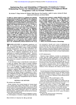
Rhesus leukapheresis procedure. To collect PBMNCs by leukapheresis from donors weighing less than 5 kg, modifications were
made to the fluid path of a CS3000 Plus Blood Cell Separator
(Baxter Healthcare Corp, Fenwal Division, Deefield L)to lower
1645
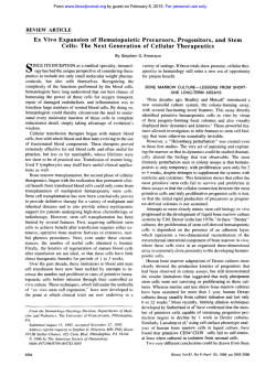
the extracorporeal volume requirement to 132 mL (see Fig 1). Collection was accomplished using a small S25A separation chamber
and a shunt chamber (Fenwal no. 710700027) in the place of a
collection chamber. A standard apheresis kit (Fenwal no. 4R2210)
was installed in the CS3000. After autoprime, the roller clamps to
the acid citrate dextrose-NIH formulation (ACD-A), saline, and vent
prime lines were closed to prevent hemodiluting the donor when
using halthigate. The return line was modified by tightly rolling
and taping a 150-mL transfer pack (Fenwal no. 4R2001) and steriledocking a male h e r to the shortened outlet line. A blood component
recipient set with a 170-pm filter and drip chamber (Fenwal no.
4C2100) was spiked into the modified 150-mL transfer pack and
connected to the packed red blood cell line using a needle lock
device (Fenwal no. 2C7831). The blood component recipient set
was connected to a 20- to 18-gauge Angiocath placed in the saphenous vein of the donor. Hemostats were placed both on the standard,
unused, return line and the inlet for the ACD line present on the
draw line. The apheresis kit was primed with autologous blood that
had been collected in citrate phosphate dextrose plus adsol 2 to 3
weeks before the leukapheresis procedure. The donor received a
dose of 100 U k g heparin immediately before the procedure. The
inlet line was connected to an indwelling 6.6 French catheter placed
in the right atrium of the heart. Blood was processed at the rate of
12 mL/min in automatic mode for a total of 2.5 times the animal’s
calculated blood volume. At the completion of the procedure, the
product was collected and 5 mL of ACD was added. The remaining
cells were salvaged and either were used to prime the CS3000 for
future leukapheresis procedures or were directly reinfused into the
animal. PBMNCs were collected by leukapheresis on day 4 and day
5 of cytokine administration. These days were selected based on
evidence in rhesus macaques (data not shown) and in baboond6 that
circulating progenitor numbers had increased by these time points.
The number of PBMNCs processed during the leukapheresis procedure was calculated by averaging the number of mononuclear cells
in the complete blood cell count of samples taken immediately before, in the middle of, and at the end of the leukapheresis procedure,
and then multiplying this average by the volume of blood processed.
Cell counts were performed on a Coulter Model S5 electronic cell
counter (Hialeah, FL) or on a Cell-Dyn3500 automated hematology
analyzer (Abbott Laboratories, Abbott Park, IL). The number of
PBMNCs processed for each cytokine mobilization group was evaluated, and the mean and standard error of the mean (SEM) was then
determined. The number of PBMNCs collected as the product was
based on the complete blood cell count of the leukapheresis product
multiplied by the total percentage of lymphocytes and monocytes
within the product and the volume of the leukapheresis product. The
PBMNCs for each cytokine mobilization group was evaluated, and
the mean and standard of deviation (SD) was then determined. The
PBMNCs collected from the leukapheresis products on day 4 and
day 5 were processed and analyzed as independent samples.
BM collection. Before the administration of hematopoietic
growth factors (day 0), 30 mL of heparinized BM was surgically
harvested from one femur. Additional BM was harvested immediately before the second leukapheresis procedure (day 5 ) from the
alternate femur. After BM harvest, the animal received a course of
buprenorphine (0.1 to 0.3 mgkg intramuscularly) for 3 days to alleviate any bone pain that may have been associated with the harvest.
Immunoselection of CD34+ cells. CD34+ cells from the BM and
PB leukapheresis product were recovered by positive immunoselection using the Ceprate LC-34-Biotin Kit (CellPro, Inc) according to
the manufacturers instructions. An accurate determination of CD34+
cell yield after immunoselection was not determined, in part,because
of the rarity of CD34+ cells inboth the BM and leukapheresis
product. No apparent difference was observed when comparing the
immunophenotype of immunoselected CD34+ cells and CD34+ cells
that were analyzed from the original blood sample. Cytospin prepara-
From www.bloodjournal.org by guest on February 6, 2015. For personal use only.
1646
DONAHUE ET AL
Fig 1. A diagram of tho rheapheresis
procedure
is
shown. R e d denotes the red
blood cell pathway; yellow, the
pathway of plasma; and blue,
the areawhere the leukapheremis product was collected.
MIS
tions of the cells were prepared to document cellular morphology.
Separate immunoselections were performed on each leukapheresis
product collected on day 4 and day 5. The CD34' cells collected
on each day were stained and analyzed independently.
Culture of immmoselected CD34+ cells. CD34+ cells were immunoselected from the leukapheresis productof animals mobilized
with SCF and G-CSF and were culturedin suspension for 3.5 days
MD)
in Dulbecco's modified Eagle's medium (Biofluids, Rockville,
plus 15% fetal calf semm (Hyclone Laboratories, Inc, Logan, UT)
supplemented with 50 n g / d of L 6 and 100 n g / d of SCF (both
cytokines provided by Amgen, Inc).
I m u n o p h e m ~ p i n gof CD34 cells from BM and PB. The BM
and PB leukapheresisCD34+cellsthatwerepositivelyselected
using the CellRo immunoadsorption system were immunophenotyped with CD3Callophycocyanin (APC) to evaluate CD34 purity
andwerealsoimmunophenotypedwithCD38-fluoresceinisothiocyanate (FITC) andThy-l-phycoerythrin
PE) to identify
CD34+CD38 and CD34+Thy-1 subpopulations. The CD34 MoAb
used (clone 563) was a gift from Dr G. Gaudernack (Institution of
Transplantation Immunology, Rikshospitalet, The National Hospital,
Oslo, Norway). Clone563 is a murineIgGl that recognizes a different CD34 epitope from that
of the Cellpro CD34 MoAb clone (clone
From www.bloodjournal.org by guest on February 6, 2015. For personal use only.
CELLS
STATUS OF CD34'
PHENOTYPE
CYCLE AND CELL
1647
120
B
A
T
20
-
F
x
f?
15
0
a
3
z
0
z
CONTROL
SCF
40
X
G-CSF
G-CSF + SCF
C
I
0
+
P
El0
10
0-
CONTROL
SCF
G-CSF
G-CSF + SCF
G-CSF
CONTROL
SCF
G-CSF+ SCF
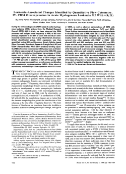
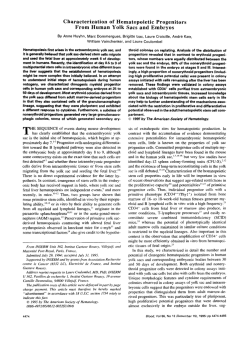
Fig 2. (AI Cytokine mobilization of WBC into PB. The average (SDI circulating WBClpL of blood from animals that had received a 4- and
5-day subcutaneous courseof either no cytokines In = 6), 200 pglkgld SCF In = 61,100 pg/kg/d of G-CSF in = 61. or the combination of SCF
and G-CSF (n = 11) before Ieukepheresis. (B) Total number of mononuclear cells processed and collected from PB by leukapheresis. The
average (SEMI number of mononuclear cells processed through the CS3000 Plus Cell Separator andthe averege (SDI number (SDI of PBMNCs
collected in the Ieukapheresisproduct from animals that received either no cytokines (n = 61, SCF (n = 61, G-CSF in = 61, or the combination
of SCF and G-CSF In = 11). (C) Absolute number of CD34+ cells obtained after immunoselectionfrom the leukapheresis product. The average
(SDI absolute number ofCD34' cells immunoselected from the Ieukapheresis product from animals thet received either no cytokines In = 6).
SCF (n = 51, GCSF (n = 6). or the combination of SCF and G-CSF (n = 6). (DI The percentage ofThy-l expressing CD34' immunoselected cells
from theIeukaphereds product or BM. The average (SDI percentage of CD34+ cells that express Thy-l from (1) the leukapheresis product of
animals that received a C and 5-day subcutaneous course of either no cytokines (n = 61, SCF (n = 51. G-CSF (n = 6). or the combination of
SCF end GCSF (n = 6); or (2) the BM of animals that received either no cytokines (n = 61, SCF (n = 61, G-CSF (n = 61, or the combination of
SCF and G-CSF (n = 5).
12.8) used in the immunoselection. Most antihuman CD34 MoAbs
either do not cross react with rhesus macaque CD34' cells or recognize the same epitope as that of the antibody used in the immunoselection. Fortunately, immunoselection for rhesus CD34+ cells with
clone 12.8 did not interfere with subsequent CD34 staining with
clone 563. For immunophenotyping, the CD34 clone 563 was directly conjugated to APC by Molecular Probes, Inc (Eugene, OR).
The CD38-FITC MoAb used was the OKTlO clone (Ortho Diagnostics, Raritan, NJ). The Thy-l -PE clone used was a gift from Dr P.
Lansdorp (Terry Fox Laboratories Vancouver, British Columbia,
Canada). To evaluate adhesion proteins CD29-FITC and CDw49dFITC antibodies were obtained from AMAC, Inc (Westbrook, ME).
Cells were incubated in the MoAbs or isotype controls for 30
minutes on ice and then washed 2 times with phosphate-buffered
saline (PBS) containing 1% bovine serum albumin. Fc receptors
were blocked by preincubating the cells for 5 minutes in 10% human
AB serum (Advanced Biotechnologies Inc, Columbia, MD) without
washing before the addition of MoAbs. Cells were fixed in I %
paraformaldehyde in PBS and stored at 4°C. All tubes were first run
to analyze for immunophenotyping patterns of CD34-APC, CD38FITC, and Thy-l-PE before the cells were processed for DNA
analysis. The remaining cells were then processed for DNA analysis
using a nuclear DNA staining procedurez7 thatwasmodified for
intact cells. The DNA-staining procedure involved taking a 300-pL
aliquot of cells and slowly adding 700 pL of-20°C cold 100%
ethanol to yield a final concentration of 70% ethanol. The cells were
From www.bloodjournal.org by guest on February 6, 2015. For personal use only.
1648
DONAHUE ET AL
put on dry ice for 5 to 10 minutes, and then 3 mL of cold PBS plus
1% fetal calf serum with 0.01 pmoVL 4',6-diamidino-2-phenylindole
(DAPI; Molecular Probes) was added and tubes were gently mixed
and centrifuged for 10 minutes at 1,000 rpm. The supernatant was
removed leaving approximately 200 pL of liquid, and the cells were
gently mixed. Cells were allowed to stain in the DAPI solution for
at least 1 hour or overnight before analysis on the Coulter Elite flow
cytometer (Hialeah, E).
Flow cytometry data for FITC, PE, APC, DAPI, forward scatter
and side scatter was collected in listmode. The DAPI fluorescence
was measured as both linear-integrated and linear-peak signals and
FITC, PE, and APC were collected as logarithmic signals. Cell
doublets were excluded by gating using either DAPI-peak versus
DAPI-integrated signals or using DAPI-peak versus ratio of DAPIpeak/-integrated signals. The pre-DNA processing immunophenotyping was compared with post-DNA immunophenotyping to assure
that fluorescence of FITC, PE, and APC did not change. The
CD34Thy-l' populations were then gated, and DNA histograms
for this population were created and imported into the Multicycle
software package by Phoenix Flow Systems (San Diego, CA) for
DNA curve-fitting and statistical analysis of the GO/Gl, S, and G2/
M phases of the cell cycle. These results were tabulated, means and
SDs determined, and, where stated, statistical analysis performed
using the Student's t-test of the differences between two means.
The absolute number of CD34'Thy- I cellslmL of either BM or
leukapheresis product was calculated by multiplying the percentage
of CD34' cells that were Thy-l+ by the absolute number of CD34'
cells collected, and dividing this number by the volume of either
BM or leukapheresis product collected for each animal. These numbers were then averaged based on group, and an SD was determined.
+
RESULTS
Rhesus apheresis of cytokine-mobilized PB cells. The
effectiveness of different human cytokines to mobilize the
release of rhesus leukocytes from BM into the PB was evaluated. White blood cell (WBC) counts measured immediately
before leukapheresis (Fig 2A) show that G-CSF alone and
the combination of G-CSF and SCF both increased the WBC
count significantly above the levels achieved with SCF alone
or with no cytokine therapy. The WBC average (SD) measured for the control group without cytokines was 4.7 (0.7)
X 103/pL (n = 6); for those treated with SCF alone, the
WBC count was 11.0 (7.1) X 103/pL (n = 6); for those
treated with G-CSF alone, the WBC count was 82.0 (30.2)
x 103/pL(n = 6); and for those treated with the combination
of G-CSF and SCF, the WBC count was 61.6 (19.3) X lo'/
pL (n = 6). No significant difference in WBC count was
observed between G-CSF-mobilized and G-CSF- and SCFmobilized animals atday 4 and day 5. This is consistent
with a similar study in baboons in which, at these early time
points, there was little difference in WBC count between the
two groups.16 The relative effectiveness of G-CSF or the
combination of G-CSF and SCF to increase mobilized PB
cells was also observed in both the PBMNC fraction processed and collected (Fig 2B). The average (SD) PBMNCs
collected after leukapheresis was 1.3 (0.3) X 10' PBMNCs
(n = 6) for the control group, 1.5 (0.2) X lo9 PBMNCs (n
= 6) for the SCF alone group, 5.5 (3.5) X IO9 PBMNCs (n
= 6) for the G-CSF alone group, and 3.6 (2.1) X 10'
PBMNCs (n = 11) for the SCF and G-CSF group (Fig 2B).
The average collection efficiency for PBMNCsusing the
CS3000 was40.4% (12.5%) (n = 24), withno apparent
difference in efficiency between mobilized andnonmobilized donors.
CD34' cell mobilization and recovery. CD34' cells
were positively selected from the leukapheresis product or
BM, and the purities of the recovered CD34' cells were
evaluated. Purities after immunoselection for CD34+ cells
were consistently better for BM than for the leukapheresis
product. Immunoselected CD34' cells from mobilized and
nonmobilized BM had CD34 purities averaging 88.7%
(7.4%) (n = 23). Leukapheresis products processed using
the same immunoadsorptive system had CD34 purities averaging 60.9% (21.8%) (n = 23). Cytokine mobilization with
G-CSF and SCF clearly increased the absolute number of
CD34' cells recovered from the leukapheresis product following the CD34 immunoselection (Fig 2C). The absolute
number of CD34' cells was greater from the leukapheresis
products obtained from animals mobilized with the combination of G-CSF and SCF (2.3 [1.3] X IO7 CD34' cells; n =
6) than the leukapheresis products obtained from animals
mobilized with G-CSF alone (0.7 [0.4] X IO7 CD34+ cells;
n = 6; see Fig 2C). Still fewer CD34' cells were collected
from leukapheresis products obtained from animals mobilized with SCF alone (0.2 [0.1] X lo7 CD34' cells; n = 5 )
and from nonmobilized animals (0.1 [O. l ] X IO7 CD34'
cells; n = 6; see Fig 2C). Leukapheresis products from day
4 and day 5 yielded similar numbers ofCD34' cells. The
absolute number of CD34' cells collected per milliliter of
BM was 2.7 (1.3) X IO5 (n = 12), 7.3 (1.7) X 10' (n = 3),
4.3 (1.3) X lo' (n = 3), and 5.7 (3.3) X los (n = 3) for the
nonmobilized, SCF-mobilized, G-CSF-mobilized, and SCF
plus G-CSF-mobilized animals, respectively. The absolute
number of CD34+ cells collected per milliliter of leukapheresis product was 0.4 (0.4) X lo5 (n = 6), 0.4 (0.2) X 10' (n
= 5 ) , 1.8 (1.2) X IO5 (n = 6), and 5.8 (3.5) X I O 5 (n = 6)
for the nonmobilized, SCF-mobilized, G-CSF-mobilized,
>
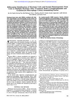
Fig 3. G-CSF and SCF mobilized CD34+ cells immunoselectedfrom arhesusmacaque, RQ826. CD34+ cells were immunoselectedfrom
rhesus macaque("26) (A) premobilization BM, (B) postmobilizationBM, or (C) leukaphererisproduct postmobilizationwith G-CSF and SCF
for 5 days. lmmunophenotypicanalysis using CD3CAPC.CDWFITC, and Thy-l-PE was performed on the cells gated on size and granularity
(outlined in green). The CD34*Thy-1+ cells were gated and color backgated (delineated in red). The cell cycle analysis shown was performed
on the gated CD34+Thy-l+ cells.
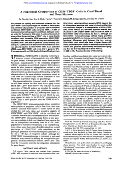
and postculture leukapheresis product CD34+Thy-1* cells from a G-CSF
Fig 5. DNA cell cycle analysis of BM, PB, leukapheresis product,
and SCF-mobilized rhesus macaque, R Q l l l l . lmmunophenotypicanalysis and DNA cycle analysis of CD34' cells immunoselectedfrom BM,
PB, and the leukapheresis product of a rhesus macaque (RQ1111)mobilized with SCF and G-CSF. Some of the leukapheresisproduct immunoselected CD34+ cells were cultured in 50 pg/mL of IL-6 and 100 pg/mL of SCF for 3.5 days and also analyzed for immunophenotype and DNA
cell cycle. The CD34+Thy-lCcells were gated (delineated in red), and cell cycle analysis
was performed on these gated CD34+Thy-l+ cells. Cells
that are CD34-Thy-lf are primate granulocytes.
From www.bloodjournal.org by guest on February 6, 2015. For personal use only.
PHENOTYPE AND CELLCYCLE STATUS OF CD34+ CELLS
1649
A. RQ826 PRE-MOBILIZATION BONE MARROW
BlLlZATlON G-CSF+SCF BONE MARROW
SIZE
THY-l
CD38-FITC
-PE CONTENTDNA
SIZE
THY-l
CD38-FITC
-PE CONTENTDNA
Fig 3.
RQ1111 POST-MOBILILIZATION G-CSF+SCF
BONE MARROW
0.1
l
%O
tQD 8 0 0 0
-PE
"W-1 -PE
THY-l
-PE
BONE MARROW
DNA CONTENT
BLOOD
0.1
l
10
0
i0
APHERESIS
'1000
0.1
i
10
0
l0
o
l0
0
CULTURED
APHERESIS
0.1
i
10
100
io00
THY-l
THY-%-PE
CULTURED
APHERESIS
APHERESIS
BLOOD
DNA CONTENT
DNA CONTENT
CONTENT
DNA
Flg 5.
From www.bloodjournal.org by guest on February 6, 2015. For personal use only.
DONAHUE ET AL
1650
and SCF plus G-CSF-mobilized animals, respectively. For
the BM, the absolute number of CD34' cells collected using
the Ceprate LC-34-Biotin column represented 1.6% (0.5%)
(n = 12), 2.0% (0.8%) (n = 3), 0.4% (0.2%) (n = 3), and
1.0% (0.4%)(n = 3) of the mononuclear cells collected
after ficoll-hypaque separation for the nonmobilized, SCFmobilized, G-CSF-mobilized, and G-CSF plus SCF-mobilized animals, respectively. For the leukapheresis product,
the absolute number of CD34' cells represented 0.09%
(0.08%) (n = 6), 0.16% (0.14%) (n = S), 0.07% (0.04%) (n
= 6), and 0.24% (0.18%) (n = 6) of the mononuclear cells
collected after ficoll-hypaque separation for the nonmobilized, SCF-mobilized, G-CSF-mobilized, and G-CSF plus
SCF-mobilized animals, respectively.
Phenotypic analysis. BM and PB CD34+ cells obtained
with or without cytokhe mobilization were examined for
differences in Thy-l and CD38 expression. All CD34+ cells
expressed some level of Thy-l. A subpopulation of CD34'
cells expressed higher levels of Thy-l and was designated
as Thy-l+ (Fig 3). CD34+ cells obtained from PB had a
greater proportion of cells expressing higher levels of Thy1 than did CD34+ cells obtained from BM, independent of
cytokine mobilization. CD34+ cells isolated from the BMof
nontreated animals were 12.1% (2.4%) Thy-l+ (n = 6). The
percentage of BM CD34' cells expressing Thy-l at higher
levels remained constant whether the animals were treated
with SCF (10.3% [1.9%]; n = 6), G-CSF (8.1% [1.4%]; n
= 6), or the combination of SCF and G-CSF (7.6% [2.8%];
n = 5; see Fig 2D). In contrast to the BM, circulating CD34+
cells isolated from the leukapheresis product had a greater
proportion of CD34' cells expressing higher levels of Thy1 than did BM ( P < .01). This difference between PB and
BM was independent of whether an animal had received a
cytokine or not (Fig2D). Circulating CD34' cells were
32.2% (11.7%) Thy-l+ from nonmobilized animals (n = 6),
38.8% (5.6%) Thy-l+ from animals treated with SCF (n =
S), 37.6% (10.3%) Thy-l+ from animals treated with G-CSF
(n = 6), and 51.6% (8.7%) Thy-l' from animals treated
with the combination of SCF and G-CSF (n = 6; see Fig
2D). The absolute number of CD34+Thy-l ' cells collected
per milliliter of BM was 2.8 (1.5) X LO4 (n = 12), 8.0 (2.0)
X lo4 (n = 3), 3.7 (0.7) X IO4 (n = 3), and 5.3 (4.3) X IO4
for the nonmobilized, SCF-mobilized, G-CSF-mobilized,
and SCF plus G-CSF-mobilized animals, respectively. The
absolute number of CD34'Thy-l' cells collected per milliliterof leukapheresis product was 0.8 (5.5) X IO4 (n = 6),
1.8 (0.5) X IO4 (n = 5 ) , 7.2 (6.5) X lo4 (n = 6), and 30.0
(21.5) X IO4 for the nonmobilized, SCF-mobilized, G-CSFmobilized, and SCF plus G-CSF-mobilized animals, respectively. Interestingly, the cellular distribution of Thy-l appears to be different between rhesus macaques and humans.
Unlike human granulocytes, which are negative for Thy-l,
granulocytes isolated from rhesus macaques strongly express
Thy-l. Two populations of CD34+Thy-l+cells were identified (Fig 3) based on their CD38 expression, CD38-bright
and CD38-dim. Using the OKTl0 clone, we have been unable to identify aCD34+CD38- cell population in rhesus
BM or leukapheresis product. For animal RQ826, 84% of
the backgated CD34'Thy-1' BM cells were CD38-bright
before cytokine mobilization, 77% of the BM CD34'Thy-
80
60
O N OCYTOKINE QSCF
BG-CSF
~SCFIG-CSF
i l
APH CD34+
BM CD34t T H Y - l +
APH
CD34+
THY-l+
Fig 4. Cell cycle analysis of cytokine-mobilized immunoselected
CD34' cells from B M or the leukapheresis product. Percentage of
cells in S+GZ/M for cells gated on CD34+ or CD34+Thy-l+ immunophenotyping. The average (SDI percentage of CD34+ or CD34+ThyI + cells in S+GZ/M was determined for BM from animals that received either no cytokines (n = 141, SCF (n = 31, G-CSF (n = 31, or
the combination of SCF and G-CSF (n = 71, and the leukapheresis
product from animals that received either no cytokines (n = 61, SCF
(n = 6), G-CSF (n = 6), or the combination of SCF and G-CSF (n =
11).
l' cells were CD38-bright after G-CSF and SCF administration, and 90% of the mobilized PB G-CSF and SCF
CD34'Thy-1' cells were CD38-bright (Fig 3). The population of cells seen in Fig 3 that were dimly staining for CD34
and Thy-l were small in size and expressed low levels of
CD38.
Because there were differences in Thy-l expression between BM and PB, we were also interested in determining
whether there were phenotypic differences in the expression
of adhesion proteins between circulating PB- and BM-derived CD34+ cells. In particular, we evaluated the expression
of the integrin a 4 p 1on CD34+ cells. This adhesion protein
has been shown to play a role in hematopoietic stem cell
and microenvironment interactions." CD34' cells isolated
from the BM and leukapheresis product were no different
in CD29 (the integrin @ lchain) and CDw49d (the VLA-a4
chain) expression (data not shown).
Cell cycle analysis. After the initial immunophenotyping, the immunoselected CD34+ cells were stained with
DAPT for simultaneous DNA analysis and immunophenotyping. An example of an animal mobilized with the combination of G-CSF and SCF and analyzed for CD34, CD38, and
Thy-l expression and DNA cell cycle status is shown in Fig
3. As shown in Fig 3 and summarized in Fig 4, circulating
PB CD34+ cells and CD34' Thy-l' cells have fewer cells
cycling in S+G2/M than their BM-derived counterparts ( P
< .01). For example, after cytokine therapy with G-CSF and
SCF, 36.3% (6.8%) of the CD34'Thy-1+ BM cells were in
S+G2/M (n = 7), whereas only 9.6% (3.2%) of the PBcirculating CD34+Thy-l'cells were in S+G2/M (n = 1 1 ;
see Fig 4). The difference in cell cycle status observed between either PB and BM CD34+ cells or CD34+ Thy-l'
HY-l+
From www.bloodjournal.org by guest on February 6, 2015. For personal use only.
1651
PHENOTYPE AND CELLCYCLE STATUS OF CD34'CELLS
had a greater percentage of cells in S+G2/M from a baseline
value of 14.3% (3.7%) and 9.1% (3.3%) to postculture values
of 39.6% (7.9%) and 39.2% (12.3%) for CD34+ cells and
CD34' Thy-l+ cells, respectively (Fig 6 ) .
60
50
B
T
40
.. . .
.. .
(I)
30
W
0
IY
g 20
l0
a
CD34r
Fig 6. Comparison of BM, PB, leukapheresis product,and postcultule leukaphenris product CD34+ cells and CD34*Thy-l+ cells. The
average (SDI percentage of CD34+ or CD34+Thy-l+ cells in S+G2/M
for BM (n = 5). PB In = 4). leukapheresis product In = 51, and after
culture of the CD34+ cells isolatedfrom theleukapheresis productin
50 pglmL of IL-6 and 100 pglmL of SCF for 3.5 days In = 4).
cells was independent of cytokine mobilization. Similar to
the G-CSF and SCF mobilization example, differences in
cell cycle status between PB and BM CD34+ cells or CD34+
Thy- 1 cells were observed without cytokine treatment, with
G-CSF treatment alone, or with SCF treatment alone. Administration of cytokines did not significantly alter the cell
cycle profiles observed in premobilization and postmobilization BM. However, there was a slight increase in the percentage of cycling PB CD34+ cells observed with SCF+G-CSF
mobilization, but this increase was not statistically significant for the data set. As one might expect, small CD34+
cells were not in the S + GUM phases of the cell cycle.
One potential explanation for the differences observed in
Thy-l expression and cell cycle status between BM-derived
and leukapheresis-derived CD34' cells was that the leukapheresis procedure itself may have selected for nondividing
cells of this particular phenotype. Counterflow centrifugation
is commonly used as a method for isolating cells in different
phases of the cell cycle. To evaluate this possibility further,
G-CSF and SCF-mobilized CD34+ cells were immunoselected both from the PB and the leukapheresis product and
compared. No significant differences in phenotype or in cell
cycle status were observed between CD34' cells immunoselected directly from the PB versus the leukapheresis PBMNC
product (Figs 5 and 6).
To evaluate whether a greater percentage of PB CD34+
cells would be in cycle after culture, immunoselected PB
CD34+ cells were placed in culture for 3.5 days in media
supplemented with SCF and L-6. Over the 3.5 days, the
cells increased 1.6-fold (0.5-fold) in number (n = 4). After
culture, the cell cycle status and immunophenotype were
reevaluated. The phenotype of the circulating CD34+ cells
was altered after culture, with a substantial loss in the percentage of CD34'Thy-I+ cells (Fig 5). In addition, both the
CD34' cells and the CD34+Thy-1+ subset following culture
+
DISCUSSION
No alterations in phenotype were observed for either the
BM or PBMNC CD34+ cells with cytokine mobilization.
The proportions of cells expressing CD34, Thy-l, and CD38
all remained fairly constant. What did change was the absolute number of CD34+ cells immunoselected from the leukapheresis product. Despite comparable numbers of
PBMNCs being collected from animals receiving G-CSF
alone and the combination of G-CSF and SCF, greater numbers of CD34+ cells were collected from animals immobilized with the combination of G-CSF and SCF. This observation may explain why there was a significant difference in
WBC count between G-CSF-mobilized and G-CSF and
SCF-mobilized baboons after approximately 1 week of cytokine therapy.I6
The immunoselected CD34+ cells obtained from the PB,
BM, and leukapheresis product were characterized for purity
and for CD38 and Thy-l expression. Unlike human CD34+
cells, which have a distinct CD38- subset,I8rhesus macaque
CD34+ cells express CD38 on all CD34+ cells. The absence
of a CD34+CD38- population of cells in rhesus macaques,
however, does not prevent multilineage reconstitution of rhesus macaques after BM transplantation, because immunoselected CD34+ cells from rhesus macaques have previously
been shown to contribute to multilineage hematopoietic reconstit~tion.~~
The distribution of Thy-l on rhesus macaque CD34+ cells
collected from the leukapheresis product and BM are quite
similar to that observed for human CD34+ cells. Human
CD34+ cells isolated from the BMand the leukapheresis
product of cancer patients treated with cytotoxic chemotherapy and hematopoietic growth factors have been found to
differ in Thy-l e x p r e s s i ~ n .These
~ ~ . ~immunophenotypic
~
differences between PB- and BM-derived CD34+ cells may
account for the accelerated hematopoietic engraftment observed in patients receiving mobilized PB when compared
withthat for those receiving
An increase inthe absolute number of CD34+ cells and CD34+Thy-l+ cells collected by leukapheresis was observed with either G-CSF
or the combination of G-CSF and SCF therapy. Thus, the
increased frequency of Thy-l+ cells in the circulation may
be caused by either a failure of a subpopulation ofBM
CD34+ cells to migrate into the circulation or a predilection
for CD34+Thy-l+cells to circulate.
In addition to differences between BM andPB CD34+
cell immunophenotype, there was a consistent difference between the cell cycle status of PB and BM CD34+ cells.
The percentage of noncycling CD34+ cells was consistently
higher for the PB or leukapheresis product than that for the
BM. Preliminary results suggest that human PB CD34' cells
may also have fewer cells in S-phase under steady state
conditions and after mobilization with chemotherapy and
cytokines than CD34+ cells isolated from BM.31Both observations are consistent with earlier studies that found that
circulating granulopoieti~~~
and erythr~poietic'~progenitor
From www.bloodjournal.org by guest on February 6, 2015. For personal use only.
1652
DONAHUE ET AL
phocytes andor monocytes, for further study. Evaluation of
cells were moreresistant to 3H-thymidinesuicide, and a more
3- to Srecent study that found that progenitor
PB
cells mobilized by cytokine-mobilized PB and BM stem cells from
kg donors may have application in pediatric and veterinary
G-CSF and other cytokines were resistant to 'H-thymidine
suicide.14 All these results suggest that there is a selective
medicine in which celltransfusion, cell therapy, and genetic
difference in cell cycle status between CD34' cells found
therapy are practiced. Future studies may examinewhy there
is a predilection for noncycling CD34' cells to circulate in
in the circulation and CD34' cells found within the BM,
despite the presenceof high levelsof circulating hematopoithe PB and may further delineatethe role of the subpopulations of CD34+ cells in hematopoiesis.
etic growth factors. Potential reasons for this difference are
that noncycling CD34+ cells may have a lower affinity for
binding to the hematopoietic microenvironment than do cyACKNOWLEDGMENT
cling CD34+ cells and,therefore, would have a greater tenThe authors are indebted toMr B. Thompson and Mr E. West for
dency to circulate, or that, for CD34+ cells to cycle, adhertheirassistance in caringfortheanimals;MrC.Carterandhis
ence tothe BM stroma isrequired. No phenotypic difference
associates in the Department of Transfusion Medicine
in the Clinical
in a 4 p l expression,however,wasobserved
between PB
Center for their help on this project; Dr S . Feldman and his staff in
CD34+ cells and
their BM counterpart. However, differences
the Laboratory of Animal Medicine and Surgery (NHLBI) for placing and removing the central line catheters; and Dr P. Rabinovitch
in a 4 p l functionor in theexpression of other adhesion
(University of Washington,Seattle, WA) and DrK. Becker (Phoenix
molecules, such as lymphocyte function-associated antigenFlow Systems, Inc, San Diego,
CA) for their help in the interpretation
1, between BM-derived and circulating CD34+ cells were
of the DNA cell cycle analysis data. We would also like to thank
not pre~luded.'~
Dr G . Gaudernack (The National Hospital, Oslo, Norway) for supFailure to cycle when exposed toa combination of hemaplyingtheCD34MoAb,clone563;DrP.Lansdorp(TerryFox
topoietic growth factors hasbeen used asa criteria for identi- Laboratory, Vancouver, Canada) for supplying the Thy-l antibody,
fying primitive, hematopoietic stem cells. Recent examples
clone 5E10, directly conjugated to PE; and Amgen, Inc for supplying
include retention of the membrane label PKH26 ona subset
all the hematopoietic growth factors used in these studies.
of CD34' cells whencultured in serum-free medium supplemented with IL-3,L - 6 , SCF, and e~ythropoietin'~ resisand
REFERENCES
tance of a subset of CD34+ cells to the antimetabolite S1. CivinCI,StraussLC,BrovallC,FacklerMJ,SchwartzJF,
fluorouracil when stimulated with IL-3 andSCF.17 Although
Shaper JH: Antigenic analysis
of hematopoiesis.111. A hematopoietic
IL-3 was not used in our study, G-CSF and the combination progenitor cell surface antigen defined by a monoclonal antibody
of G-CSF and SCF have been
used in mice, dogs, nonhuman raised against KG-la cells. J Immunol 133:157, 1984
primates, and humans to mobilize progenitors that contain
2. Baumhueter S , Singer M, Henzel W, Hemmerich S , Renz M,
BM repopulating cell^.'^^'^"^ Thissuggests thatmobilized
Rosen S , Lasky LA: Binding of L-selectin to the vascular sialomucin
CD34. Science 262:436, 1993
CD34' cells contain a population of primitive cells that are
3. Fina L, Molgaatd HV, Robertson D, Bradley NJ, Monoghan
not in cycle in the presence of G-CSF and SCF and have
P, Delia E, Sutherland DR, Baker MA, Greaves M F : Expression of
the capacity to repopulate the BM of a myeloablated host.
the CD34 gene in vascular endothelial cells. Blood 752417, 1990
Because cellularreplication is required for efficient retroviral
4. Andrews RG, Singer J W , Bernstein ID Human hematopoietic
infe~tion,'~ itwould appear that immunoselectedCD34+
precursors in long-term culture: Single
CD34' cells that lack detectcells from the leukapheresis product would be more
difficult
able T cell, B cell, and myeloid cell antigens produce multiple colto transducethan BM.However, this does not appearto
ony-forming cells when cultured with marrow stromal cells. J Exp
be the case in that immunoselected CD34+ cells from the
Med172:355,1990
leukapheresis product could beinduced to proliferate in sus5. Bender JG, UnverzagtKL, Walker DE, Lee W, Van EppsDE,
pension culture, and in that the absolute number of CD34'
SmithDH, Stewart CC, To LB:Identificationandcomparisonof
CD34-positive cells and their subpopulations from normal peripheral
cells that were collected per milliliter of the leukapheresis
bloodandbonemarrowusingmulticolorflowcytometry.Blood
product from the SCF and G-CSF-mobilized animals was
77:2591,1991
greater than that collected from BM. Theseobservations are
6. Bender JG, Unverzagt K: Flow cytometric analysis of periphconsistent with murine studies that have shown thatretrovieral blood stem cells. J Hematother 2:421, 1993
rus transduction efficiency was higher for PB from splenec7. Bender JG, Unverzagt K, Walker DE, Lee W, Smith S , Wiltomized mice mobilized with G-CSF and SCF than for BM
liams S , Van Epps DE: Phenotypic analysis and characterization of
from micetreated with S - f l u o r ~ u r a c i land
, ~ ~ that the culturing CD34+ cells from normal human bone marrow, cord blood, periphof murine BM cellsin IL-3, E - 6 , and SCF enhancesretrovieral blood, and mobilized peripheral blood from patients undergoing
autologous stem cell transplantation. Clin Immunol Immunopathol
ral transduction efficiencies in murine hematopoietic cells.40
7010, 1994
The leukapheresis procedure developed has permitted the
collection of large numbersof PB leukocytes from 3- to 5-kg 8. Kessinger A, Armitage JO, SmithDM, Landmark JD, Bierman
PJ, Weisenburger DD: High-dose therapy and autologous peripheral
donors without the need for exposing the donorallogeneic
to
blood stem cell transplantation for patients with lymphoma. Blood
blood productsin priming theinstrument. This prevents allo74:1260,1989
sensitization before myelosuppression, thus minimizing the
9. Gianni AM, Siena S , Bregni M, Tarella C, SternA, Bonadonna
opportunity for developing antibodies to erythrocyte,leukoG: Granulocyte-macrophage colony-stimulating factor
to harvest circyte,and platelet antigensandsubsequent
destruction of
culatinghaematopoieticstemcellsforautotransplantation.Lancet
transfusedbloodproducts
because of antibodyformation.
2580, 1989
10. Sheridan WP, Begley CG, Juttner CA, SzerJ, To LB. Maher
In addition, this leukapheresis procedure has permitted the
D, McGrath KM, Morstyn G, Fox RM: Effect of peripheral-blood
collection of large quantities of other PB cells, suchas lym-
From www.bloodjournal.org by guest on February 6, 2015. For personal use only.
PHENOTYPE AND CELLCYCLE STATUS
OF CD34' CELLS
progenitor cells mobilized by filgrastim (G-CSF) on platelet recovery
after high-dose chemotherapy. Lancet 339540, 1992
11. Brugger W, Bross K, Frisch J, Dem P,Weber B, Mertelsmann
R, Kanz L: Mobilization of peripheral blood progenitor cells by
sequential administration of interleukin-3 and granulocyte-macrophage colony-stimulating factor following polychemotherapy with
etoposide, ifosfamide, and cisplatin. Blood 79: 1193, 1992
12. McNiece IK, Briddell RA, Hartley CA, Andrews RC: The
role of stem cell factor in mobilization of peripheral blood progenitor
cells: Synergy with G-CSF. Stem Cells 11:83, 1993
13. McNiece IK, Briddell RA, Hartley CA, Smith KA, Andrews
RG: Stem cell factor enhances in vivo effects of granulocyte colony
stimulating factor for stimulating mobilization of peripheral blood
progenitor cells. Stem Cells 11:36, 1993
14. Bodine DM, Seidel NE, Gale MS, Nienhuis AW, Orlic D:
Efficient retrovirus transduction of mouse pluripotent hematopoietic
stem cells mobilized into the peripheral blood by treatment with
granulocyte colony-stimulating factor and stem cell factor. Blood
84:1482, 1994
15. de Revel T, Appelbaum FR, Storb R, Schuening F, Nash R,
Deeg J, McNiece I, Andrews R, Graham T: Effects of granulocyte
colony-stimulating factor and stem cell factor, alone and in combination, on the mobilization of peripheral blood cells that engraft lethally
irradiated dogs. Blood 83:3795, 1994
16. Andrews RC, Briddell RA, Knitter CH, Opie T, Bronsden M,
Myerson D, Appelbaum FR, McNiece IK: In vivo synergy between
recombinant human stem cell factor and recombinant human granulocyte colony-stimulating factor in baboons: Enhanced circulation
of progenitor cells. Blood 84:800, 1994
17. Andrews RC, Briddell RA, Knitter CH, Rowley SD, Appelbaum FR, McNiece I K Rapid engraftment by peripheral blood progenitor cells mobilized by recombinant human stem cell factor and
recombinant human granulocyte colony-stimulating factor in nonhuman primates. Blood 85:15, 1995
18. Terstappen LW",
Candour D, Huang S, Lund-Johansen F,
Manion K, Nguyen M, Mickaels R, Olweus J, Topker S: Assessment
of hematopoietic cell differentiation by multidimensional flow cytometry. J Hematother 2:431, 1993
19. Terstappen LWMM, Huang S, Safford M, Lansdorp PM, Loken MR: Sequential generations of hematopoietic colonies derived
from single nonlineage-committed CD34'38- progenitor cells.
Blood 77:1218, 1991
20. Craig W, Kay R, Cutler RL, Lansdorp PM: Expression of
Thy-l on human hematopoietic progenitor cells. J Exp Med
177:1331, 1993
21. Baum CM, Weissman IL, Tsukamoto AS, Buckle AM, Peault
B: Isolation of a candidate human hematopoietic stem-cell population. Proc Natl Acad Sci USA 89:2804, 1992
22. Mayani H, Lansdorp PM: Thy-l expression is linked to functional properties of primitive hematopoietic progenitor cells from
human umbilical cord blood. Blood 83:2410, 1994
23. Murray L, Chen B, Galy A, Chen S, Tushinski R, Uchida N,
Negrin R, Tricot G, Jagannath S, Vesole D, Barlogie B, Hoffman
R, Tsukamoto A: Enrichment of human hematopoietic stem cell
activity in the CD34'Thy- 1 +Lin- subpopulation from mobilized peripheral blood. Blood 85:368, 1995
24. Howard M, Grimaldi JC, Bazan JF, Lund FE, Santos-Argumedo L, Parkhouse RME, Walseth TF, Lee HC: Formation and
hydrolysis of cyclic ADP-ribose catalyzed by lymphocyte antigen
CD38. Science 262:1056, 1993
25. Reems JA, Torok-Storb B: Cell cycle and functional differ-
1653
ences between CD34+/CD38"' and CD34'138'" human marrow cells
after in vitro cytokine exposure. Blood 85:1480, 1995
26. Gore SD, Amin S, Weng L-J, Civin CI: Steel factor supports
the cycling of isolated human CD34' cells in the absence of other
growth factors. Exp Hematol 23:413, 1995
27. Otto F DAPI staining of fixed cells for high-resolution flow
cytometry of nuclear DNA, inDarzynkiewicz Z, Crissman HA (eds):
Methods in Cell Biology, v01 33 Flow Cytometry. San Diego, CA,
Academic, 1990, p 105
28. Williams DA, Rios M, Stephens C, Patel V: Fibronectin and
VLA-4 in haematopoietic stem cell-microenvironment interactions.
Nature 352:438, 1991
29. Bodine DM, Moritz T, Donahue RE, LuskeyBD, Kessler
SW, Martin DIK, Orkin SH, Nienhuis AW, Williams DA: Longterm in vivo expression of a murine adenosine deaminase gene in
rhesus monkey hematopoietic cells of multiple lineages after retroviral mediated gene transfer into CD34+ bone marrow cells. Blood
82:1975, 1993
30. Haas R, Mohle R, Pforsich M, Fruehauf S, Witt B, GoldSchmidt H, Hunstein W: Blood-derived autografts collected during
granulocyte colony-stimulating factor-enhanced recovery are enriched with early Thy-]+ hematopoietic progenitor cells. Blood
85:1936, 1995
31. Danova M, Rosti V, Mazzini G, Locatelli F, Comoli P, Riccardi A, Cazzola M, Ascari E: Proliferative activity of bone marrow
versus peripheral blood CD34-positive cells on steady state and
following chemotherapy plus G-CSF. Blood 84:23a, 1994 (abstr,
SUPPI 1)
32. Tebbi K, Rubin S, Cowan DH, McCulloch EA: A comparison
of granulopoiesis in culture from blood and bone marrow cells of
nonleukemic individuals and patients with acute leukemia. Blood
48:235, 1976
33. Ogawa M, Crush OC, O'Dell W, Hara H, MacEchern MD:
Circulating erythropoietic precursors assessed in culture: Characterization in normal men and patients with hemoglobinopathies. Blood
50:1081, 1977
34. Roberts AW, Metcalf D: Noncycling state of peripheral blood
progenitor cells mobilized by granulocyte colony-stimulating factor
and other cytokines. Blood 86:1600, 1995
35. Mohle R, Haas R, Hunstein W: Expression of adhesion molecules and c-kit on CD34' hematopoietic progenitor cells: Comparison of cytokine-mobilized blood stem cells with normalbone marrow
and peripheral blood. J Hematother 2:483, 1993
36. Lansdorp PM, Dragowska W, Mayani H: Ontogeny-related
changes in proliferative potential of human hematopoietic cells. J
Exp Med 178:787, 1993
37. Berardi AC, Wang A, Levine JD, Lopez P, Scadden DT:
Functional isolation and characterization ofhuman hematopoietic
stem cells. Science 267: 104, 1995
38. Miller DC, Adam MA, Milter AD: Gene transfer by retrovirus
vectors occurs only in cells that are actively replicating at the time
of infection. Mol Cell Biol 10:4239, 1990
39. Bodine DM, Seidel NE, Gale MS, Nienhuis AW, Orlic D:
Efficient retrovirus transduction of mouse pluripotent hematopoietic
stem cells mobilized into the peripheral blood by treatment with
granulocyte colony-stimulating factor and stem cell factor. Blood
84: 1482, 1994
40. Luskey BD, Rosenblatt M, Zsebo K, Williams DA: Stem cell
factor, interleukin-3, and interleukin-6 promote retroviral-mediated
gene transfer into murine hematopoietic stem cells. Blood 80:396,
1992
From www.bloodjournal.org by guest on February 6, 2015. For personal use only.
1996 87: 1644-1653
Peripheral blood CD34+ cells differ from bone marrow CD34+ cells in
Thy- 1 expression and cell cycle status in nonhuman primates
mobilized or not mobilized with granulocyte colony-stimulating factor
and/or stem cell factor
RE Donahue, MR Kirby, ME Metzger, BA Agricola, SE Sellers and HM Cullis
Updated information and services can be found at:
http://www.bloodjournal.org/content/87/4/1644.full.html
Articles on similar topics can be found in the following Blood collections
Information about reproducing this article in parts or in its entirety may be found online at:
http://www.bloodjournal.org/site/misc/rights.xhtml#repub_requests
Information about ordering reprints may be found online at:
http://www.bloodjournal.org/site/misc/rights.xhtml#reprints
Information about subscriptions and ASH membership may be found online at:
http://www.bloodjournal.org/site/subscriptions/index.xhtml
Blood (print ISSN 0006-4971, online ISSN 1528-0020), is published weekly by the American
Society of Hematology, 2021 L St, NW, Suite 900, Washington DC 20036.
Copyright 2011 by The American Society of Hematology; all rights reserved.
© Copyright 2026