
Characterization of the Monocyte-Specific Esterase (MSE
Characterization of the Monocyte-Specific Esterase (MSE) Gene
C o r d C . U p h o f f , Z h e n - B o H u , S u z a n n e M . G i g n a c , Weill* M a , Fred A . R a i n e y , M a r i n a K r e u t z , W o l f - D i e t e r L u d w i g ,
1
1
1
1
2
3
and Hans G . Drexler'
' D S M , G e r m a n C o l l e c t i o n of M i c r o o r g a n i s m s a n d C e l l Cultures, Department of H u m a n a n d A n i m a l C e l l Cultures, Braunschweig,
-'Universitat R e g e n s b u r g , M e d i z i n i s c h e K l i n i k , D e p a r t m e n t o f Internal M e d i c i n e I, R e g e n s b u r g , *Freie U n i v e r s i t a t B e r l i n , U n i v e r s i t a t s k l i n i k u m
Rudolf Virchow, Department of M e d i c a l O n c o l o g y and A p p l i e d Molecular Biology, Berlin, G e r m a n y
Carboxylic esterases are widely distributed in hematopoietic
cells. Monocytes express the esterase isoenzyme (termed
'monocyte-specific esterase', MSE) that can be inhibited by
NaF in the a-naphthyl acetate cytochemical staining. We examined the expression of MSE in normal cells and primary and
cultured leukemia-lymphoma cells. The MSE protein was demonstrated by isoelectric focusing (IEF); MSE mRNA expression
was investigated by Northern blotting and reverse transcriptase-polymerase chain reaction (RT-PCR). The following
samples were positive for MSE protein and Northern mRNA
expression: 20/24 monocytic, 4/32 myeloid, and 1/20 erythroidmegakaryocytic leukemia cell lines, but none of the 112 lymphoid leukemia or lymphoma cell lines; of the normal purified cell
populations only the monocytes were positive whereas, T, B
cells, and granulocytes were negative; of primary acute (myelo)
monocytic leukemia cells (CD14-positive, FAB M4/M5
morphology) 14/20 were Northern mRNA and 11/14 IEF protein
positive. RT-PCR revealed MSE expression in 29/49 Northernnegative lymphoid leukemia-lymphoma cell lines. The RT-PCR
signals in monocytic cell lines were on average 50-fold
stronger than the mostly weak trace expression in lymphoid
specimens. On treatment with various biomodulators, only alltrans retinoic acid significantly upregulated MSE message and
protein levels but could not induce new MSE expression in several leukemia cell lines; lipopolysaccharide and interferon-y
increased MSE expression in normal monocytes. Analysis of
DNA methylation with sensitive restriction enzymes showed no
apparent regulation of gene expression by differential methylation; the MSE gene is evolutionarily conserved among mammalian species; the half-life of the human MSE transcripts was
about 5-6 h. The extent of MSE expression varied greatly
among different monocytic leukemia samples. However, the
MSE overexpression in a significant number of specimens was
not associated with gene amplification, gross structural
rearrangements or point mutations within the cDNA region.
Taken together, the results suggest that MSE expression is not
absolutely specific for, but strongly associated with cells of the
monocytic lineage; MSE is either not expressed at all or
expressed at much lower levels in cells from other lineages.
The biological significance, if any, of rare MSE messages in
lymphoid cells detectable only by the hypersensitive RT-PCR
remains unclear. Further studies on the regulation of this gene
and on the physiological function of the enzyme will no doubt
be informative with respect to its striking overexpression in
some malignant cells and to a possible role in the pathobiology
of monocytic leukemias.
INTRODUCTION
Esterases represent a d i v e r s e s p e c t r u m of e n z y m e s w i t h a n
u b i q u i t o u s tissue d i s t r i b u t i o n that share certain features
Received February 15, 1994. A c c e p t e d April 2 2 , 1994.
Correspondence to: D r H a n s G . D r e x l e r , M D , P h D , D S M , G e r m a n
C o l l e c t i o n of M i c r o o r g a n i s m s a n d C e l l Cultures, M a s c h e r o d e r W e g
1 B, D - 3 8 1 2 4 B r a u n s c h w e i g , G e r m a n y .
LEUKEMIA
©
1994 Macmillan
1510
Press Ltd
r e g a r d i n g substrate s p e c i f i c i t y (1). T h e s e esterases b e l o n g to
the class of serine h y d r o l a s e s that are d e f i n e d as functionally
related h y d r o l y t i c e n z y m e s c o n t a i n i n g a serine residue in their
a c t i v e site (2). T h i s e n z y m e class c o m p r i s e s the serine protease
m u l t i g e n e f a m i l y as w e l l as v a r i o u s c a r b o x y l - , c h o l i n - , aryl-/
acetyl- a n d a c e t y l c h o l i n e s t e r a s e s (3).
T h e c a r b o x y l e s t e r a s e s (EC 3.1.1.1) are a heterogeneous
g r o u p of c e l l u l a r e n z y m e s c a p a b l e of h y d r o l y z i n g a variety ot
a l i p h a t i c o r a r o m a t i c esters u n d e r a c i d i c o r neutral c o n d i t i o n s
(4). In h e m a t o l o g y these e n z y m e s are k n o w n as non-specific
esterases, a c t i n g most e f f i c i e n t l y o n short-chain (acetate and
butyrate) esters. T h e e n z y m a t i c activity c a n b e i n h i b i t e d by
s o d i u m f l u o r i d e (NaF) in m o n o c y t i c c e l l s , but not in cells
of the g r a n u l o c y t i c series (5); h o w e v e r , it s h o u l d b e noted
that NaF-resistance o r sensitivity is here c l e a r l y a relative
p h e n o m e n o n (6).
N e v e r t h e l e s s , the u n i q u e substrate a n d i n h i b i t o r specificity
o f the esterase f o u n d in m o n o c y t e s i n d i c a t e d early o n that the
h i g h activity in m o n o c y t e s m i g h t b e d u e to e n z y m e variants
that are not present in other l e u k o c y t e s (7). This n o t i o n was
strengthened b y data f r o m e l e c t r o p h o r e t i c analyses of e n z y m e
extracts, first b y p o l y a c r y l a m i d e g e l e l e c t r o p h o r e s i s a n d later
by i s o e l e c t r i c f o c u s i n g (IEF) ( r e v i e w e d in (8)). T h e s e z y m o g r a m
IEF studies of n o r m a l a n d m a l i g n a n t m y e l o i d c e l l s h a v e c o n sistently d e m o n s t r a t e d the e x i s t e n c e of t w o m a i n g r o u p s ot
esterase b a n d s . O n e g r o u p of IEF b a n d s is c o m m o n to all
m y e l o i d c e l l s (termed common
esterase, C o m E s t , b y Scott et
al. (9)) a n d o n e is a d d i t i o n a l l y d e t e c t e d in c e l l s of m o n o c y t i c
o r i g i n (termed monocyte-specific
esterase, M S E ) . In the IEF
a n a l y s i s the C o m E s t g r o u p appears as a series of b a n d s w i t h
isoeletric p o i n t s (pl) r a n g i n g f r o m 6 . 3 - 7 . 9 w h i l e M S E c o m prises 1-5 b a n d s ( d e p e n d i n g o n the IEF system used) w i t h a
n a r r o w p l range 5.5-6.2 (3,9).
For a l o n g t i m e it w a s not k n o w n w h e t h e r C o m E s t a n d M S E
b e l o n g to a m u l t i g e n e f a m i l y representing post-transcript i o n a l l y m o d i f i e d variants o f the s a m e e n z y m e o r w h e t h e r they
are c l e a r l y distinct at the m o l e c u l a r a n d g e n e t i c l e v e l , b e i n g
related o n l y in their substrate s p e c i f i c i t y (9). R e c e n t e x p e r i m e n t a l e v i d e n c e supports the s e c o n d v i e w , n a m e l y that C o m Est a n d M S E are u n r e l a t e d e n z y m e s p e c i e s : o n e a p p e a r s to be
a m o n o m e r i c acetylesterase a n d the other a t r i m e r i c c a r b o x y l esterase, r e s p e c t i v e l y (1,9).
W e recently d e m o n s t r a t e d the s p e c i f i c i t y of M S E as
e x p r e s s i o n at the m R N A ( e x a m i n e d b y N o r t h e r n blotting) a n d
p r o t e i n level (by IEF) w e r e c l e a r l y restricted to c e l l s c o m m i t t e d
to the m o n o c y t e - m a c r o p h a g e l i n e a g e (10). In o r d e r to s u b stantiate these c o n c l u s i o n s in a c o m p r e h e n s i v e survey a n d to
further c l a r i f y the nature o f M S E , w e a n a l y z e d a greatly
extended panel of primary and continuously cultured leukem i a c e l l s a p p l y i n g e x t r e m e l y sensitive d e t e c t i o n m e t h o d s ,
e x a m i n e d the g e n e e x p r e s s i o n u n d e r in vivo a n d m a n i p u l a t e d
in vitro c o n d i t i o n s , a n d c h a r a c t e r i z e d the M S E g e n e in
further d e t a i l .
LEUKEMIA,
Vol 8, N o 9 (September), 1994: pp 1510-1526
MATERIAL AND METHODS
Primary
Normal
and Malignant
Cell
Material
Fresh l e u k e m i a c e l l s w e r e taken f r o m p e r i p h e r a l b l o o d (PB)
or b o n e m a r r o w ( B M ) f r o m patients w i t h m y e l o i d l e u k e m i a o f
the m o r p h o l o g i c a l M 4 o r M 5 subtypes a c c o r d i n g to t h e
F r e n c h - A m e r i c a n - B r i t i s h (FAB) c l a s s i f i c a t i o n . S a m p l e s w e r e
sent to the reference l a b o r a t o r y o f a u t h o r W . D . L for i m m u n o p h e n o t y p e a n a l y s i s . P B o r B M m o n o n u c l e a r c e l l s w e r e separated b y standard F i c o l l - H y p a q u e density gradient centrifugation ( L y m p h o p r e p , N y c o m e d , O s l o , N o r w a y ) . A l l s a m p l e s
Were e x a m i n e d w i t h the f o l l o w i n g p a n e l o f surface markers
using f l o w c y t o m e t r y : C D 2 , C D 3 , C D 4 , C D 7 , C D 1 0 , C D 1 3 ,
C D 1 4 , C D 1 5 , C D 1 9 , C D 3 3 , C D 3 4 , C D 4 1 , C D w 6 5 , glycop h o r i n A , H L A - D R . C e l l s w e r e p e l l e t e d a n d f r o z e n in l i q u i d
nitrogen.
N o r m a l P B m o n o n u c l e a r c e l l s taken d i r e c t l y b y venup u n c t u r e o f l a b o r a t o r y staff o r f r o m buffy coats (generously
p r o v i d e d b y the G e r m a n R e d C r o s s B l o o d T r a n s f u s i o n C e n t e r ,
Springe, G e r m a n y ) w e r e isolated b y standard F i c o l l - H y p a q u e
density gradient c e n t r i f u g a t i o n . T h e c e l l s w e r e separated f r o m
the vast majority o f t h r o m b o c y t e s b y repeated l o w - s p e e d s e d i m e n t a t i o n s ( 2 0 0 x g for 7 m i n ) . M o n o n u c l e a r c e l l s w e r e
adjusted t o 2 - 2 0 x 1 0 cells/ml w i t h m a c r o p h a g e - s e r u m free
m e d i u m ( M - S F M ; G i b c o B R L , Eggenstein, G e r m a n y ) . Tissue
culture dishes (Nunc, W i e s b a d e n , G e r m a n y ) containing 1 0 15 m l o f this c e l l s u s p e n s i o n w e r e i n c u b a t e d for 1 h at 3 7 ° C
in a h u m i d i f i e d i n c u b a t o r w i t h 5 % C 0 . N o n - a d h e r e n t c e l l s
w e r e s u b s e q u e n t l y r e m o v e d b y w a s h i n g t h e c u l t u r e dishes
repeatedly w i t h w a r m phosphate-buffered s a l i n e (PBS) c o n taining 0 . 5 % M-SFM. For R N A preparation cells were washed
f r o m the d i s h e s w i t h g u a n i d i n i u m i s o t h i o c y a n a t e . T c e l l s w e r e
e n r i c h e d b y s h e e p r e d b l o o d c e l l rosetting ( I C N F l o w , M e c k e n h e i m , G e r m a n y ) . G r a n u l o c y t e s w e r e c o l l e c t e d f r o m the b o t t o m o f the F i c o l l gradient a n d separated f r o m erythrocytes b y
a d e x t r a n gradient ( D e x t r a n T - 5 0 0 ; P h a r m a c i a , F r e i b u r g ,
Germany). N o r m a l B cells were obtained from surgically
r e m o v e d tonsils after s h e e p red b l o o d c e l l rosetting o f t h e
m o n o n u c l e a r c e l l p r e p a r a t i o n . T h e purities o f the n o r m a l c e l l
populations were verified by immunostaining a n d flow cytom e t r i c analysis ( F A C S c a n ; B e c t o n D i c k i n s o n , H e i d e l b e r g ,
G e r m a n y ) : T c e l l s w e r e e n r i c h e d t o 9 7 % ( C D 3 + ), B c e l l s t o
9 7 % ( C D 1 9 + ), m o n o c y t e s t o 8 6 % ( C D 1 4 + a n d p o s i t i v e i n
the a-naphthyl acetate esterase c y t o c h e m i c a l staining), a n d
g r a n u l o c y t e s to > 9 5 % ( m o r p h o l o g i c a l analysis). In o r d e r t o
increase the q u a n t i t y a n d the purity of a v a i l a b l e n o r m a l m o n o cytes, m o n o c y t e s w e r e isolated f r o m m o n o n u c l e a r c e l l s (after
F i c o l l - H y p a q u e centrifugation) b y counter-current e l u t r i a t i o n
(JGM-E B e c k m a n c e n t r i f u g e ; B e c k m a n , M u n c h e n , G e r m a n y )
u s i n g a l a r g e - v o l u m e c h a m b e r (50 ml) a n d a JE-5 rotor at 2 5 0 0
r.p.m. a n d a f l o w rate o f 1 1 0 m l / m i n in H a n k ' s b a l a n c e d salt
s o l u t i o n s u p p l e m e n t e d w i t h 2 % h u m a n a l b u m i n . Elutriated
m o n o c y t e s w e r e > 9 5 % pure as d e t e r m i n e d b y m o r p h o l o g y
and antigenic phenotype.
6
2
Culture
of Cell
Lines
and In V i t r o
Stimulation
A l l h u m a n c e l l lines w e r e d e r i v e d f r o m patients w i t h l e u k e m i a
or l y m p h o m a (11). T h e c o n t i n u o u s h u m a n a n d a n i m a l c e l l
lines w e r e either taken f r o m t h e stock o f the c e l l b a n k ( D S M ,
G e r m a n C o l l e c t i o n o f M i c r o o r g a n i s m s a n d C e l l Cultures) (12)
or w e r e g e n e r o u s l y m a d e a v a i l a b l e for this study b y t h e o r i g i n a t i n g investigators. C e l l s w e r e g r o w n u n d e r o p t i m a l c o n d i t i o n s in 5 0 m l o r 2 6 0 m l tissue c u l t u r e flasks o r 2 4 - w e l l
plates ( N u n c ) in their a p p r o p r i a t e m e d i a ( R P M I 1 6 4 0 ,
M c C o y ' s 5 A , L e i b o w i t z ' s L-15, Iscove's M D M , D u l b e c c o ' s
M E M , o r M E M a l p h a ; G i b c o BRL) s u p p l e m e n t e d w i t h 5 - 2 0 %
heat-inactivated (at 5 6 ° C for 4 5 m i n ) fetal b o v i n e s e r u m
(Sigma, D e i s e n h o f e n , G e r m a n y ) at 3 7 ° C in a h u m i d i f i e d
a t m o s p h e r e o f 5 % C 0 in air. T h e c e l l s w e r e e x a m i n e d d a i l y
in t h e c u l t u r e flasks u n d e r a n inverted m i c r o s c o p e . C u l t u r e s
w e r e i n c u b a t e d w i t h o u t a n t i b i o t i c s in o r d e r to a v o i d s u b l i m a l
b a c t e r i a l i n f e c t i o n . O n l y m y c o p l a s m a - f r e e cultures w e r e u s e d ;
f r e e d o m of m y c o p l a s m a c o n t a m i n a t i o n w a s c h e c k e d r o u t i n e l y
by D A P I s t a i n i n g a n d c u l t i v a t i o n o n agar. T h e c e l l s w e r e
harvested in their l o g a r i t h m i c g r o w t h phase w i t h v i a b i l i t i e s
exceeding
9 0 % as d e t e r m i n e d
b y trypan
blue d y e
e x c l u s i o n . C e l l pellets w e r e kept f r o z e n at - 2 0 ° C o r p r o cessed immediately.
2
In vitro s t i m u l a t i o n o f c e l l lines w a s c a r r i e d o u t w i t h t w o
protein kinase C (PKC) activators, t h e p h a r m a c o l o g i c a l 1 2 - 0 t e t r a d e c a n o y l p h o r b o l 13-acetate (TPA; Sigma) a n d t h e natural
Bryostatin 1 (Bryo 1; k i n d l y p r o v i d e d b y Prof G . R . Pettit,
T e m p e , A Z , U S A ) , the v i t a m i n A - a n a l o g u e a\\-trans r e t i n o i c
a c i d ( A T R A ; Sigma), a n d the c a l c i u m transport regulator 1,25d i h y d r o x y v i t a m i n D (Vit. D 3 ; Sigma). T h e i n d u c e r s w e r e first
d i s s o l v e d in e t h a n o l o r D M S O at 1 0 ~ * M a n d then further
d i l u t e d in R P M I 1 6 4 0 m e d i u m s o that the final c o n c e n t r a t i o n s
of the solvent w e r e m a x i m a l l y 0 . 0 1 % in the e x p e r i m e n t s . T h e
c e l l s w e r e e x p o s e d t o 1 0 ~ M s o l u t i o n s o f the reagents for u p
to 4 days. P B m o n o c y t e s a n d s o m e c e l l lines w e r e s t i m u l a t e d
w i t h 1 0 0 ng/ml l i p o p o l y s a c c h a r i d e (LPS; Sigma) a n d 2 0 0 U/ml
interferon-y (IFN-y; B o e h r i n g e r
Mannheim,
Mannheim,
G e r m a n y ) for u p to 2 4 h.
3
7
RNA
Isolation
and Northern
Blotting
Total c e l l u l a r R N A w a s isolated u s i n g the g u a n i d i n i u m isot h i o c y a n a t e - c e s i u m c h l o r i d e m e t h o d (13). N o r t h e r n blots
w e r e p r e p a r e d b y separating 10 /xg o f total R N A in a n agarose
gel c o n t a i n i n g 1 % f o r m a l d e h y d e . T h e R N A w a s transferred
to a n y l o n m e m b r a n e ( N y t r a n , S c h l e i c h e r a n d S c h u l l , D a s s e l ,
G e r m a n y ) a n d cross-linked w i t h 1 2 0 0 J ultraviolet light ( U V
Stratalinker 1 8 0 0 ; Stratagene, H e i d e l b e r g , G e r m a n y ) . After 2 h
of p r e - h y b r i d i z a t i o n t h e filters w e r e h y b r i d i z e d w i t h a nicktranslated ( G i b c o BRL) o r r a n d o m p r i m e d ( U S B , B a d H o m b u r g , G e r m a n y ) [«- P]dCTP-labeled HMSE-1 p r o b e o v e r n i g h t
at 6 2 ° C . T h e filters w e r e then w a s h e d stringently a n d e x p o s e d
for a u t o r a d i o g r a p h y t o X-ray films (Fuji RX) w i t h intensifying
screens at - 8 0 ° C . Filters w e r e r e h y b r i d i z e d w i t h a h o u s e k e e p i n g gene as the c o n t r o l .
}2
Probes
A 1 7 4 6 - b p EcoRI fragment c l o n e d into p U C 1 9 c o n t a i n i n g the
partial c o d i n g s e q u e n c e ( n u c l e o t i d e s - 1 0 to 1 5 1 2 f r o m the
3'-end o f t h e H M S E gene) w a s k i n d l y p r o v i d e d b y D r F.
Z s c h u n k e ( G o t t i n g e n , G e r m a n y ) (2). A 2 3 8 - b p fragment o f the
HMSE-1 c D N A f r o m the 5'-end (nt - 1 0 t o 2 2 8 , o b t a i n e d after
BamHl d i g e s t i o n a n d e x t r a c t i o n f r o m t h e gel) w a s e m p l o y e d
in s o m e e x p e r i m e n t s . T h e /3-actin p r o b e p A c t - 1 , a 1.25-kb Pst\
fragment ( c l o n e d i n p B R 3 2 2 ) f r o m t h e c D N A o f hamster j8a c t i n (obtained f r o m D r J . W . G . Janssen, U l m , G e r m a n y ) w a s
used as c o n t r o l .
Determination
of RNA
Half-Life
T h e half-life o f the M S E m R N A w a s d e t e r m i n e d b y e x p o s u r e
of the c e l l s t o 10/Ltg/ml a c t i n o m y c i n D (Sigma), a n i n h i b i t o r
of t r a n s c r i p t i o n , for 0 - 2 4 h b e f o r e harvest of the c e l l s a n d s u b sequent i s o l a t i o n of R N A .
4 h with
air c o o l i n g .
Subseqently
the gel w a s
dried
and
e x p o s e d to an X-ray f i l m for a u t o r a d i o g r a p h y o v e r n i g h t at
room temperature.
Reverse
PCR)
Transcriptase-Polymerase
Chain
Reaction
(RTDNA
Five m i c r o g r a m s of total R N A w a s used as a t e m p l a t e for first
strand c D N A synthesis u s i n g a reverse transcriptase p r e a m p l i f i c a t i o n system kit ( S u p e r s c r i p t ; G i b c o BRL) in a final v o l u m e
of 2 0 /il P C R buffer ( c o n t a i n i n g 2 0 m M Tris-HCI of p H 8.4,
5 0 m M K C I , 2.5 m M M g C I , 0.1 mg/ml BSA) s u p p l e m e n t e d
w i t h 0.5 /Ag of o l i g o d T p r i m e r . After h e a t i n g the m i x t u r e at
7 0 ° C for 10 m i n , 2 0 0 U of M o l o n e y m u r i n e l e u k e m i a virus
reverse transcriptase a n d 1 /LLI of 1 0 m M d N T P m i x w e r e a d d e d
to the r e a c t i o n s y s t e m . T h e r e a c t i o n m i x t u r e w a s t h e n i n c u bated at 4 2 ° C for 5 0 m i n , at 9 0 ° C for 5 m i n a n d t h e n q u i c k l y
c h i l l e d o n i c e . After brief c e n t r i f u g a t i o n , 2 U R N a s e H w a s
a d d e d to the r e a c t i o n m i x t u r e for 2 0 m i n at 3 7 ° C . R N A
s a m p l e s f r o m s o m e c e l l lines w e r e treated w i t h D N a s e I
(RNase-free f r o m B o e h r i n g e r M a n n h e i m ) p r i o r to reverse t r a n s c r i p t i o n . T h e r e a c t i o n w a s i n c u b a t e d at 3 7 ° C for 1 h a n d then
at 9 5 ° C for 5 m i n a n d i m m e d i a t e l y c o o l e d o n i c e . F i v e
m i c r o l i t r e s of the reverse transcriptase r e a c t i o n m i x t u r e c o n t a i n i n g the first strand c D N A w a s d i l u t e d w i t h P C R buffer
(10 X : 5 0 0 m M K C I , 15 m M M g C I , 1 0 0 m M Tris-HCI p H 8 . 3 ,
0 . 0 0 1 % gelatin) c o n t a i n i n g 2 0 p m o l of e a c h u p s t r e a m a n d
d o w n s t r e a m p r i m e r , 10 n m o l of d N T P m i x a n d 1.25 U of T a q
DNA
polymerase
(Amersham-Buchler,
Braunschweig,
G e r m a n y ) . T h e p r i m e r s used in the e x p e r i m e n t w e r e d e s i g n e d
a c c o r d i n g to s e q u e n c e d a t a p u b l i s h e d p r e v i o u s l y (2): sense 5'G G C A G T T A C T C T C A G A G C T A - 3 ' (sequence nucleotides 9 2 1 1 1 , MSE-P1) a n d antisense 5 ' - C T T C C A C A G G A G T G A C A T G G C - 3 ' ( s e q u e n c e n u c l e o t i d e s 9 6 0 - 9 4 0 , MSE-P2). O l i g o n u c l e o t i d e p r i m e r s w e r e p r e p a r e d o n an a u t o m a t e d D N A s y n t h e s i z e r ( C y l c o n e Plus, M i l l i p o r e , E s c h b o r n , G e r m a n y ) . T h e
P C R w a s t h e n p e r f o r m e d w i t h a D N A t h e r m a l c y c l e r (Perkin
E l m e r C e t u s , H e i d e l b u r g , G e r m a n y ) for 3 2 c y c l e s u n d e r the
f o l l o w i n g c o n d i t i o n s : 3 0 s at 9 4 ° C for d e n a t u r a t i o n , 3 0 s at
5 5 ° C for a n n e a l i n g , a n d 2 m i n at 7 2 ° C for e x t e n s i o n . T h e
a m p l i f i e d P C R p r o d u c t s w e r e e l e c t r o p h o r e s e d in 1 . 2 % a g a rose gels, s t a i n e d w i t h e t h i d i u m b r o m i d e a n d o b s e r v e d u n d e r
u l t r a v i o l e t light. G e l s w e r e b l o t t e d o n t o n y l o n filters u s i n g the
S o u t h e r n t e c h n i q u e as d e s c r i b e d b e l o w . In o r d e r to assess the
q u a l i t y of reverse t r a n s c r i b e d R N A a n d s u c c e s s f u l P C R - a m p l i f i c a t i o n , a l i q u o t s f r o m the s a m e p r o d u c t s o b t a i n e d f r o m
reverse t r a n s c r i p t i o n w e r e a m p l i f i e d in p a r a l l e l u s i n g the f o l l o w i n g t w o j3-actin p r i m e r s : sense 5 ' - A T G G A T G A T G A T A T C G C C G C G - 3 ' a n d antisense 5 ' - C T A G A A G C A T T T G C G G T GGAC-3'.
2
2
Polymerase
Conformation
Chain
Reaction
Polymorphism
and
Single-Strand
(PCR-SSCP)
P C R - S S C P a n a l y s i s w a s m o d i f i e d f r o m the p r e v i o u s d e s c r i p t i o n (14). In brief, 6 /Ltl reverse t r a n s c r i b e d c D N A w a s a m p l i fied b y P C R (30 c y c l e s ; the s a m e c o n d i t i o n s as u n d e r RT-PCR)
in the p r e s e n c e o f 10 fid [ a - P ] d C T P u s i n g the t w o p r i m e r s
MSE-P1 a n d M S E - P 2 (see a b o v e ) . F r o m the a m p l i f i c a t i o n p r o d u c t 4 /LLI w e r e d i l u t e d 1 : 2 5 w i t h 0 . 1 % S D S , 10 m M E D T A
a n d heated at 9 5 ° C after a d d i n g 1 v o l u m e s e q u e n c i n g stop
solution ( 9 5 % formamide, 20 mM EDTA, 0 . 0 5 % bromophenol
b l u e , 0 . 0 5 % x y l e n e c y a n o l , 2 0 m M N a O H ) . T h e n , 2-4 /il of
this m i x t u r e w a s l o a d e d o n t o a 6 % n o n - d e n a t u r i n g p o l y a c r y l a m i d e gel c o n t a i n i n g 9 0 m M Tris-borate, 2 m M E D T A ( p H 8),
a n d 1 0 % g l y c e r o l . E l e c t r o p h o r e s i s w a s p e r f o r m e d at 2 5 W for
32
Sequencing
R T - P C R p r o d u c t s w e r e p u r i f i e d u s i n g Jetsorb ( G e n o m e d , Bad
O e y n h a u s e n , G e r m a n y ) . RT-PCR p r o d u c t s w e r e s e q u e n c e d
u s i n g the T a q D y e D e o x y ™ T e r m i n a t o r C y c l e S e q u e n c i n g Kit
( A p p l i e d B i o s y s t e m s , W e i t e r s t a d t , G e r m a n y ) . T h e primers
used w e r e MSE-P1 as sense, antisense 5 ' - G G T T C T T G G C C A A T G G A G A C A - 3 ' ( s e q u e n c e n u c l e o t i d e s 5 2 6 - 5 0 6 , MSE-P3),
antisense
5'-GGCTGG ATCTTCATTCACAGC-3'
(sequence
n u c l e o t i d e s 1 5 2 6 - 1 5 0 6 , MSE-P4), a n d M S E - P 2 as antisense at
a c o n c e n t r a t i o n of 4 p m o l per s e q u e n c e r e a c t i o n . T h e s e q u e n c i n g w a s p e r f o r m e d w i t h the D N A t h e r m a l c y c l e r for 25
c y c l e s u n d e r the f o l l o w i n g c o n d i t i o n s : 3 0 s at 9 6 ° C for d e n a t u r a t i o n , 15 s at 5 0 ° C for a n n e a l i n g , a n d 4 m i n at 6 0 ° C for
e x t e n s i o n . T h e s e q u e n c e r e a c t i o n s w e r e p u r i f i e d as d e s c r i b e d
by the m a n u f a c t u r e r . T h e s e q u e n c e r e a c t i o n s w e r e e l e c t r o p h o r e s e d u s i n g the A p p l i e d B i o s y s t e m ' s 3 7 3 A D N A S e q u e n c e r .
Southern
Blotting
H i g h m o l e c u l a r w e i g h t D N A w a s e x t r a c t e d f r o m f r o z e n PBSw a s h e d l e u k e m i c c e l l s u s i n g s t a n d a r d m e t h o d s (13). For the
different e x p e r i m e n t s 2 0 /ig of D N A w a s d i g e s t e d o v e r n i g h t
w i t h 1 0 0 U of o n e of the restriction e n z y m e s
HindlW
( P h a r m a c i a , F r e i b u r g , G e r m a n y ) , # a m H I , EcoRI, Pst\ ( G i b c o
BRL), M s p l , /-/pall, o r Cfo\ ( B o e h r i n g e r M a n n h e i m ) . T o 1/10
of the r e a c t i o n m i x 2 0 0 n g l a m b d a D N A ( P h a r m a c i a ) w a s
a d d e d as c o n t r o l . T h e d i g e s t e d D N A w a s e x t r a c t e d w i t h p h e n o l , p r e c i p i t a t e d in e t h a n o l a n d separated in a 0 . 7 % agarose
g e l . T h e D N A w a s f i x e d w i t h 0.2 N H C I a n d t h e n d e n a t u r e d
w i t h 0.5 M N a O H / 1 . 5 M N a C I . After n e u t r a l i z a t i o n w i t h 0.5 M
Tris-HCI ( p H 7.0)/3 M N a C I the D N A w a s b l o t t e d w i t h
2 0 x S S C o n n y l o n m e m b r a n e s a n d then treated
and
h y b r i d i z e d as d e s c r i b e d a b o v e u n d e r N o r t h e r n blot a n a l y s i s .
Pulsed
Field
Gel
Electrophoresis
P u l s e d f i e l d gel e l e c t r o p h o r e s i s (PFGE) is an e l e c t r o p h o r e t i c
t e c h n i q u e alternative to standard S o u t h e r n a n a l y s i s that uses
a l t e r n a t i n g pulses of current d i r e c t e d at a n g l e s t h r o u g h an a g a rose gel to separate large D N A restriction fragments. D i g e s t e d
g e n o m i c D N A w a s separated o n a 1 . 5 % agarose gel u s i n g the
C h e f - D r II P F G E system (Bio-Rad, M u n c h e n , G e r m a n y )
a c c o r d i n g to the r e c o m m e n d a t i o n s of the m a n u f a c t u r e r . In
brief, the gel w a s run for 5 h at 14°C a p p l y i n g a 2 0 0 V
i n v e r t i n g f i e l d . T h e s w i t c h i n g c y c l e i n c r e a s e d f r o m 1 to 4 s
o v e r these 5 h. F o l l o w i n g P F G E the gel w a s treated s i m i l a r l y
to a s t a n d a r d S o u t h e r n b l o t .
Isoelectric
Focusing
E n z y m e e x t r a c t i o n , s e p a r a t i o n b y IEF a n d v i s u a l i z a t i o n of
esterase i s o e n z y m e s h a v e b e e n d e s c r i b e d in d e t a i l e l s e w h e r e
(10). In brief, e n z y m e s w e r e e x t r a c t e d b y repeated c y c l e s of
f r e e z i n g - t h a w i n g a n d s o l u b i l i z e d b y a d d i t i o n of T r i t o n X 1 0 0
(Serva, H e i d e l b e r g , G e r m a n y ) . After c e n t r i f u g a t i o n , a l i q u o t s of
supernatant c o n t a i n i n g the e n z y m e p r e p a r a t i o n (extracted
f r o m e q u a l n u m b e r s o f cells) w e r e separated b y a n a l y t i c a l IEF
o n h o r i z o n t a l thin-layer p o l y a c r y l a m i d e gels ( 4 . 8 % a c r y l /
b i s a c r y l a m i d e , p H range 2-11 of the a m p h o l y t e Servalyt;
Serva) u s i n g an L K B M u l t i p h o r system ( P h a r m a c i a ) . Isoe n z y m e s w e r e v i s u a l i z e d o n the gels by s u b m e r s i o n in a s t a i n ing s o l u t i o n c o n t a i n i n g a - n a p h t h y l acetate (Sigma) as substrate
a n d Fast B l u e RR (Serva) as the c o u p l i n g d i a z o n i u m salt.
A d d i t i o n of 4 0 m M N a F to the s t a i n i n g bath i n h i b i t e d s e l e c tively the M S E b a n d at a b o u t p H 6.0 (3). B a n d s o n a u t o r a d i o g r a p h e d R N A films after N o r t h e r n blot analysis o r RT-PCR
S o u t h e r n b l o t t i n g a n d o n d r i e d IEF gels w e r e q u a n t i f i e d densit o m e t r i c a l l y (LKB U l t r o s c a n D e n s i t o m e t e r ; P h a r m a c i a ) .
RESULTS
Expression
of
MSE
Detection
of Protein by IEF and of mRNA
by Northern
Blotting.
M S E p r o t e i n w a s d e m o n s t r a t e d by IEF o n p o l y a c r y l a m i d e gels as a d i s t i n c t b a n d w i t h an isoeletric p o i n t of a b o u t
p H 6 . 0 . T h i s b a n d c o u l d be s e l e c t i v e l y i n h i b i t e d by N a F . In
the N o r t h e r n blot a n a l y s i s a s i n g l e b a n d c o r r e s p o n d i n g to an
M S E transcript of 2.0 k b w a s d e t e c t e d . E x p r e s s i o n of M S E p r o tein a n d m R N A w e r e e x a m i n e d in all s a m p l e s , i.e. n o r m a l c e l l
p o p u l a t i o n s , p r i m a r y l e u k e m i a c e l l s , a n d l e u k e m i a c e l l lines,
by IEF a n d N o r t h e r n b l o t t i n g , r e s p e c t i v e l y (Tables 1 a n d 2;
Figures 1 a n d 2).
O f the p u r i f i e d n o r m a l c e l l p o p u l a t i o n s {n= 10 f r o m different i n d i v i d u a l s ) , o n l y the PB m o n o c y t e p r e p a r a t i o n s c o n s i s t -
Table 1
e n t l y d i s p l a y e d M S E p r o t e i n a n d m R N A , w h e r e a s PB T c e l l s ,
PB g r a n u l o c y t e s , a n d t o n s i l l a r B c e l l s w e r e all negative.
T w e n t y - t w o PB or B M acute ( m y e l o ) m o n o c y t i c l e u k e m i a
s a m p l e s , d i a g n o s e d as s u c h o n m o r p h o l o g i c a l - c y t o c h e m i c a l
(FAB M 4 or M 5 ) a n d i m m u n o p h e n o t y p i c a l g r o u n d s ( C D 1 4 + ) ,
w e r e tested. m R N A e x p r e s s i o n w a s f o u n d in 14/20 cases
tested; 11/14 cases a n a l y z e d s h o w e d the M S E b a n d in the IEF.
M S E e x p r e s s i o n w a s e x a m i n e d in 1 8 8 c e l l lines (Table 2).
A l l 112 l y m p h o i d l e u k e m i a a n d l y m p h o m a c e l l lines w e r e
negative at the N o r t h e r n m R N A a n d p r o t e i n level (29 pre Bc e l l l e u k e m i a , 17 B-cell l e u k e m i a , 21 T-cell l e u k e m i a , 13
m y e l o m a , 8 Burkitt, 8 H o d g k i n , a n d 16 n o n - H o d g k i n l y m p h o m a c e l l lines). In the c a t e g o r y ' m y e l o i d l e u k e m i a c e l l
l i n e s ' {n = 32) four a n d five c e l l lines w e r e M S E - p o s i t i v e in the
IEF a n d N o r t h e r n a n a l y s i s , r e s p e c t i v e l y . O n e out of 2 0 c e l l
lines w i t h m e g a k a r y o c y t i c and/or e r y t h r o i d features s h o w e d
the M S E b a n d s in the IEF a n d N o r t h e r n gels. T h e M S E isoe n z y m e a n d the M S E transcript w e r e f o u n d in 20/24 c e l l lines.
Detection
of mRNA
by RT-PCR.
T o assess the d e t e c t i o n
sensitivity of the IEF, N o r t h e r n a n d RT-PCR a n a l y s i s , p o s i t i v e
c e l l s w e r e d i l u t e d w i t h M S E - n e g a t i v e c e l l s . In p r e l i m i n a r y
e x p e r i m e n t s w e s h o w e d that it w a s p o s s i b l e w i t h IEF to v i s u a l i z e a n M S E - p o s i t i v e p o p u l a t i o n in a m i x t u r e c o n s i s t i n g of
1 % p o s i t i v e c e l l s (cell l i n e T H P - 1 ) a n d 9 9 % negative c e l l s
(RC-2A). T h e d i l u t i o n e x p e r i m e n t s p e r f o r m e d here r e v e a l e d
that N o r t h e r n b l o t t i n g h a d a m a x i m a l sensitivity of 2 . 5 % posi-
E x p r e s s i o n o f M S E m R N A a n d P r o t e i n in P r i m a r y H u m a n L e u k e m i a C e l l s
Patient no.
Surface Marker E x p r e s s i o n
CD13
CD14
CD15
CD33
F A B Subtype
3
CDw65
M S E Expression
Protein IEF
b
mRNA
mRNA
Northern
1
2
3
4
5
6
7
8
9
10
11
12
13
14
15
16
17
18
19
20
21
22
70
10
44
50
75
24
24
12
82
71
37
6
34
34
30
37
82
34
82
21
14
16
56
5
75
70
60
34
40
52
55
85
34
44
69
47
55
62
70
60
47
19
33
57
31
75
5
70
37
20
30
44
47
11
26
34
16
20
12
0
13
14
48
32
77
23
90
35
79
90
76
76
60
20
81
85
83
56
75
80
80
36
70
90
71
73
82
50
80
70
66
87
60
20
68
80
49
83
63
56
76
86
80
52
45
72
35
66
87
70
M5
M5
M4
M5
M4eo
M4
M4
M5a
M5
M4
M4
M5
M5a
M5b
M4
M4
M4eo
M4eo
M4eo
M4/M5
M5
M5
(+)
+
-
[0.71X]*
[0.95x]
+
(+)
(+)
[1.01x]
[0.59x]
[0.70x]
+
++
(+)
[1.19x]
[4.43x]
[0.37x]
(+)
[0.61x]
++
++
[6.22x]
[5.74x]
-
++
[2.89x]
+
[0.55x]
+
(+)
[1.24x]
[0.1 Ox]
+
(+)
(+]
[1.06x]
[0.40x]
[0.25x]
(+)
(+)
+
(+)
(+)
[0.24x]
[0.36x]
[0.48x]
[0.1 Ox]
[0.08x]
++
+
[5.61x]
[0.18x]
-
-
-
RT-PCR
5
C
d
+
-
+
+
+
+
+
E x p r e s s i o n of these surface markers (percentage of positive cells) was examined by indirect immunofluorescence staining and either
microscopic or flow cytometric analysis using the monoclonal antibodies My7 (CD13), VIM-13 or U C H M 1 (CD14), VIM-D5 (CD15), My9
(CD33), VIM-2 (CDw65); other markers examined are not listed but confirmed the diagnosis.
"Intensity of bands on IEF gels and Northern gels; the intensity of expression (thickness of the band) was g r a d e d in comparison with those
from normal control monocyte samples: -, negative; ( + ), weaker; +, same intensity; + + , overexpression (stronger band),
intensity of bands on Southern gels hybridized with the HMSE-1 probe after RT-PCR: -, negative; +, positive.
i n t e n s i t y of expression (thickness of bands in IEF and Northern) analyzed densitometrically in comparison with normal unstimulated monocytes; data are given as X-fold of the normal values.
Cell Line
Origin
Protein
3
IEF"
Pre-B
1
2
3
4
5
6
7
8
9
10
11
12
13
14
15
16
17
18
19
20
21
22
23
24
25
26
27
28
29
Leukemia Cell
1E8
207
380
697
ALL-1
BAY-91
BV-173
EU-1
HAL-01
HPB-NULL
KARPAS-353
KOPN-8
LAZ-221
LILA-1
LK-63
MIK-ALL
NALM-1
NALM-6
NALM-16
OM9;22
PC-53
PRE-ALP
RCH-ACV
REH
RS4;11
SUP-B15
SUP-B27
TAHR-87
TOM-1
Lines
Pre B-ALL
Pre B-ALL
ALL
Pre B-ALL
Pre B-ALL
ALL
CML-BC
ALL
Pre B-ALL
ALL
Pre B-ALL
ALL
ALL
Pre B-ALL
Pre B-ALL
Pre B-ALL
CML-BC
ALL
ALL
ALL
Pre B-ALL
Pre B-ALL
Pre B-ALL
ALL
ALL
Pre B-ALL
Pre B-ALL
AUL
Pre B-ALL
-
B Leukemia Cell Lines
1
BALL-1
2
BALM-1
BALM-6
3
4
BALM-8
5
BONNA-12
6
EH
7
EHEB
ESKOL
8
9
HAIR-M
HC-1
10
11
HK
12
JVM-2
JVM-3
13
14
JVM-13
15
KARPAS-231
16
MN-60
17
WIEN-133
ALL
B-ALL
B-ALL
B-ALL
HCL
HCL
B-CLL
HCL
HCL
HCL
HCL
B-PLL
B-PLL
B-PLL
B-ALL
B-ALL
B-ALL
-
T Leukemia Cell Lines
1
BE-13
2
CCRF-CEM
CML-T1
3
4
DU-528
5
HPB-ALL
6
JURKAT
7
KE-37
8
LOUCY
MDS
9
10
MKB-1
11
MOLT-3
12
MOLT-13
13
MOLT-15
14
MOLT-16
MOLT-17
15
MO-T
16
T-ALL
ALL
CML-BC
T-ALL
T-ALL
T-ALL
T-ALL
T-ALL
CMMoL
AML
ALL
T-ALL
AMoL
T-ALL
T-ALL
HCL
mRNA
Northern"
-
-
-
-
-
-
-
-
RT-PCR
+
(+)
-
-
-
-
-
(+)
-
-
-
-
-
-
-
-
—
-
—
-
-
-
-
-
(+)
+
+
—
-
-
-
-
-
-
(+)
++
-
-
—
-
++
+
-
-
-
-
-
(+)
-
(+)
--
C
Cell Line
Origin
Protein
3
IEP
T Leukemia Cell Lines
(Continued,)
17
MT-1
18
P12/ICHIKAWA
19
PEER
20
PF-382
21
SKW-3
ATL
ALL
T-ALL
ALL
CLL
Myeloma Cell Lines
1
EJM
2
IM-9
3
KARPAS-620
4
KARPAS-707
5
L-363
6
LP-1
7
MM-1
8
MM-S1
9
NCI-H929
10
OPM-2
11
U-266
12
U-1958
13
U-1996
Myeloma
Myeloma
Plasma cell leukemia
Myeloma
P l a s m a cell leukemia
Myeloma
Myeloma
Myeloma
Myeloma
Myeloma
Myeloma
Plasma cell leukemia
Myeloma
Burkitt
1
2
3
4
5
6
7
8
-
Hodgkin Lymphoma
1
CO
2
HDLM-1
3
HDLM-2
4
HDLM-3
5
KM-H2
6
L-428
7
L-540
8
SUP-HD1
Cell Lines
Hodgkin
Hodgkin
Hodgkin
Hodgkin
Hodgkin
Hodgkin
Hodgkin
Hodgkin
Non-Hodgkin Lymphoma
1
DEL
2
DOHH-2
3
HBL-1
4
HT-58
5
KARPAS-299
6
KARPAS-422
7
MC-116
8
MH-1
9
PFI-285
10
RL
11
SCC-3
12
ST-4
13
SUP-T1
14
U-698-M
15
WSU-NHL
16
WSU-WM
Myeloid Leukemia
1
EM-2
2
EM-3
3
EOL-1
4
EOL-3
5
GDM-1
6
GF-D8
Cell Lines
CML-BC
CML-BC
AML-eosino
AML-eosino
CML-BC
A M L M1
-
(+)
-
-
-
+
-
-
-
-
-
-
-
-
-
-
-
-
-
-
-
-
—
—
-
-
-
-
-
-
-
-
-
-
-
-f
-
-
-
(+)
-
-
-
-
-
-
-
-
-
-
-
Cell Lines
Malignant histiocytosis
B N H L (immunoblastic)
B N H L (diffuse large cell)
B N H L (centroblastic)
T N H L (histiocytic)
B NHL
B N H L (undifferentiated)
Malignant histiocytosis
T NHL
B N H L (undifferentiated)
N H L (diffuse large cell)
T N H L (convoluted type)
T N H L (lymphoblastic)
B N H L (lymphoblastic)
B N H L (histiocytic)
Waldenstrom
RT-PCR
-
-
(nodular sclerosis)
(nodular sclerosis)
(nodular sclerosis)
(nodular sclerosis)
(mixed cellularity)
(nodular sclerosis)
(nodular sclerosis)
(nodular sclerosis)
Northern"
-
-
Lymphoma Cell Lines
BJAB
Burkitt
CA-46
Burkitt
DAUDI
Burkitt
DG-75
Burkitt
EB-1
Burkitt
NAMALWA
Burkitt
NAMALWA-IPN.45 Burkitt
ROS-17
Burkitt
mRNA
(+)
+
+
(+)
-
+
(+)
+
(+)
++
(+)
-
-
-
—
+
-
+
(+)
+
(+)
+
+
+
+
-
-
++
—
-
-
-
+
-
C
Cell Line
Origin
Myeloid Leukemia Cell Lines (Continued,)
7
AML
GM-153
8
GM/SO
CML-BC
9
HL-60
A M L M2
10
Mast cell leukemia
HMC-1
11
KASUMI-1
A M L M2
12
KBM-7
CML-BC
KCL-22
CML-BC
13
14
AML
KG-1
15
KG-1A
AML
16
KMT-2
Umbilical cord blood
17
KOPM-28
CML-BC
18
KU-812
CML-BC
19
KU-812F
CML-BC
20
KY-821
AML
21
CML-BC
KYO-1
22
CML-BC
MOLM-6
23
MOLM-7
CML-BC
24
CML-BC
MOLM-8
25
MR-87
AML
26
NB-4
A M L M3
27
OCI-AML-5
AML
28
PL-21
AML M3
29
TI-1
AML M2
30
TS9;22
CML-BC
31
UCSD/AML-1
AML
32
YS9;22
CML-BC
Monocytic Leukemia Cell Lines
1
CTV-1
AML M5
2
DD
Histiocytic lymphoma
3
JOSK-I
A M L M4
4
JOSK-K
AML M5
5
JOSK-M
CML-BC
6
A M L M5
JOSK-S
7
KBM-3
A M L M4
8
KBM-5
CML-BC
ML-2
A M L M4
9
10
MOBS-1
AMoL
11
MONO-MAC-6
AML M5
12
MV4-11
A M L M5
13
NOMO-1
AMoL
14
OCI-AML-1
AML M4
15
OCI-AML-2
A M L M4
16
OCI-AML-3
A M L M4
17
PLB-985
A M L M4
18
RC-2A
A M L M4
RW-LEU-4
CML-BC
19
20
SKM-1
AMoL
THP-1
AML M5
21
22
TK-1B
A M L M4
23
U-937
Histiocytic lymphoma
24
X-376
AML
Megakaryocytic-Erythroid
1
CHRF-288-11
2
CMK
DAMI
3
4
F-36P
F-36EGM
5
6
HEL
7
K-562
8
KMOE-02
9
LAMA-84
10
M-07e
11
MB-02
12
MEG-01
Protein
3
Leukemia Cell Lines
A M L M7
A M L M7
A M L M7
AML M6
A M L M6
A M L M6
CML-BC
A M L M6
CML-BC
A M L M7
A M L M7
CML-BC
mRNA
IEF"
Northern"
RT-PCR
-
-
-
-
(+)
(+)
+
(+)
-
-
-
-
-
-
-
+++
++
++
+++
+
(+)
(+)
+
+++
(+)
+++
+++
++
++
+++
+++
+++
(+)
+++
+++
(+)
+++
+
+
+
++
+++
+
+
++
+++
+++
(+)
+++
+
+++
+++
++
(+)
++
-
-
-
-
-
-
-
-
-
-
—
(+)
(+)
++
-
(+)
(+)
(+)
++
+
+++
+++
++
+
(+)
++
-
+
+
+
+ -»+++
+++
++
++
+++
+++
+++
+++
+++
+
+++
+++
+++
+++
+++
+++
++
+++
+
++
++
(+)
+
(+)
+++
++
(+)
C
Cell Line
Megakaryocytic-Erythroid
13
MEGAL
14
MKPL-1
15
MOLM-1
16
0CI-M1
17
0CI-M2
18
T-33
19
TF-1
20
UT-7
Origin
Protein
3
Leukemia Cell Lines
A M L M7
A M L M7
CML-BC
A M L M6
A M L M6
CML-BC
A M L M6
A M L M7
(Continued)
mRNA
IEF"
Northern"
RT-PCR
-
-
-
-
-
-
++
+
++
+
+
+++
C
+
+++
A L L , acute lymphoblastic leukemia; A M L , acute myeloid leukemia; A M o L , acute (myelo)monocytic leukemia;
ATL, adult T-cell leukemia; A U L , acute undifferentiated leukemia; C L L , chronic lymphocytic leukemia; CMLB C , chronic myeloid leukemia in blast crisis; C M M o L , chronic myelomonocytic leukemia; H C L , hairy cell
leukemia; NHL, non-Hodgkin's lymphoma; PLL, prolymphocytic leukemia.
C e l l lines were assigned to the respective categories b a s e d on their origin and their phenotypic and functional characteristics (e.g. surface markers, receptor gene rearrangements, expression of hemoglobin, etc.);
original diagnoses (types and subtypes of malignancies) are given as far as indicated in the original publications (see also (11)).
"Intensity of bands on IEF gels and Northern gels; the intensity of expression (thickness of the band) was
g r a d e d in comparison with those from normal control monocyte samples: -, negative; ( + ), weaker; +,
same intensity, + + , overexpression (stronger band); + + + , strong overexpression (very strong band),
intensity of bands on Southern gels hybridized with the HMSE-1 probe after RT-PCR; -, negative; ( + ),
weakly positive; +, + + , + + + , different degrees of positivity.
a
tive c e l l s (PLB-985) in a b a c k g r o u n d of 9 7 . 5 % negative c e l l s
(KG-1). RT-PCR w a s 5 0 - 2 5 0 times m o r e sensitive than N o r t h ern as the RT-PCR p r o d u c t of M S E w a s r e p r o d u c i b l y detecta b l e at a 1 : 2 0 0 0 to 1 : 10 0 0 0 d i l u t i o n . T h e s e results c o n f i r m
s i m i l a r levels of sensitivity in d i l u t i o n e x p e r i m e n t s d o n e to
detect the e n z y m e m y e l o p e r o x i d a s e ( M P O ) : 1 - 3 % for N o r t h ern a n d 0 . 0 5 - 0 . 1 % for RT-PCR (15). T h e m o r e sensitive RTP C R m e t h o d w a s c a r r i e d out, b o t h to c o n f i r m the results
o b t a i n e d by N o r t h e r n b l o t t i n g , a n d to further c h a r a c t e r i z e a n y
trace e x p r e s s i o n of c e l l lines negative at the p r o t e i n a n d
N o r t h e r n m R N A l e v e l . T h e RT-PCR generated a u n i q u e a n d
a c c u r a t e l y s i z e d p r o d u c t of 8 6 8 b p . T h e identity of P C R p r o d u c t s f r o m the c e l l lines w a s c o n f i r m e d by S o u t h e r n blots of
the P C R p r o d u c t s (Figure 3). T h e intensity of e x p r e s s i o n s h o w n
by RT-PCR is, of c o u r s e , a gross estimate a n d not an exact
q u a n t i f i c a t i o n (since w e d i d not p e r f o r m q u a n t i t a t i v e P C R ) ,
but p r o v i d e s nevertheless a c l e a r a n d r e p r o d u c i b l e p i c t u r e of
M S E trace e x p r e s s i o n .
O n e h u n d r e d a n d fourteen c e l l lines w e r e e x a m i n e d for
M S E m R N A e x p r e s s i o n by RT-PCR u s i n g p r i m e r s MSE-P1
a n d -P2, f o l l o w e d by S o u t h e r n b l o t t i n g a n d h y b r i d i z a t i o n w i t h
the HMSE-1 p r o b e (Table 2). Seventy-two c e l l lines w e r e p o s i t i v e ; u p o n s i g n i f i c a n t l y longer e x p o s u r e of the f i l m s to the
r a d i o a c t i v e filter a further 17 of the 4 2 i n i t i a l l y negative c e l l
lines s h o w e d w e a k b a n d s . O f the 4 9 N o r t h e r n - n e g a t i v e
l y m p h o i d l e u k e m i a a n d l y m p h o m a c e l l lines, 2 9 ( 5 9 % ) w e r e
p o s i t i v e , most of t h e m w e a k l y or v e r y w e a k l y . S y s t e m i c c o n t a m i n a t i o n s w e r e e x c l u d e d as negative c o n t r o l c e l l lines a n d
negative c o n t r o l s ( H 0 ) w e r e c o n s i s t e n t l y negative in all
e x p e r i m e n t s ; f u r t h e r m o r e , the results w e r e c o n f i r m e d in 2 4
c e l l lines u s i n g the p r i m e r pair MSE-P1 a n d -P3. N o p a r t i c u l a r
pattern of p o s i t i v i t y or negativity w a s seen a m o n g the v a r i o u s
c a t e g o r i e s . Eighteen of 22 N o r t h e r n - n e g a t i v e m y e l o i d l e u k e m i a c e l l lines d i s p l a y e d b a n d s in the RT-PCR S o u t h e r n blots.
A l l 21 m o n o c y t i c c e l l lines investigated w e r e RT-PCR-positive
i n c l u d i n g four c e l l lines that w e r e b o t h IEF- a n d N o r t h e r n 2
negative. W i t h o n e e x c e p t i o n a l l s a m p l e s (A? = 1 8 ) f r o m c e l l
c u l t u r e s w i t h m e g a k a r y o c y t i c - e r y t h r o i d features w e r e N o r t h ern-negative, but RT-PCR-positive. S c a n n i n g d e n s i t o m e t r y of
the RT-PCR S o u t h e r n blots i n d i c a t e d that M S E m R N A w a s
a p p r o x i m a t e l y 1 0 - 5 0 - f o l d a n d 3-10-fold m o r e a b u n d a n t in
m o n o c y t i c c e l l lines (e.g. JOSK-I, )OSK-K, j O S K - S , U-937)
than in l y m p h o i d l e u k e m i a (e.g. J V M - 1 3 , N A L M - 6 , REH) a n d
e r y t h r o i d - m e g a k a r y o c y t i c c e l l lines (e.g. F-36P, L A M A - 8 4 ,
O C I - M 1 ) , respectively.
Overexpression
of
MSE
P r e v i o u s l y , w e noted a striking v a r i a b i l i t y in the s t a i n i n g i n t e n sity of the M S E p r o t e i n b a n d in the IEF a m o n g different m o n o c y t i c l e u k e m i a s a m p l e s w h i c h , h o w e v e r , w a s not e v i d e n t in
n o r m a l c o n t r o l s . C o m p a r e d w i t h the b a n d s f r o m n o r m a l PB
m o n o c y t e p o p u l a t i o n s a c e r t a i n p e r c e n t a g e of the l e u k e m i c
s p e c i m e n s s h o w e d i n c r e a s e d e x p r e s s i o n of the i s o e n z y m e . A s
c e l l extracts w e r e p r e p a r e d f r o m the s a m e n u m b e r of c e l l s
a n d e q u a l a m o u n t s of m R N A (10 /ig) w e r e a p p l i e d in the gels,
w e compared
the intensity of M S E , IEF, a n d N o r t h e r n
b a n d s b e t w e e n n o r m a l a n d l e u k e m i c s a m p l e s by densitometric s c a n n i n g .
Leukemia
Cell Lines.
T h e extent of M S E m R N A a n d p r o tein e x p r e s s i o n v a r i e d greatly a m o n g the 2 6 p o s i t i v e c e l l lines.
N o r t h e r n blot a n d IEF a n a l y s i s s h o w e d that the steady-state
levels of M S E transcripts a n d i s o e n z y m e b a n d s w e r e e l e v a t e d
in 16/26 ( 6 2 % ) a n d in 9/25 ( 3 6 % ) c e l l lines, r e s p e c t i v e l y , r e l a tive to n o r m a l p u r i f i e d m o n o c y t e s (Figures 1, 2, 4, a n d 5). W e
c o n s i d e r e d a n y a b u n d a n t M S E e x p r e s s i o n as ' o v e r e x p r e s s i o n '
w h e n cases s h o w e d in the d e n s i t o m e t r i c s c a n n i n g at least
t w i c e the a m o u n t of N o r t h e r n m R N A or IEF p r o t e i n f o u n d in
the n o r m a l u n s t i m u l a t e d m o n o c y t e s . A c c o r d i n g to these c r i teria 16 c e l l lines o v e r e x p r e s s e d M S E m R N A a n d n i n e c e l l
I
I
2
I
4
H
s
i
UT-7
U-937
ML-2
SKM-1
RW-LEU-4
NB-4
PL-21
OCI-AML-3
Monos
JOSK-I
Figure 1
A n a l y s i s o f M S E m R N A e x p r e s s i o n in a p a n e l o f l e u k e m i a c e l l l i n e s a n d n o r m a l PB m o n o c y t e s ( M o n o s ) . T o t a l c e l l u l a r R N A f r o m
the c e l l l i n e s i n d i c a t e d w a s a n a l y z e d b y N o r t h e r n b l o t t i n g a n d h y b r i d i z e d w i t h t h e H M S E - 1 c D N A p r o b e . T h e M S E m e s s a g e has a s i z e o f
2.0 k b . I n s p e c t i o n o f t h e e t h i d i u m b r o m i d e - s t a i n e d g e l c o n f i r m e d integrity o f the R N A . H y b r i d i z a t i o n w i t h the /3-actin p r o b e w a s u s e d as a n
i n t e r n a l c o n t r o l for the l o a d i n g o f s i m i l a r a m o u n t s o f R N A
lines o v e r e x p r e s s e d the p r o t e i n . T h e p r o t e i n levels w e r e not
c l o s e l y related to m R N A levels in s o m e c e l l lines. T h e levels
of p r o t e i n e x p r e s s i o n in seven c e l l lines o v e r e x p r e s s i n g R N A
message fell into the range of 8 0 - 2 0 0 % of that of n o r m a l
m o n o c y t e s . T h e intensities of the IEF b a n d s of 16 c e l l lines
w e r e in the range of 5 0 - 1 7 0 % of those of n o r m a l m o n o c y t e s .
L o w M S E e x p r e s s i o n m i g h t be c a u s e d by d e f i c i e n c i e s at v a r i o u s levels, but are m o r e l i k e l y b a s e d o n the relative i m m a turity of the c e l l s , i.e. c e l l s w e r e arrested at d e v e l o p m e n t a l
stages w h e r e p h y s i o l o g i c a l l y this g e n e p r o d u c t is not yet
expressed.
Primary
Leukemia
Cells.
T w o out of the 14 ( 1 4 % )
mRNA-
p o s i t i v e cases e x p r e s s e d M S E at levels that w e r e 2 . 8 - 5 . 6 - f o l d
h i g h e r than the M S E m R N A levels f o u n d in n o r m a l m o n o c y t e s
(Table 1). A b n o r m a l l y h i g h M S E p r o t e i n levels w e r e d e t e c t e d
in 3/11 ( 2 7 % ) M S E - p o s i t i v e l e u k e m i c s a m p l e s (range 4 . 4 - 6 . 2 -
fold) (Table 1).
Induced
Expression
of
MSE
Leukemia
Cell Lines.
In o r d e r to a n a l y z e w h e t h e r M S E
e x p r e s s i o n c a n be m o d u l a t e d in vitro, p o s i t i v e a n d negative
c e l l lines w e r e s t i m u l a t e d w i t h A T R A , T P A , V i t . D 3 or B r y o 1
a n d LPS + IFN-y. A T R A u p r e g u l a t e d steady-state levels of
M S E transcripts in p o s i t i v e c e l l lines (n= 3); T P A w a s not very
effective a n d either up- or d o w n r e g u l a t e d , albeit o n l y slightly,
the m R N A a n d p r o t e i n levels in p o s i t i v e c e l l lines (n = 4)
(Table 3; Figures 6 a n d 7). V i t . D 3 a n d B r y o 1 w e r e not effective in a l t e r i n g the M S E e x p r e s s i o n , neither up- nor d o w n r e g u l a t i o n in p o s i t i v e c e l l lines nor n e w l y i n d u c e d e x p r e s s i o n
in negative c e l l lines (HL-60, NB-4). LPS + IFN-y c a u s e d o n l y
m i n o r , i n s i g n i f i c a n t c h a n g e s ( < 2 5 % ) in M S E m R N A a n d p r o tein e x p r e s s i o n in seven l e u k e m i a c e l l lines. N e i t h e r A T R A ,
T P A , nor LPS + IFN-y i n d u c e d n e w M S E transcripts or p r o t e i n
in the i n i t i a l l y negative c e l l lines H L - 6 0 , K G - 1 , a n d TI-1. T h e
m o r p h o l o g i c a l a p p e a r a n c e of c e l l lines in w h i c h
MSE
e x p r e s s i o n c o u l d not be i n d u c e d or altered w a s , nevertheless,
c l e a r l y m o d i f i e d attesting to the e f f i c i e n c y of the i n d u c e r s
Figure 2
E x p r e s s i o n o f M S E p r o t e i n in p r i m a r y m o n o c y t i c l e u k e m i a s a m p l e s (a) a n d in l e u k e m i a c e l l l i n e s (b) in c o m p a r i s o n w i t h n o r m a l
PB m o n o c y t e s ( M o n o s ) . E n z y m e s w e r e e x t r a c t e d , s o l u b i l i z e d , s e p a r a t e d b y IEF o n p o l y a c r y l a m i d e gels a n d v i s u a l i z e d b y a - n a p h t h y l a c e t a t e
s t a i n i n g . T h e M S E i s o e n z y m e is f o c u s e d as a d i s t i n c t s i n g l e o r d o u b l e b a n d at a b o u t p H 6 . 0 ( i n d i c a t e d b y a r r o w s ) . T h i s b a n d c o u l d b e s e l e c t i v e l y
i n h i b i t e d b y N a F (not s h o w n ) . T h e g r o u p o f IEF b a n d s w i t h h i g h e r i s o e l e c t r i c p o i n t s ( p H 6 . 3 - 7 . 9 , the u p p e r b a n d s in t h e figure) h a v e b e e n
t e r m e d common
esterases ( C o m Est) (9), but a r e e x p r e s s e d i n d e p e n d e n t l y o f the M S E i s o e n z y m e ; c o m p a r e for e x a m p l e i s o e n z y m e patterns o f
E O L - 1 , E O L - 3 , j O S K - l , a n d j O S K - K a n d p a t i e n t s #8, #12, a n d # 1 3 . E M - 3 a n d p a t i e n t s #8 a n d #13 are c l e a r l y M S E - n e g a t i v e . A l i q u o t s o f e n z y m e
e x t r a c t s f r o m e q u a l n u m b e r s o f c e l l s w e r e s e p a r a t e d b y IEF. T h u s , t h e i n t e n s i t y o f t h e M S E b a n d s c o u l d b e c o m p a r e d r e l a t i v e t o t h e l e v e l o f
expression by normal monocytes
u s e d : a d h e r e n c e to the plastic flask in T P A - o r B r y o 1-exposed
c u l t u r e s ; e n l a r g e m e n t of the c y t o p l a s m a in TPA-, B r y o 1- or
A T R A - t r e a t e d c e l l s ; c e l l u l a r aggregation in c u l t u r e s w i t h LPS.
Normal Monocytes.
E x p o s u r e of n o r m a l m o n o c y t e s to LPS
+ IFN-y e n h a n c e d b o t h M S E m R N A a n d p r o t e i n levels b y a
factor of 1.5-2. C u l t u r e in serum-free m e d i u m (M-SFM) w i t h
o r w i t h o u t A T R A s h o w e d a d o w n r e g u l a t i o n of M S E m R N A
e x p r e s s i o n in n o r m a l m o n o c y t e s to 2 0 % at 4 8 h, 1 % at 72 h
a n d 0 . 3 % at 9 6 h of the c o n t r o l v a l u e s at 0 h, r e s p e c t i v e l y .
T h u s A T R A w a s not effective.
Characterization
Half-Life
of MSE
of the MSE
mRNA.
Gene
T h e half-life (f*) of the M S E t r a n -
script w a s d e t e r m i n e d by i n c u b a t i n g the JOSK-l,
NOMO-1
a n d P L B - 9 8 5 c e l l s in the p r e s e n c e of the t r a n s c r i p t i o n i n h i b i t o r
a c t i n o m y c i n D. T h e h of M S E m R N A w a s a b o u t 5-6 h.
Single Gene and Gene Amplification.
G e n o m i c D N A of
the c e l l lines JOSK-l, K G - 1 , M O B S - 1 , N B - 4 , N O M O - 1 , O C I A M L - 2 , OCI-AML-3, PLB-985, SKM-1, T A H R - 8 7 , THP-1, and
UT-7 w e r e d i g e s t e d w i t h the restriction e n z y m e s EcoRI,
H/ndlll or Psfl w h i c h h a v e c l e a v a g e sites in the h u m a n M S E
g e n e (16) a n d p r o b e d w i t h the HMSE-1 c D N A o r w i t h a 2 3 8 b p fragment f r o m the 5'-end (nt - 1 0 - 2 2 8 , o b t a i n e d after
Bamhl
d i g e s t i o n a n d e x t r a c t i o n f r o m the gel) of the HMSE-1
c D N A . G e n o m i c D N A f r o m different c e l l lines cut w i t h the
s a m e e n z y m e (HindWl) s h o w e d i d e n t i c a l b a n d i n g patterns in
S o u t h e r n blots (Figure 8); the intensities of the v a r i o u s b a n d s
w e r e e q u a l or v e r y s i m i l a r in all c e l l lines despite the fact that
these c e l l lines t r a n s c r i b e d q u i t e different a m o u n t s of m R N A .
3
82- 1 1 1
1
wmmmm
1727
MSE-PI
MSE
H H H H
506-526
940-960
CDNA
MSE-P4
1506-1526
PCR p r o d u c t
868
bp
t
H
J] J)
5 5
u S
S o -
^ r» co ^ _ ^ 5
x>oPFHOiSSooa<)4SoSl2S§uuoSu^
MSE
-
- 8 6 8
-2000
- 1600
- 1000
A c t i n -*
00
1
S; > 6 o
so
*
a.
41
9
s
1"
1/3
O
t
R
-
j
S
9
<
I
S
e
OS
2
x
rfS
MSE
MSE
•
Figure 3
RT-PCR analysis of MSE transcripts in leukemia-lymphoma cell lines using ethidium bromide staining and hybridization after Southern blotting with the HMSE-1 c D N A probe, (a) Schematic diagram of the HMSE-1 c D N A showing the primers MSE-P1 and -P2 used for RTPCR detection of MSE m R N A and the primers MSE-P1 through -P4 employed for D N A sequencing, (b) Agarose gel electrophoresis of the PCR
products (868 bp) amplified using primer pair P1/P2 in a 32-cycle PCR. Molecular size markers were run in the outside lane (denoted as 1 kb
ladder). Lane marked H 0 without template R N A represents the negative control. PCR products of the same RT reaction using /3-actin primers
were used as controls for the quality of the R N A and successful PCR amplification. The cell lines OCI-AML-3 and RW-LEU-4 are here negative
which might have been caused by insufficient reverse transcription and/or amplification (see the weak actin band); on repeated experiments
these two cell lines were clearly positive, (c) The products were transferred to nylon membranes by the method of Southern and hybridized
with the HMSE-1 probe. Compare the weak expression in lymphoid cell lines (e.g. EHEB, REH) vs. the strong signals in monocytic cell lines
(e.g. RW-LEU-4)
2
\JOSK-
o
s ^
Figure 4
O v e r e x p r e s s i o n o f M S E m R N A i n t h e c e l l l i n e j O S K - l a n d t h e p a t i e n t #1 i n c o m p a r i s o n w i t h n o r m a l P B m o n o c y t e s ( M o n o s ) (left).
P a t i e n t #8 is c l e a r l y n e g a t i v e . T h e filter w a s r e h y b r i d i z e d w i t h a j3-actin c D N A p r o b e . Laser d e n s i t o m e t r i c s c a n n i n g a l l o w e d f o r q u a n t i f i c a t i o n
of t h e results (right)
EM-3
ML-2
NB-4
EOL-3
U-937
64
MV4-11
PL-21
72
ioo
Monos
EOL-1
M
52
108
100
119
12a
PLB-985
RW-LEU-4
TK-1B
JOSK-M
OCI-AML-2
X-376
MONO-MAC-6
MOBS-1
KBM-5
JOSK-K
THP-1
JOSK-S
KBM-3
SKM-1
OCI-AML-3
UT-7
JOSK-l
NOMO-1
% of normal Monos
% of normal Monos
Figure 5
O v e r e x p r e s s i o n o f M S E m R N A a n d p r o t e i n i n l e u k e m i a c e l l l i n e s . T h e i n t e n s i t y o f t h e b a n d s o n N o r t h e r n b l o t s a n d IEF gels w e r e
m e a s u r e d b y laser d e n s i t o m e t r y a n d r e l a t e d to t h e v a l u e s f o u n d f o r n o r m a l P B m o n o c y t e s ( M o n o s ) w h i c h w e r e set as 1 0 0 % . O v e r e x p r e s s i o n
w a s d e f i n e d as 2 0 0 % o r h i g h e r o f t h e m R N A o r p r o t e i n l e v e l s o f t h e M o n o s . T h u s , 1 6 / 2 6 a n d 9/25 l e u k e m i a c e l l l i n e s o v e r e x p r e s s e d M S E
m R N A a n d p r o t e i n , r e s p e c t i v e l y (EM-3 w a s m R N A + , b u t p r o t e i n - n e g a t i v e ) . T h e l e v e l s o f s i g n a l s f r o m d i f f e r e n t b l o t s o r gels w e r e n o t c o m p a r e d
w i t h t h e l e v e l s f r o m o t h e r e x p e r i m e n t s as d i f f e r e n t e x p o s u r e t i m e s o r s t a i n i n g p e r i o d s m i g h t h a v e b e e n u s e d ; i n s t e a d , p o s i t i v e c o n t r o l s t a n d a r d s
(jOSK-1 a n d M o n o s ) w e r e i n c l u d e d t o w h i c h t h e d e n s i t o m e t r y d a t a w e r e r e l a t e d . (•), m R N A ; (•), p r o t e i n
Treatment
Cell L\nes
C h a n g e s in E x p r e s s i o n "
a
m R N A (%)
Protein
(%)
ATRA
HL60, KG-1 TI-1
KBM-3
MONO-MAC-6
NB-4
Remained negative
+82
+ 241
+427
Remained negative
+ 158
+43
+310
TPA
HL-60, KG-1, TI-1
EOL-1, M O N O - M A C - 6 , NB-4
KBM-3
Remained negative
< + 25
-37
Remained negative
< + 25
-53
LPS + IFN-y
normal monocytes
EM-3
HL-60
JOSK-S, ML-2, M O N O - M A C - 6 , NB-4, THP-1, U-937
+ 111
-25
Remained negative
<±25
ND
Remained negative
Remained negative
<±25
ND, not done.
^Cells were e x p o s e d to 10
M A T R A , 10 M TPA or 100 ng/ml L P S + 200 U/ml IFN-y; ATRA- or TPAtreated cells were cultured for 96 h; R N A and protein of LPS-treated cells were harvested after 4 h and
24 h, respectively. There were no c h a n g e s in M S E expression in cell lines cultured continuously in their
respective media; however, culture of normal monocytes for 96 h led to a clear downregulation of M S E
m R N A expression.
" C h a n g e s in M S E expression c o m p a r e d with the respective untreated control cells as a s s e s s e d by densitometric scanning of the b a n d s on the Northern blots or IEF gels.
7
7
ATRA
TPA
LPS + IFN-7
Figure 6
E x p r e s s i o n o f M S E m R N A d u r i n g t r e a t m e n t o f NB-4 w i t h 1 0 " M A T R A , o f M O N O - M A C - 6 w i t h 10
M T P A , a n d o f U-937 w i t h
100 ng/ml L P S + 200 U / m l I F N - y for t h e t i m e c o u r s e s i n d i c a t e d . N o t e t h e M S E u p r e g u l a t i o n in NB-4 (an a b o u t 5-fold i n c r e a s e ) o v e r 72 h o f
A T R A - t r e a t m e n t a n d t h e l a c k o f a n y s i g n i f i c a n t c h a n g e s i n M O N O - M A C - 6 a n d U-937. L o n g e r e x p o s u r e to L P S + I F N - y (up to 10 h) a l s o d i d
not p r o d u c e a n y m a j o r c h a n g e s
7
7
T h e fragments seen after restriction w i t h EcoRl o r H/ndlll w e r e
the m e t h y l a t i o n - s e n s i t i v e
c o n s i s t e n t w i t h the g e n e m a p p u b l i s h e d p r e v i o u s l y (16). T h e s e
schizomer
d a t a i n d i c a t e the p r e s e n c e of a s i n g l e c o p y M S E g e n e in the
i=Cfo\).
h u m a n g e n o m e a n d a r g u e against a n y g e n e a m p l i f i c a t i o n as
o n l y if the internal c y t i d i n e is u n m e t h y l a t e d , Msp\ w i l l cut the
causing high M S E m R N A expression.
s a m e s e q u e n c e i r r e s p e c t i v e of the m e t h y l a t i o n status of the
enzyme
Msp\
enzyme
and
/-/pall, its i n s e n s i t i v e iso-
the
W h e r e a s H p a l l a n d Hha\
sensitive
enzyme
cut the s e q u e n c e
n u c l e o t i d e s . N o d i f f e r e n t i a l restriction patterns of HpaU,
Hha\
CCGG
Msp\
o r Hha\ w e r e d e t e c t e d b e t w e e n the three c e l l lines 6 9 7 , HLDNA
Methylation.
determined
in
D i f f e r e n c e s in D N A m e t h y l a t i o n w e r e
cell
lines
with
different
degrees
of
e x p r e s s i o n : 6 9 7 (negative in R T - P C R , N o r t h e r n , IEF);
(RT-PCR + , N o r t h e r n - a n d lEF-negative);
MSE
HL-60
6 0 , and PLB-985
suggesting that d e m e t h y l a t i o n of c y t i d i n e
residues in the s e q u e n c e C p G of the M S E g e n e is not c o r r e lated w i t h its e x p r e s s i o n .
P L B - 9 8 5 (RT-PCR + ,
N o r t h e r n + , IEF + ). For S o u t h e r n blots a n d h y b r i d i z a t i o n w i t h
the H M S E - 1 p r o b e , D N A w a s restricted to c o m p l e t i o n w i t h
Expression
in Various
Species.
T o detect s e q u e n c e s h o m -
2
u
2
2
2
<
<
<
u
JZ
SO
OS
9
Figure 7
E x p r e s s i o n o f M S E p r o t e i n in the N B - 4 l e u k e m i a c e l l l i n e e x p o s e d to 1 0
N B - 4 c e l l s w i t h A T R A u p r e g u l a t e d the M S E p r o t e i n e x p r e s s i o n (arrow) a b o u t 4-fold
o l o g o u s to M S E in other o r g a n i s m s , S o u t h e r n blot a n a l y s i s of
h u m a n , hamster, p o r c i n e , b o v i n e , fish, a n d insect D N A w a s
p e r f o r m e d u s i n g H/ndlll as restriction e n z y m e a n d the H M S E 1 c D N A as p r o b e (Figure 9). W h i l e u n d e r c o n d i t i o n s of h i g h
s t r i n g e n c y a n u m b e r of discrete b a n d s w e r e seen in all m a m m a l i a n s a m p l e s , n o h y b r i d i z a t i o n to fish or insect D N A w a s
d e t e c t e d i n d i c a t i n g that the M S E s e q u e n c e s are c o n s e r v e d in
the m a m m a l i a n g e n o m e s .
Rearrangements
and Point Mutations.
G i v e n the apparent
M S E g e n e o v e r e x p r e s s i o n in a n u m b e r of m o n o c y t e - d e r i v e d
c e l l lines w e used P C R - S S C P in o r d e r to detect a n y g e n o m i c
gross alterations or p o i n t m u t a t i o n s . P C R - S S C P w a s p e r f o r m e d
o n s a m p l e s f r o m n o r m a l PB m o n o c y t e s a n d f r o m the c e l l lines
JOSK-l, KB-3-1, N B - 4 , N O M O - 1 , a n d U - 9 3 7 . T h e H E L A d e r i v a t i v e c e l l l i n e KB-3-1 w a s e x a m i n e d as w e f o u n d p r e v i o u s l y that these c e l l s e x p r e s s e d a s i g n i f i c a n t l y shorter t r u n c ated transcript (about 1.4 k b vs. n o r m a l l y 2.0 kb) (10). T h e
s a m e m i g r a t i o n patterns of the m a j o r b a n d s w e r e f o u n d for
the six s a m p l e s . In the region of the m i n o r b a n d s the PB m o n o cytes a n d the KB-3-1 c e l l s r e v e a l e d differently m i g r a t i n g fragments. T o e l u c i d a t e the nature of these S S C P patterns in m o r e
detail w e s e q u e n c e d the c D N A o b t a i n e d by reverse t r a n s c r i p t i o n u s i n g an a u t o m a t e d s e q u e n c e r a n d the four primers MSEP1 to -P4 (Figure 3a). N o p o i n t m u t a t i o n s w e r e d e t e c t e d
w i t h i n the first 1 5 2 6 nt of the o p e n r e a d i n g f r a m e . A c o m p a r i son of the s e q u e n c e s p u b l i s h e d by other investigators f r o m the
l e u k e m i a c e l l l i n e U - 9 3 7 (2) a n d a l v e o l a r m a c r o p h a g e s (17)
w i t h o u r data s h o w e d a differential o c c u r r e n c e of a n u c l e o t i d e
triplet in the v a r i o u s s a m p l e s . T h i s triplet e n c o m p a s s e d nt
8 9 2 - 8 9 4 ( C A G c o d i n g for the neutral p o l a r a m i n o a c i d
g l u t a m i n e ) . W h i l e the c e l l l i n e U-937 s t u d i e d e l s e w h e r e (2)
a n d o u r KB-3-1 l a c k e d the triplet, these n u c l e o t i d e s w e r e p r e sent in the a l v e o l a r m a c r o p h a g e s (17), the PB m o n o c y t e s , the
c e l l lines JOSK-l, N B - 4 , N O M O - 1 , a n d in o u r U - 9 3 7 .
7
M A T R A for the t i m e p e r i o d s i n d i c a t e d . T r e a t m e n t o f
DISCUSSION
It has b e e n asserted that h u m a n M S E s h o u l d be o n e of the
f e w e x a m p l e s of a h e m a t o p o i e t i c lineage-specific e n z y m e (9).
H e r e , w e s h o w e d that M S E e x p r e s s i o n w a s i n d e e d restricted
to c e l l s d e r i v e d f r o m the m o n o c y t i c l i n e a g e at the p r o t e i n a n d
m R N A level w h e n u s i n g IEF a n d standard N o r t h e r n b l o t t i n g ,
respectively. A p p l y i n g the e x t r e m e l y sensitive m e t h o d of P C R ,
m o r e than half of the l y m p h o i d l e u k e m i a a n d l y m p h o m a c e l l
lines that w e r e negative in the N o r t h e r n analysis d i s p l a y e d
M S E e x p r e s s i o n as w e l l .
P C R - a m p l i f i e d p r o d u c t s w e r e e x a m i n e d by direct v i s u a l i z a t i o n in e t h i d i u m b r o m i d e s t a i n i n g after gel e l e c t r o p h o r e s i s .
Southern blotting, h y b r i d i z a t i o n w i t h the HMSE-1 p r o b e (thus
c o n f i r m i n g the s p e c i f i c i t y of the P C R products), a n d e x t e n d e d
a u t o r a d i o g r a p h i c e x p o s u r e a d d e d a n o t h e r o r d e r of m a g n i t u d e
of sensitivity. T h u s , the use of current RT-PCR t e c h n o l o g y
reflecting an e x t r e m e l y e l e v a t e d level of t e c h n i c a l sensitivity
is necessary to detect M S E message in l y m p h o i d c e l l s . Possibly, u s i n g nested p r i m e r s a n d a s e c o n d r o u n d of P C R might
s h o w e v e n m o r e M S E - p o s i t i v e l y m p h o i d c e l l lines. D e s p i t e
the frequent e x p r e s s i o n of M S E by l y m p h o i d c e l l lines, 'truly
m o n o c y t i c ' c e l l lines c o u l d be r e a d i l y d i s t i n g u i s h e d by their
1 0 - 5 0 - f o l d h i g h e r message intensity. m R N A trace e x p r e s s i o n
(defined as RT-PCR + , Northern-negative) w a s never p a r a l leled by protein e x p r e s s i o n , at least not o n e that w a s v i s i b l e
in the IEF g e l . Future studies o n the r e g u l a t i o n of this gene
m i g h t e l u c i d a t e the r o l e , if any, of M S E m R N A
trace
e x p r e s s i o n in the a b s e n c e of a n y s i g n i f i c a n t p r o t e i n p r o duction.
T h e lack of M S E p r o t e i n or m R N A (Northern) e x p r e s s i o n in
s o m e m o n o c y t e - d e r i v e d c e l l lines a n d p r i m a r y s a m p l e s might
be e x p l a i n e d as f o l l o w s : (i) an i n h e r i t a b l e m o n o c y t e esterase
d e f i c i e n c y w i t h an a u t o s o m a l d o m i n a n t m o d e of t r a n s m i s s i o n
has b e e n reported r e c e n t l y ; the i n c i d e n c e s w e r e 0 . 8 % , 1 . 7 % ,
a n d 3 . 9 % for n o r m a l i n d i v i d u a l s a n d for patients w i t h either
n o n - m a l i g n a n t or m a l i g n a n t diseases, respectively (18); (ii) a
differentiation ( C F U - M , monoblast, p r o m o n o c y t e ,
monocyte)
at w h i c h the c e l l s b e g i n to t r a n s c r i b e the M S E g e n e p h y s i o logically.
Previous
90/146 (62%)
which
o
(78%)
studies d e t e c t e d
the M S E
IEF
band
p r i m a r y M 4 a n d M 5 cases ( r e v i e w e d in
is s i m i l a r to the i n c i d e n c e s f o u n d here,
at the p r o t e i n a n d 14/20 ( 7 0 % )
respectively.
i.e.
at the m R N A
in
(3))
11/14
level,
In a series of e x p e r i m e n t s w e sought to d e t e r m i n e w h e t h e r
the M S E p r o t e i n a n d m R N A e x p r e s s i o n c o u l d be m o d u l a t e d
in several c e l l lines u s i n g a p a n e l of p h a r m a c o l o g i c a l
and
p h y s i o l o g i c a l b i o r e g u l a t o r s , i.e. the P K C activators T P A
and
Bryo 1, the vitamin analogues A T R A a n d D 3 , a n d L P S + IFN-y.
T h e results c a n be s u m m a r i z e d as f o l l o w s : (i) n o n e of these
reagents c o u l d i n d u c e p r o t e i n or m R N A e x p r e s s i o n in a n y of
the M S E - n e g a t i v e c e l l l i n e s ; (ii) A T R A u p r e g u l a t e d M S E t r a n scription and
p r o t e i n synthesis
in p o s i t i v e c e l l
lines;
(iii)
neither T P A or Bryo 1 had significant effects; (iv) L P S + IFN-y
s t i m u l a t e d M S E e x p r e s s i o n in n o r m a l PB m o n o c y t e s , but d i d
not c a u s e s i g n i f i c a n t c h a n g e s
in g e n e e x p r e s s i o n in
some
s e v e n c e l l lines. Further studies o n the effect of A T R A o n M S E
e x p r e s s i o n w i l l n o d o u b t be i n f o r m a t i v e w i t h respect to the
regulatory m e c h a n i s m s of this g e n e .
A s d o c u m e n t e d p r e v i o u s l y , there is a c o n s i d e r a b l e v a r i a -
3.0
1.6
b i l i t y in the s t a i n i n g intensities of M S E in l e u k e m i c c e l l s (19).
T h e c o m p a r i s o n of b a n d s f r o m n o r m a l PB m o n o c y t e
—
popu-
lations w i t h laser d e n s i t o m e t r i c s c a n n i n g s h o w e d that a c e r -
tain p e r c e n t a g e of the m o n o c y t i c l e u k e m i a s a m p l e s e x p r e s s e d
i n c r e a s e d levels of this i s o e n z y m e . W e e x t e n d e d these o b s e r -
v a t i o n s at the m R N A a n d p r o t e i n l e v e l ; N o r t h e r n blot a n a l y s i s
c o n f i r m e d that steady-state
levels of M S E
transcripts
were
e l e v a t e d relative to n o r m a l m o n o c y t e s (in 1 4 % a n d 6 2 % of
the
Figure 8
R e s t r i c t i o n p a t t e r n o f the M S E g e n e in h i g h m o l e c u l a r
w e i g h t D N A i s o l a t e d f r o m s e v e r a l M S E m R N A - p o s i t i v e a n d -negative
l e u k e m i a c e l l l i n e s . A f t e r e n d o n u c l e a s e d i g e s t i o n w i t h HindUl
the
D N A f r a g m e n t s w e r e s e p a r a t e d b y p u l s e d f i e l d gel e l e c t r o p h o r e s i s ,
S o u t h e r n b l o t t e d , a n d h y b r i d i z e d to the H M S E - 1 p r o b e . T h e s i z e s o f
the b a n d s are s p e c i f i e d in t h e f i g u r e . T h e b a n d s f r o m a l l c e l l l i n e s
h a v e a p p r o x i m a t e l y t h e s a m e i n t e n s i t y ; t h e r e w e r e n o gross a l t e r a t i o n s
o f the r e s t r i c t i o n patterns
primary
and
continuously
cultured
leukemias,
r e s p e c t i v e l y ) ; IEF s h o w e d that a large f r a c t i o n of M S E - p o s i t i v e
l e u k e m i a s w e r e d i s t i n g u i s h e d , in q u a n t i t a t i v e terms, by
o v e r a b u n d a n c e of M S E
vivo
an
p r o t e i n (in 2 7 % a n d 3 6 % of the in
a n d in vitro l e u k e m i a s , respectively). A s all the e n z y m e
extracts w e r e p r e p a r e d f r o m the s a m e n u m b e r of c e l l s a n d as
i d e n t i c a l q u a n t i t i e s of total R N A w e r e a n a l y z e d , c l e a r l y
o v e r e x p r e s s i o n of the M S E g e n e a p p e a r s to o c c u r .
an
T o d e t e r m i n e w h e t h e r g e n o m i c alterations might be r e s p o n -
s i b l e for h i g h e x p r e s s i o n , g e n o m i c D N A of negative c e l l lines
a n d c e l l lines e x p r e s s i n g n o r m a l or o v e r a b u n d a n t
levels of
M S E w e r e s u b j e c t e d to Sothern blot a n a l y s i s . N o gross alter-
m i s d i a g n o s i s (i.e. a c u t e m y e l o i d l e u k e m i a w i t h o u t a m o n o cytic
component
vs.
acute
myelomonocytic/monocytic
l e u k e m i a ) of the cases f r o m w h i c h the c o n t i n u o u s c e l l lines
h a d b e e n e s t a b l i s h e d a n d of o u r fresh l e u k e m i a s a m p l e s c a n not be e n t i r e l y e x c l u d e d ; r e g a r d i n g o u r p r i m a r y l e u k e m i a
s p e c i m e n s the e l i g i b i l i t y c r i t e r i a i n c l u d e d o n l y cases that w e r e
C D 1 4 + a n d b e l o n g e d m o r p h o l o g i c a l l y to the F A B categories
M 4 or M 5 ; w i t h regard to the c e l l lines w e w e r e left to rely o n
the o r i g i n a l p u b l i c a t i o n s d e t a i l i n g the features of the s e e d i n g
m a t e r i a l ; (iii) s o m e c e l l lines a p p e a r to h a v e lost c h a r a c t e r i s t i c
features still f o u n d o n the o r i g i n a l c e l l s , e.g. most c o n t i n u o u s
m o n o c y t e - d e r i v e d c e l l lines are n o l o n g e r C D 1 4 + d e s p i t e
strong e x p r e s s i o n of this surface m a r k e r o n the initial p o p u lation (12); s i g n i f i c a n t d i f f e r e n c e s b e t w e e n c e l l lines a n d the
r e s p e c t i v e p r i m a r y c e l l s w e r e reported for other surface a n t i gens, m o r p h o l o g i c a l a p p e a r a n c e , etc. (11); (iv) this 'dediffere n t i a t i o n ' d u r i n g the e s t a b l i s h m e n t of the c e l l lines m i g h t h a v e
led to a n arrest at a m o r e i m m a t u r e stage of d i f f e r e n t i a t i o n
a n d s o m e of the freshly e x p l a n t e d l e u k e m i c c e l l s m i g h t c o r r e s p o n d to rather i m m a t u r e n o r m a l c o u n t e r p a r t s ; it w i l l be interesting to assign by in situ h y b r i d i z a t i o n the p r e c i s e stage of
ations of the M S E g e n e w e r e d e t e c t e d in these s a m p l e s , a r g u -
i n g against a p o s s i b l e a m p l i f i c a t i o n or rearrangement of the
g e n e a n d suggesting, instead, i n c r e a s e d e x p r e s s i o n of a struc-
turally u n a l t e r e d g e n e . A n a l y s i s of M S E g e n o m i c D N A , reverse
transcribed D N A ,
mRNA,
and protein revealed
identically
s i z e d b a n d s a n d the s a m e i s o e l e c t r i c p o i n t s , r e s p e c t i v e l y , for
all n o r m a l a n d fresh or c u l t u r e d m a l i g n a n t c e l l s i n d i c a t i n g that
a n y a b n o r m a l i t i e s d i d not result f r o m d i f f e r e n c e s in the gross
D N A , R N A , or p r o t e i n structures.
T h e n , a two-step strategy b a s e d o n P C R - S S C P a n d
PCR-
d i r e c t e d s e q u e n c i n g w a s used to d e f i n e a n y m i n u t e alterations
or p o i n t m u t a t i o n s of the g e n e . W h i l e n o p o i n t
could
be
detected
in
the
cDNA,
sequence
mutations
comparison
b e t w e e n p u b l i s h e d data a n d o u r results u n v e i l e d the p r e s e n c e
of a base triplet ( C A G at nt 8 9 2 - 8 9 4 c o d i n g for g l u t a m i n e ) in
o u r n o r m a l a n d m a l i g n a n t s p e c i m e n s a n d the lack of these
n u c l e o t i d e s in the U-937 d e r i v e d HSSE-1 s e q u e n c e (2) a n d in
a H e L a - s u b c l o n e aberrantly e x p r e s s i n g this g e n e . T h e s i g n i f i -
c a n c e of this f i n d i n g is u n c l e a r at the present t i m e ; it might
possibly
represent
an
insignificant
p o l y m o r p h i s m . In
any
event, in o u r U - 9 3 7 these n u c l e o t i d e s w e r e c l e a r l y present.
Figure 9
C o n s e r v a t i o n o f the M S E g e n e in m a m m a l i a n s p e c i e s . G e n o m i c D N A f r o m c e l l l i n e s o f d i f f e r e n t s p e c i e s w a s c u t w i t h H/ndlM, sizes e p a r a t e d b y a g a r o s e g e l e l e c t r o p h o r e s i s , S o u t h e r n b l o t t e d a n d h y b r i d i z e d w i t h the H M S E - 1 p r o b e . T h e c e l l l i n e s u s e d w e r e as f o l l o w s : A M C 6 S C 8 ( p o r c i n e k i d n e y ) , B H K - 2 1 (hamster k i d n e y c e l l s ) , D-11 ( r a i n b o w trout liver), K N R - 0 2 8 ( b o v i n e e m b r y o n a l k i d n e y ) , M B - 0 2 0 (insect,
c a b b a g e m o t h larvae), O C I - A M L - 2 ( h u m a n A M L M 4 )
Precisely what the m e c h a n i s m s are that led to overabundant
levels of M S E m R N A a n d protein is o p e n to further experiments,
but our preliminary analysis has already e x c l u d e d s o m e possibilities. It remains to be determined whether the high proportion
of blast cells overexpressing M S E is related to an a b n o r m a l regulation of the M S E gene d u e to the l e u k e m i c process (possibly
caused by alterations, e.g. point mutations, in regulatory
elements c o n t r o l l i n g the level of gene expression) or is triggered
by a m o r e p h y s i o l o g i c a l m e c h a n i s m already present in normal
cells. In other w o r d s , does the M S E overexpression reflect an
abnormality or a process that is normal for a particular stage of
m o n o c y t e differentiation or activation?
Cross-reactivity w a s f o u n d w i t h other m a m m a l i a n s p e c i e s
suggesting that the M S E gene is e v o l u t i o n a r i l y c o n s e r v e d . T h e
lack of e x p r e s s i o n in m a n y other h u m a n c e l l types (urinary
b l a d d e r , k i d n e y , breast, s k i n , c e r v i x , o v a r y , a n d c o l o n ) (10)
favors an e x c l u s i v e role in the h e m a t o p o i e t i c system. A n a l y s i s
of D N A m e t h y l a t i o n of the M S E gene d i d not s h o w differential
restriction
patterns
of
methylation-sensitive
restriction
e n z y m e s b e t w e e n M S E - p o s i t i v e a n d -negative c e l l lines sug-
g e s t i n g that d e m e t h y l a t i o n of c y t i d i n e r e s i d u e s is not c o r r e lated w i t h M S E e x p r e s s i o n .
W h i l e a great d e a l of i n f o r m a t i o n a b o u t the e x p r e s s i o n of
MSE
has b e e n a c c u m u l a t e d in the present a n d p r e v i o u s s t u d -
ies, o n l y l i m i t e d i n f o r m a t i o n has b e e n g a t h e r e d a b o u t (i) the
c e l l u l a r f u n c t i o n of M S E , a n d (ii) its r e g u l a t i o n in c e l l s . W i t h
respect
to the
first i s s u e , p r o g r e s s
has
been
slow,
b e c a u s e the p h y s i o l o g i c a l substrate for this e n z y m e
in
part
remains
unknown.
Further s t u d i e s o n t h e m e c h a n i s m s b y w h i c h this g e n e is
regulated
should
macrophage
lead
to
differentiation
new
insights
and
function.
r e g u l a t o r s , e.g. t r a n s c r i p t i o n f a c t o r s , o f t h e
into
monocyte-
Identification
MSE
gene
of
is a
p r i m e o b j e c t i v e of f u t u r e s t u d i e s . O v e r e x p r e s s i o n o f this g e n e
b y t r a n s f e c t i o n s h o u l d e n a b l e us to p r o v i d e further a n s w e r s
o n the up- a n d d o w n r e g u l a t i o n of M S E
mRNA
and
protein
during activation, proliferation, and differentiation of m o n o c y t e s - m a c r o p h a g e s . T h e next o b v i o u s step w o u l d b e to i d e n t ify a n d c h a r a c t e r i z e t h e g e n o m i c r e g i o n s for p r o m o t e r s and/or
other regulatory e l e m e n t s of the M S E
g e n e ; to this e n d
the
thus far e l u s i v e 5 ' - r e g i o n o f t h e g e n e m u s t b e c l o n e d .
Acknowledgements.
ander
von
Dr Z.B.
Hu
was
supported
by the
Alex-
Humboldt-Foundation.
REFERENCES
1. P a t e l D , S c o t t C S . I n h i b i t o r s t u d i e s o f p u r i f i e d h a e m o p o i e t i c
(myeloid) c e l l esterases: e v i d e n c e for the existence of distinct
e n z y m e species. B i o c h e m Pharmacol 1991;42:1577-1585.
2. Z s c h u n k e F, S a l m a s s i A , K r e i p e H , B u c k F, P a r w a r e s c h M R , R a d zun
Hj. c D N A
c l o n i n g a n d characterization of
human
m o n o c y t e / m a c r o p h a g e serine esterase-1. B l o o d 1 9 9 1 ; 7 8 : 5 0 6 512.
3. D r e x l e r H G , G i g n a c S M , P a t e l D , S c o t t C S . L i n e a g e - s p e c i f i c
m o n o c y t i c esterase, a d i s t i n c t m a r k e r f o r l e u k e m i a s o f m o n o c y t i c
o r i g i n : c y t o c h e m i c a l , i s o e n z y m a t i c a n d b i o c h e m i c a l features.
Leuk L y m p h o m a 1991;4:295-312.
4 . Li C Y , L a m K W , Y a m L T . Esterases i n h u m a n l e u k o c y t e s , j H i s t o c h e m C y t o c h e m 1973;21:1-12.
5. F l a n d r i n G , D a n i e l M T , C r o c k a r d A . C y t o c h e m i s t r y i n t h e c l a s s i f i c a t i o n o f l e u k e m i a s . In: C a t o v s k y D , e d . T h e l e u k e m i c c e l l , 2 n d
edn. Edinburgh: C h u r c h i l l Livingstone, 1991:23-46.
6. Scott C S , H o u g h D , B y n o e A G , J o n e s D B , R o b e r t s B E . F r a c t i o n ation a n d further c h a r a c t e r i z a t i o n of g r a n u l o c y t i c a n d m o n o c y t i c
a - n a p h t h y l a c e t a t e ( A N A E ) esterases. J H i s t o c h e m C y t o c h e m
1984;32:579-584.
7. L a m W K W , C h e n J, Taft E, Y a m L T . B i o c h e m i c a l c h a r a c t e r i z a t i o n
o f a n a r y l a c e t i c ester h y d r o l a s e i s o l a t e d f r o m h u m a n m o n o c y t e s .
Clin C h e m 1978;24:1177-1181.
8. S c o t t C S , D r e x l e r H G . I s o e n z y m e s t u d i e s o f n o r m a l a n d l e u k a e m i c h a e m o p o i e t i c c e l l s . In: S c o t t C S , e d . L e u k a e m i a c y t o c h e m i s t r y - p r i n c i p l e s a n d p r a c t i c e . C h i c h e s t e r : Ellis H o r w o o d ,
1989;295-341.
9. S c o t t C S , P a t e l D , K e e n j N . P u r i f i c a t i o n o f h u m a n m o n o c y t e - s p e c ific esterase ( M S E ) : m o l e c u l a r a n d k i n e t i c c h a r a c t e r i s t i c s . Br )
Haematol 1992;81:470-479.
1 0 . U p h o f f C C , G i g n a c S M , M e t g e K, Z s c h u n k e F, R a d z u n H J , D r e x l e r
H G . E x p r e s s i o n o f t h e m o n o c y t e - s p e c i f i c esterase g e n e i n l e u k e m i a - l y m p h o m a cell lines. L e u k e m i a 1993;7:58-62.
1 1 . D r e x l e r H G , G i g n a c S M , M i n o w a d a J. H e m a t o p o i e t i c c e l l l i n e s .
In: H a y RJ, P a r k J G , G a z d a r A , e d s . A t l a s o f h u m a n t u m o r c e l l
l i n e s . O r l a n d o : A c a d e m i c Press, 1 9 9 4 : 2 1 3 - 2 5 0 .
1 2 . D r e x l e r H G , M a c L e o d R A F , Q u e n t m e i e r H , S t e u b e K, e d s .
D S M catalogue of h u m a n a n d a n i m a l cell lines, 4th e d n .
B r a u n s c h w e i g , 1994.
1 3 . M a n i a t i s T , F r i t s c h EF, S a m b r o o k J, e d s . M o l e c u l a r c l o n i n g , a l a b oratory m a n u a l . C o l d Spring Harbor: C o l d Spring Harbor Laborat o r y Press, 1982.
1 4 . O r i t a M , S u z u k i Y , S e k i y a T , H a y a s h i K. R a p i d a n d s e n s i t i v e
detection of point mutations a n d D N A p o l y m o r p h i s m s using the
polymerase chain reaction. G e n o m i c s 1989;5:874-879.
15. M a W , H u Z B , D r e x l e r H G . Sensitivity of different m e t h o d s for
the d e t e c t i o n o f m y e l o p e r o x i d a s e in l e u k e m i a c e l l s . L e u k e m i a
1994;8:336-342.
1 6 . S h i b a t a F, T a k a g i Y , K i t a j i m a M , K u r o d a T, O m u r a T. M o l e c u l a r
c l o n i n g a n d characterization of a h u m a n carboxylesterase gene.
G e n o m i c s 1993;17:76-82.
1 7 . M u n g e r JS, S h i G P , M a r k E A , C h i n D T , G e r a r d C , C h a p m a n H A .
A s e r i n e esterase r e l e a s e d b y h u m a n a l v e o l a r m a c r o p h a g e s is
c l o s e l y r e l a t e d to l i v e r m i c r o s o m a l c a r b o x y l - e s t e r a s e . J B i o l C h e m
1991;266:18832-18838.
1 8 . M a r k e y G M , M c C o r m i c k JA, M o r r i s T C M , A l e x a n d e r H D , N o l a n
L, M o r g a n L M , R e y n o l d s M E , E d g a r S, B e l l A L , M c C a i g u e M D ,
R o b e r t s o n J H . M o n o c y t e esterase d e f i c i e n c y i n m a l i g n a n t n e o plasia. J C l i n Pathol 1990;45:282-286.
1 9 . Scott C S , L i n c h D C , B y n o e A G , A l l e n C , H o g g N , A i n l e y M J ,
H o u g h D, Roberts BE. Electrophoretic a n d c y t o c h e m i c a l c h a r a c t e r i z a t i o n o f a - n a p h t h y l a c e t a t e esterase i n a c u t e m y e l o i d l e u k e mia. Relationships with m e m b r a n e receptor a n d monocyte-specific a n t i g e n e x p r e s s i o n . B l o o d 1 9 8 4 ; 6 3 : 5 7 9 - 5 8 7 .

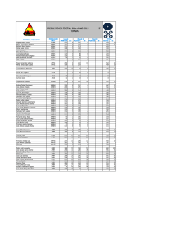
© Copyright 2026