
PDF (568 kB) - Neurologic Clinics
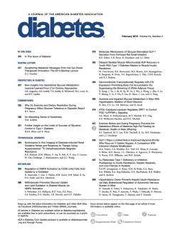
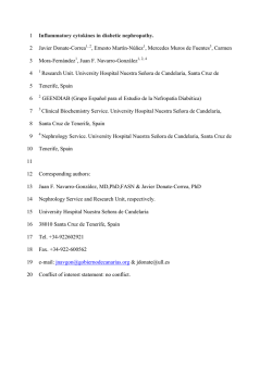
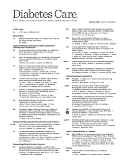
D i a b e t i c N e u ro p a t h y Pa r t 2
Proximal and Asymmetric Phenotypes
Mamatha Pasnoor, MD*, Mazen M. Dimachkie,
Richard J. Barohn, MD
MD,
KEYWORDS
Diabetic asymmetric neuropathies Diabetic amyotrophy
Focal diabetic neuropathy Diabetic lumbosacral radiculoplexopathy
Symmetric diabetic amyotrophy
KEY POINTS
Diabetic neuropathy presents with varied manifestations, including proximal and asymmetric types.
Except for entrapments, diabetic amyotrophy is the most common form of asymmetric
diabetic neuropathy.
Diabetic amyotrophy can present asymmetrically or symmetrically, with a rapid or insidious onset.
Symmetric form of diabetic amyotrophy can be indistinguishable from chronic inflammatory demyelinating polyneuropathy.
Treatment of diabetic amyotrophy with intravenous immunoglobulin or immunosuppressive drugs is controversial.
Truncal radiculopathy can cause abdominal muscle weakness.
Patients with diabetes can develop third, fourth, sixth, and seventh cranial nerve palsies.
Patients with diabetes are more susceptible to compression mononeuropathies than
those who are not diabetic.
Muscle infarction can also be seen in diabetic individuals and is clinically distinct from diabetic amyotrophy.
Treatment is mostly strict diabetic control and supportive in most of these conditions.
INTRODUCTION
Distal symmetric polyneuropathy is most common type of neuropathy associated with
diabetes; however, many subtypes of diabetic neuropathies were defined even as
early as in the 1800s.1–4 Included in these descriptions are patients with proximal
Department of Neurology, University of Kansas Medical Center, 3599 Rainbow Boulevard,
Mail-Stop 2012, Kansas City, KS 66160, USA
* Corresponding author.
E-mail address: [email protected]
Neurol Clin 31 (2013) 447–462
http://dx.doi.org/10.1016/j.ncl.2013.02.003
neurologic.theclinics.com
0733-8619/13/$ – see front matter Ó 2013 Elsevier Inc. All rights reserved.
448
Pasnoor et al
diabetic neuropathy, truncal neuropathy, limb mononeuropathies, and cranial neuropathies (Box 1). Bruns5 focused further on the entity of proximal diabetic neuropathy.
Various theories have been proposed for the pathogenesis of these neuropathies.
Treatment in most cases is tight and stable glycemic control and pain management.
A practical approach to the diagnosis and management of asymmetric and focal
diabetic neuropathies is reviewed in this article.
ASYMMETRIC/FOCAL NEUROPATHIES
Diabetic Lumbosacral Radiculoplexopathy (Diabetic Amyotrophy, Bruns-Garland
Syndrome, Proximal Diabetic Neuropathy)
The most common, and often misdiagnosed, multifocal asymmetric diabetic neuropathy is the lumbosacral radiculoplexopathy syndrome (DLSRP).6 This disorder has
been referred to by many names, including proximal diabetic neuropathy, ischemic
mononeuropathy multiplex, femoral or femoral-sciatic neuropathy, and most often,
diabetic amyotrophy and the “Bruns-Garland syndrome,” after the 2 physicians who
first reported this entity.5–21 Bruns5 first described diabetic patients with asymmetric
proximal weakness and pain in 1890. Later, Garland,13 in the 1950s, used the term
diabetic amyotrophy because of the muscle atrophy in the thighs. But this term can
falsely imply that the primary lesion is on the muscle and therefore we use the term
DLSRP.
Clinical presentation
DLSRP syndrome affects an older group of diabetic individuals, more frequently men,
usually older than 50, but occasionally we have seen the syndrome in middle-aged
diabetic individuals. Most patients have type 2 diabetes mellitus; however, this can
occur even in individuals with type 1 diabetes. In a series of 27 patients reported by
Coppack and Watkins,22 24 patients had type 2 diabetes mellitus and 3 had type 1
diabetes mellitus. The development of this neuropathy is often unrelated to glucose
control or the duration of glucose intolerance. DLSRP can be the presenting manifestation leading to the initial diagnosis of diabetes.
This neuropathy begins with severe unilateral pain in the back, hip, or thigh, which
subsequently spreads to the other side within weeks to months.6,19,22 Patients are
frequently misdiagnosed as having a compressive lumbosacral radiculopathy. Some
patients undergo unnecessary lumbar surgery despite minor changes on lumbar
magnetic resonance imaging (MRI) scan. Given the associated weight loss, patients
are often suspected to have a pelvic tumor. Within days to weeks of the pain onset,
patients develop weakness in typically proximal and, to a lesser extent, distal leg
muscles.
Box 1
Clinical classification of asymmetric diabetic neuropathies
Asymmetric/Focal and Multifocal Diabetic Neuropathies:
Diabetic lumbosacral radiculoplexopathy (Bruns-Garland syndrome; diabetic amyotrophy;
proximal diabetic neuropathy)
Truncal neuropathies (thoracic radiculopathy)
Cranial neuropathies
Limb mononeuropathies
Diabetic Neuropathy Part 2
On examination, there is weakness of hip flexors, adductors and extensors, knee
flexors and extensors, and ankle dorsiflexors and plantar flexors of varying degree.
Profound atrophy of the thigh and at times distal lower extremity muscles develops.
Weakness usually encompasses multiple root or plexus levels and is rarely isolated
to an individual root or peripheral nerve. Thus, in cases in which knee extension weakness is prominent and the possibility of a diabetic “femoral neuropathy” is considered,
if one looks closely at other L2–L4 muscles either on the neurologic examination or
with needle electromyography (EMG), the disease process can usually be found in
these adjacent areas. Similarly, if there is a significant foot drop, there is also usually
evidence of involvement in tibial or other L5 innervated muscles, and the process is
actually not confined to the peroneal nerve. There is usually distal sensory loss, but
this is often indistinguishable from the sensory abnormalities of distal symmetric polyneuropathy (DSPN), which often is present before the development of the radiculoplexopathy. Loss of knee and ankle reflexes is common.
Although the condition usually begins in one leg, spread to the other leg within
weeks or months is rather frequent. The disorder worsens in a gradually progressive
or stepwise manner. Cases have been documented in which there is worsening for
18 months.6 Eventually, the process stabilizes and gradually improves, although the
recovery may take many months. In many cases, some degree of permanent weakness may persist (Fig. 1).6
In about one-third of the cases, weakness spreads to proximal arm muscles and is
attributed to cervicobrachial radiculoplexopathy.11,19,23 Approximately 12% of
patients develop thoracic radiculopathy, leading to radiating pain in the chest or
abdomen and intercostal muscle weakness.24,25 Respiratory weakness has also
been described with this neuropathy.26
Diagnostic workup
Electrophysiologically, nerve conduction study findings may not differentiate DLSRP
neuropathy from DSPN, except for an asymmetric reduction of the femoral compound
muscle action potential amplitudes in unilateral cases. However, the needle EMG
reveals abundant fibrillation potentials in weak proximal and distal leg muscles, as
well as in the lumbosacral paraspinous muscles.6 The cerebrospinal fluid (CSF)
protein is often elevated, usually between 60 and 100 mg/dL, but occasionally
as high as 400 mg/dL. The erythrocyte sedimentation rate may be elevated as well,
but is usually less than 50 mm/h. MRI with gadolinium may show nerve root
enhancement.27
Sural nerve biopsy is not essential to the diagnosis of DLSRP syndrome. When done
to exclude mimics, it shows significant fiber loss, often in an asymmetric fashion within
and between fascicles, resembling focal ischemia (Fig. 2).6,9–11 An ischemic pathogenesis was documented by Raff and colleagues7,8 in an autopsy study showing
infarcts of proximal nerve trunks of the leg and lumbosacral plexus. Asymmetric fiber
loss in sural nerve may support this theory; however, it should be remembered that
even patients with typical DSPN may show this multifocal pattern at times. The presence of occasional thinly myelinated fibers on plastic-embedded sections or short,
thin segments on teased nerve fiber preparations should not lead the physician to
diagnose a demyelinating neuropathy, as these findings can be seen in DSPN as
well. However, it should be emphasized that the electrophysiologic, biopsy, and laboratory features are often not particularly helpful, and the diagnoses of DLSRP is
primarily clinically based on the history and neurologic examination.
There is probably a small subset of diabetic patients with amyotrophy who develop
a painless, symmetric proximal neuropathy involving the lower extremities. Asbury,21
449
450
Pasnoor et al
Fig. 1. Course of muscle strength in 6 patients showing progressive leg weakness. Broken
lines represent the period before the initial evaluations. Solid lines represent the course
during our serial examinations. The average muscle score, shown in the table adjacent to
each graph, is based on a modified Medical Research Council scale expanded to a 10-point
scale (see text). The scores for leg strength are those at the nadir of the illness. AD, ankle
dorsiflexion; AE, ankle eversion; AL, ankle inversion; AP, ankle plantar flexion; HA, hip
abduction; HE, hip extension; HF, hip flexion; KE, knee extension; KF, knee flexion; L, left;
R, indicates right. (From Barohn RJ, Sahenk Z, Warmolts JR, et al. The Bruns-Garland
syndrome (diabetic amyotrophy): revisited 100 years later. Arch Neurol 1991;48:1130–5;
with permission.)
favoring the term proximal diabetic neuropathy, considered that there was a spectrum
ranging from asymmetrical cases with a rapid onset to patients with symmetric proximal weakness of insidious onset. Chokroverty and colleagues16,17 emphasized insidious bilateral onset of proximal weakness. Pascoe and colleagues11 published a Mayo
clinic series of 44 patients with symmetric proximal weakness that has a more
restricted distribution and seems to be monophasic and self-limiting, differentiating
from chronic inflammatory demyelinating neuropathy (CIDP). However, the symmetric
presentation seems to be uncommon and tends to occur more often in young type 1
diabetic individuals who are having poor glycemic control. Therefore, we have divided
the proximal diabetic amyotrophies (DAM) into 2 forms: DAM-1 and DAM-2
(Table 1).28
Diabetic Neuropathy Part 2
Fig. 2. Cross sections of sural nerve fascicles, 1-mm thick. Nonrandom fiber loss is more apparent
and more severe in the left than in the right. (From Barohn RJ, Sahenk Z, Warmolts JR, et al.
The Bruns-Garland syndrome (diabetic amyotrophy): revisited 100 years later. Arch Neurol
1991;48:1130–5; with permission.)
If asymmetric diabetic neuropathies occur in only about 1% of the diabetic population, we think the painless, symmetric form of diabetic amyotrophy (DAM-1) is even
more uncommon. This form superficially resembles idiopathic CIDP. We believe that
for every 10 to 20 patients with asymmetric/painful amyotrophy (DAM-2) seen at
a tertiary care neuromuscular centers, only one DAM-1 patient will be seen. Whether
or not the pathogenesis of DAM-1 is different from DAM-2 and is more in line with
metabolic dysfunction is unknown.
Management
Treatment is centered around pain control and strict glycemic control. Both groups
spontaneously improve over a period of months. Physical therapy can assist in
improving functional mobility.
Controversy involving DLSRP is whether or not there is an immune-mediated pathogenesis component and if patients respond favorably to immunomodulating therapy.
This concept was first introduced by Bradley and colleagues29 in 1984 in their report of
6 patients with a painful lumbosacral plexopathy, elevated sedimentation rate, mild
Table 1
Diabetic amyotrophy: 2 presentations
DAM-1
DAM-2
Type of DM
Type 1 >2
Type 2 >1
Onset in legs and progression
Bilateral/Insidious
Chronic
Unilateral/Acute
Stepwise/Goes to other leg
Distribution
Symmetric proximal
Asymmetric proximal and distal
Pain
No
Yes
Sensory symptoms
No
Yes
Poor DM control
Yes
Yes
Weight loss
Yes
Yes
Spontaneous improvement
Yes
Yes
Frequency
Very, very rare
Uncommon: w1%
Spread to arm
Yes: ?Common
Yes: 10%
Abbreviations: DAM, diabetic amyotrophy; DM, diabetes mellitus.
451
452
Pasnoor et al
perivascular inflammation on sural nerve biopsy, and asymmetric nerve fiber loss. Five
were treated with immunosuppressive drugs (prednisone alone or prednisone and
cyclophosphamide) and 4 improved or stopped progressing. They did not believe
the diabetic individuals had typical DLSRP because “they continued to deteriorate
and to have pain for several months despite careful control of the diabetes, and
only began to improve following treatment with prednisone, although this therapy
worsened their diabetes.” Thus, Bradley and colleagues29 felt the diabetes in their
patients was incidental. Of course, others have also reported idiopathic lumbosacral
plexitis in nondiabetic patients (analogous to idiopathic brachial plexitis).30,31 Interestingly, in the earlier reports of idiopathic lumbosacral plexitis, patients improved spontaneously. Verma and Bradley32 advocated the use of intravenous immunoglobulin
(IVIG) for idiopathic lumbosacral plexitis.
Krendel and colleagues33 (in 1995) reported their experience using immunotherapy in 21 patients with diabetic neuropathy. They divided their patients into
2 groups: Group A consisted of 15 patients who had “multifocal axonal inflammatory
vasculopathy” and most of these patients seemed to correspond to what we
described previously as DLSRP. Group B patients consisted of 6 diabetic individuals
who had both arm and leg involvement, and although in 3 patients the process was
asymmetric, the investigators stated this group had “demyelinating” neuropathy by
electrophysiologic criteria. Group A patients had perivascular inflammation on nerve
biopsy and group B patients had “onion bulbs” but no inflammation. All patients
received some form of immunomodulatory therapy (15, IVIG; 13, prednisone;
5, cyclophosphamide; 3, plasma exchange; 1, azathioprine) in various combinations,
and all improved. Krendel and colleagues’33 conclusion was that there are 2 forms of
immune-mediated neuropathy in diabetic patients that responds to treatment.
Younger and colleagues34 reported their experience finding evidence of inflammation in 20 patients with diabetic neuropathy: 4 had DSPN, 12 had “proximal diabetic
neuropathy,” and 4 had mononeuropathy multiplex. All patients with proximal diabetic neuropathy and mononeuropathy multiplex had asymmetric features and we
would currently consider them as having DLSRP. As noted in the pathogenesis
section see the article by Pasnoor and colleagues elsewhere in this issue, 12 of
20 had some evidence of inflammatory cells, including 2 with DSPN. Younger and
colleagues treated 8 patients with IVIG (2 DSPN, 1mononeuropathy multiplex, 5
proximal diabetic neuropathy), all of whom had perivascular inflammation, and
they reported that all improved.
In the Mayo Clinic group series by Pascoe and colleagues,11 3 of 9 patients undergoing sural nerve biopsy had a multifocal distribution of fiber loss, and 2 had perivascular mononuclear inflammatory infiltrates. Twelve were treated with IVIG, and 9
improved. Of the 29 untreated patients, 17 spontaneously improved. They concluded
that the “efficacy of immunotherapy is unproven but such intervention may be considered in the severe progressive cases or ones associated with severe neuropathic pain.”
The experience of the French group led by Gerard Said is important to note. In an
article published in 1994, they reported inflammatory and ischemic lesions in nerve
biopsy specimens of the intermediate cutaneous nerve of the thigh in patients with
DLSRP.9 Three patients with “severe and prolonged painful disability” improved
dramatically with corticosteroid treatment. In a subsequent report in 1997 of 4 patients
with DLSRP, Said and colleagues10 described patients who had symptoms for 4, 6,
12, and 18 months before biopsy. Although all patients showed perivascular inflammation on nerve biopsy, to the investigators’ surprise, all became pain free with subsequent improvement of their weakness shortly following the biopsy. They concluded
that despite the treatment with prednisone they used in their initial article, that
Diabetic Neuropathy Part 2
DLSP “is self-limited and does not require the use of corticosteroids or immunomodulators.” In a series by Dyck and colleagues,35 all 33 patients with DLSRP had some
evidence of microvasculitis on nerve biopsy, and nearly all improved spontaneously. A
report by the group from Houston found 18 of 19 patients with DLSRP had substantial
improvement without immunomodulating therapy.36
Despite many months of persistent symptoms or progression in some patients with
DLSRP, eventually all patients spontaneously have resolution of pain and slow
improvement of weakness (see Fig. 1).6 Treatment with IVIG or other immunosuppressive drugs is controversial. In a prospective case series, 5 patients with severe pain
received IVIG after having no response to symptomatic therapy for pain and corticosteroids.37 Four had a decrease in pain. In contrast, Zochodne and colleagues38 in
2003 reported a patient who developed DLSRP while on immunosuppressive regimen
consisting of cyclosporine and myophenolate mofetil for an allograft cardiac transplant and 2 patients with DLSRP who did not respond to IVIG treatment, arguing
that immunosuppressive therapy did not prevent onset of DLSRP. In our opinion,
we do not believe IVIG should be used in patients with DLSRP. At this time, we are
not convinced that this form of immunomodulating therapy is indicated. Perhaps
this question can be resolved with a controlled trial, but such a trial will be difficult,
as each center sees a handful of patients annually and it will be difficult to get the pharmaceutical industry and the Food and Drug Administration to support a large multicenter IVIG trial in this rare disorder.
On the other hand, the experience of Said and Bradley with the improvement of pain
with prednisone should not be ignored. According to a recent Cochrane review of
immunotherapy for diabetic amyotrophy (DA) only one completed controlled trial using
IV methyprednisolone in DA was found.39,40 High doses of corticosteroids may lead to
improvement of severe pain in some patients with DLSRP, and this may be analogous
to the improvement of neuropathic pain in patients who are believed to have reflex
sympathetic dystrophy.41,42 Perhaps breaking the pain syndrome in this manner
may subsequently allow patients to begin moving their weak extremities easier. Presently, there is no convincing evidence from randomized trial to support any recommendation on the use of any immunotherapy treatment in DLSRP.39
We believe one should be cautious about jumping to the conclusion that finding
mild perivascular inflammation on biopsy, or demyelination features on either electrophysiology or pathology suggest that DLSRP is a disease that is primarily
immune-mediated and will respond to immunomodulating therapy. We cautioned
previously about the danger of heavily relying on electrophysiologic evidence of
demyelination on nerve conduction studies (NCS) of diabetic patients, as some
will fulfill research electrophysiologic criteria for CIDP even though the clinical
pattern does not correspond to CIDP, but actually is that of DLSRP. Similar caution
should be used with data from nerve biopsies of patients with DLSRP in concluding
these patients have either vasculitis or a demyelinating neuropathy. In routine clinical
practice, we do not recommend either nerve biopsy or immunomodulating therapy in
patients with typical DLSRP. Finally, we would also caution clinicians about splitting
patients with otherwise typical DLSRP because of nerve root enhancement on
lumbar MRI scan.27 If other etiologies are excluded by CSF analysis, the mere
finding of root enhancement on MRI in DLSRP should not necessarily lead to the
initiation of immunomodulating therapy.
Other DLSRP caveats
Cervical brachial radiculoplexopathy Although cervical/brachial plexus involvement
is uncommon, it does occur.11,23 In the classic early Mayo Clinic series of “diabetic
453
454
Pasnoor et al
polyradiculopathy” reported by Bastron and Thomas19 in 1981 of 105 patients, 81 had
lower extremity involvement, 15 had upper extremity involvement, and 12 had
thoracic/abdominal involvement. Obviously, a few patients had involvement of more
than one region. As mentioned previously, in the Mayo Clinic series of Pascoe and
colleagues,11 all 44 patients had (by definition) leg weakness, and 12 of these also
had arm weakness (7 bilateral, 5 unilateral). Occasionally, patients with DLSRP
develop arm pain and weakness days to weeks after the initial leg symptoms.43,44
The arm involvement is usually proximal and distal, similar to the pattern of weakness
seen in the legs. Interestingly, the arm symptoms can begin or continue to progress
after the leg symptoms have plateaued or begun to improve.
Thus, whereas cervical/brachial root and plexus involvement has not been emphasized a great deal in diabetic radiculoplexopathy, the clinician should be aware of this
possibility. We do believe that in this setting, a more extensive workup probably is to
exclude other disease entities. All of these patients should have a CSF examination for
infectious and neoplastic diseases, and nerve biopsy is probably warranted to exclude
true vasculitic neuropathy.
CIDP in diabetic patients Diabetic patients can develop typical CIDP but, as
mentioned previously, there is no increased risk of CIDP in diabetic patients.45–48
The clinical features of gradually progressive, usually painless, proximal and distal
symmetric weakness and numbness in the arms and legs should be sufficient to
distinguish CIDP from the typical symmetric and asymmetric diabetic neuropathies;
however, the laboratory results in this setting may not be particularly helpful, especially
the CSF protein. If there is an underlying diabetic DSPN on NCS and needle EMG,
electrophysiologic results can also be relatively unhelpful unless clear-cut and ample
features of an acquired, markedly demyelinating neuropathy are present. In addition,
nerve biopsy in many diabetic neuropathies can show thinly myelinated fibers, and
therefore we usually do not pursue nerve biopsy in this setting. Diagnosis is usually
based on the clinical presentation and it is reasonable to proceed with immunomodulating therapy when CIDP is strongly suspected.
True mononeuritis multiplex in diabetes: does it exist? Finally, a comment should
be made regarding “mononeuritis multiplex” in diabetic patients. We suspect that
most of these patients have diabetic radiculoplexopathy, usually lumbosacral, but
rarely cervical-brachial. It is uncommon for diabetic patients to develop a true mononeuritis multiplex in which individual distal peripheral nerves (eg, femoral, peroneal,
tibial, ulnar, median or radial) are “picked-off” in a subacute or acute fashion. It is difficult to find good documentation of this in the literature. Although the early articles by
Raff and colleagues7,8 use the term “mononeuropathy multiplex,” if one reads of clinical description of their 7 cases, they all had typical DLSRP with proximal and distal
involvement not confined to an individual nerve. If a diabetic patient develops a true
mononeuritis multiplex, the usual causes need to be pursued (vasculitis, infectious,
and hereditary). We believe that if one wants to include diabetes mellitus in the differential diagnosis of true mononeuritis multiplex, it should be at the bottom of such
a list.
OTHER ASYMMETRIC NEUROPATHIES
Truncal Radiculopathy
Another common focal form of diabetic radiculopathy involves isolated thoracic
roots.49–52 This is presumably a focal diabetic radiculopathy that is similar to DLSRP
except for the location on the trunk, thorax, or abdomen.
Diabetic Neuropathy Part 2
Clinical presentation
Patients develop abrupt pain over days to weeks with severe dysesthesias in a dermatomal pattern. In some patients, the pain may not radiate entirely around the trunk in
a full radicular pattern, but the symptoms and signs may occur in smaller, restricted
regions that imply damage of the dorsal or ventral rami or their medial or lateral
branches.25 Multiple thoracic dermatomes can be involved. Although most cases
are unilateral at onset, it can evolve bilaterally, much like DLSRP. In fact, in our experience, it is not uncommon during the evaluation of a patient with typical DLSRP to
uncover that at some point over the preceding year or 2 the patient had an episode
of truncal pain and dysesthesias, or was diagnosed with truncal “shingles” without
a rash. Patients can occasionally develop weakness of the rectus abdominus muscles.
Some patients may present with pseudohernia caused by weakening of the abdominal
musculature.49,53
On the other hand, although most patients do not demonstrate obvious motor
involvement, needle EMG can reveal abnormalities in the paraspinous or abdominal
wall muscles.
Diagnostic workup
Nerve conductions may reveal abnormalities related to distal symmetric polyneuropathy. Needle EMG findings include fibrillations in the paraspinous or abdominal wall
muscles.51,54,55
Management
Treatment is directed at symptomatic pain management, see the article by Pasnoor
and colleagues elsewhere in this issue on DSPN, in this volume. Truncal radiculopathy
should be distinguished from the wedge-shaped midline area of symmetric truncal
sensory loss that can occur in advanced DSPN50 and from rare discogenic thoracic
radiculopathy.
Prognosis
The natural history is similar to DLSRP, with persistence of sensory symptoms for
weeks to months, with gradual resolution.
Cranial Neuropathies
Diabetic patients can suddenly develop a unilateral third, fourth, sixth, or seventh
cranial nerve palsy. The oculomotor nerve was found to be most frequently affected
in one study by Greco and colleagues56 looking at 61 patients with diabetic cranial
nerve palsies. The hallmark of diabetic third nerve palsy is pupillary sparing in most
cases.
Clinical presentation
Retro-orbital pain accompanies about half of the cases. Sparing of the pupil in diabetic
third nerve palsies is due to sparing of axons at the periphery of the nerve involved in
pupillary function. Pathologic evidence supports the concept that the process is probably attributable to an ischemic watershed phenomenon in the central part of the
nerve.57–60
It has been suggested that patients with diabetes are more likely to develop
a seventh cranial nerve palsy.61 However, Bell palsy is a common event and it is difficult to substantiate if it is indeed more prevalent in diabetes.62 It is interesting to note
that in the Rochester Diabetic Neuropathy Study, neither cranial mononeuropathies
nor truncal radiculopathies were more common in diabetic patients compared with
control subjects.63
455
456
Pasnoor et al
Diagnostic workup
Imaging studies may be necessary to rule out stroke in some cases; however, history
alone without additional testing is sufficient in most of these patients.
Management and prognosis
The main risk factors for the development of cranial neuropathies are duration of diabetes and the patient’s age.64 Treatment should be mainly focused on management of
diabetes.
Most patients make a full recovery, with some early evidence of improvement within
2 to 3 months.
Isolated Mononeuropathies
Clinical presentation
It is generally believed and established in studies that diabetic individuals are more
susceptible to compression injuries compared with nondiabetic individuals.65 This
would include the median nerve at the carpal tunnel, ulnar nerve at the elbow, the peroneal (fibular) nerve at the fibular head, and perhaps the lateral cutaneous femoral nerve
(meralgia paresthetica) at the hip. In the early study by Mulder and colleagues66 in 103
cases of diabetes, 16 had mononeuropathies affecting 29 nerves as follows: common
peroneal, 13; median nerve (carpal tunnel), 9; ulnar nerve, 5; lateral femoral cutaneous
nerve, 1; and femoral nerve, 1, the latter being likely due to DLSRP. Meralgia paresthetica (mononeuropathy of the lateral femoral cutaneous nerve) is associated with
diabetes mellitus irrespective of obesity and advanced age.67
In a study from Rochester, Minnesota, there was evidence that carpal tunnel
syndrome is more common in diabetes mellitus than in the general population.68
In another Rochester Diabetic Neuropathy Study, approximately one-quarter of
patients had subclinical carpal tunnel syndrome on NCS, but only 7.7% were
symptomatic.63
Diagnostic workup
Diagnosis is usually established with electrophysiologic testing; however, electrophysiologic diagnosis of carpal tunnel or other mononeuropathies is sometimes difficult in individuals with diabetic polyneuropathy. One study showed that segmental
and comparative median nerve conduction tests in combination with standard nerve
conduction resulted in more accurate diagnosis of carpal tunnel syndrome in patients
with diabetic polyneuropathy.69
DIABETIC MUSCLE INFARCTION
In the context of discussing the various diabetic neuropathies, it is relevant to review
another neuromuscular complication of diabetes in which the muscle itself is the target
organ rather than the nerve. It is an underdiagnosed complication of long-standing
diabetes.70
Clinical Presentation
Diabetic muscle infarction (DMI) begins with the abrupt onset of thigh pain, tenderness, and swelling.71–76 Over a period of days, a firm mass develops in nearly half
of cases. The muscles most frequently involved are the vastus lateralis and medialis,
thigh adductors, and biceps femoris. Calf involvement is reported in up to 20% of
cases and bilateral involvement in 8% of cases.77 Compared with 130 cases, there
are 5 case reports of DMI affecting muscles of the upper limb of patients, particularly
in patients with type 2 diabetes with end-stage renal disease.78 Edema from the
Diabetic Neuropathy Part 2
swelling can extend to the knee and mimic a joint effusion.70 DMI tends to occur in
younger, poorly controlled diabetic patients with other end-organ complications.
There are no associated systemic symptoms or signs indicative of infection and no
skin discoloration suggesting cellulitis or thrombophlebitis. The painful mass persists
for weeks, occasionally with exacerbation of symptoms, and then spontaneously
resolves over weeks to several months. Contralateral involvement of the other thigh
can occur, even after the initial episode resolves. Up to 50% of cases will recur, mostly
involving previously unaffected muscle groups.77
Diagnostic Workup
Creatine kinase can be normal or modestly elevated. Needle EMG demonstrates fibrillation potentials in the involved muscles with a loss of voluntary motor unit potentials in
the most affected areas. Remaining motor unit potentials may be brief and short,
reflecting fragmentation of the motor unit. MRI scan of the limb muscle reveals
increased signal on T2-weighted images in the involved thigh muscles, indicative of
marked muscle edema extending into the perifascicular and subcutaneous tissues
(Fig. 3).72,73,79 Gadolinium contrast administration is contraindicated in those with
renal impairment. Radionuclide imaging with Technetium-99 demonstrates radiopharmaceutical accumulation and muscle ultrasound shows hyperechoic signal in the
mass.73
Biopsy of the region consists of large confluent areas of muscle necrosis and
edema, with loss of the normal architectural pattern. A muscle biopsy is often not
needed because it may prolong recovery and is indicated only when the presentation
is atypical, response is poor, or diagnosis is uncertain. Biopsy, when performed,
Fig. 3. Sagittal view of left thigh by T2-weighted MRI scan. There is diffuse high signal in the
biceps femoris, semimembra-nosus, and semitendinosus muscles, whereas the bone and
anterior compartment muscles appear normal. (From Barohn RJ, Kissel JT. Case-of-themonth: painful thigh mass in a young woman: diabetic muscle infarction. Muscle Nerve
1992;15:850–5; with permission.)
457
458
Pasnoor et al
Table 2
DLSRP versus DMI
DLSRP
DMI
Pain
1
1
Focally tender
–
1
Swelling/mass
–
1
Progression
1
1
Bilaterally
1
1
Atrophy
1
–
Distal weakness
1
Sensory symptoms
–
MRI muscle
Normal
Abnormal
EMG
Neuropathic
Myopathic
Abbreviations: DLSRP, diabetic lumbosacral radiculoplexopathy; DMI, diabetic muscle infarction;
EMG, electromyogram; MRI, magnetic resonance imaging; 1, present; , absent; , maybe present
or absent.
shows pale muscle on gross examination and areas of muscle necrosis and edema
surrounded by muscle fibers in various stages of degeneration and regeneration,
with hyalinosis and thickening of arterioles.71
The differential diagnosis of DMI includes, in addition to DLSRP, infection (abscess,
pyomyositis, necrotizing fasciitis), focal myositis, venous thrombosis, and tumor. Both
DMI and DLSRP syndromes begin with the abrupt onset of lower extremity pain that
can ultimately involve the opposite side. In DLSRP the pain is usually localized to the
low back, hip, or buttocks with radiation into the thigh; whereas, in DMI, the pain is
more focal and associated with swelling and a firm mass. Patients with DLSRP
develop dramatic weakness and atrophy in proximal, and usually distal lower
extremity muscles. Sensory symptoms (other than pain) do not result from DMI unless
there is a prior distal symmetric polyneuropathy. Whereas the imaging studies of the
thigh will be focally abnormal with swelling and infarction in DMI, they may show
diffuse denervating changes and atrophy on T2 sequences of the hip, thigh, and leg
muscles in DLSRP. The EMG in DLSRP is different in that it is characterized by widespread fibrillations in many muscles (usually including the paraspinous muscles), with
long-duration, polyphasic motor unit potentials and decreased recruitment pattern.
NCS may not be helpful in distinguishing the disorders, as both may show evidence
of a distal symmetric polyneuropathy.
Although there are obvious differences between DMI and DLSRP (Table 2), the
abrupt onset of both syndromes, and pathologic evidence for probable focal ischemia
in the muscle (DMI) and nerve (DLSRP) supports the theory that both entities have
a primary vascular microangiopathic etiology.
Management and Prognosis
The treatment of DMI is supportive. No evidence-based recommendations are available on management of this condition; however, a retrospective analysis supports
conservative management with bed rest, leg elevation, and adequate analgesia.
Activities should be avoided to avoid increasing the pain. There is no evidence to
support the use of corticosteroids or surgery. Short-term prognosis is good, but
the recurrence rate is high (40%), and recurrences may not affect the same muscle
group.80
Diabetic Neuropathy Part 2
Case study
A 62-year-old diabetic man on an oral hypoglycemic treatment developed sudden severe pain
that started in the lower back and radiated to the right leg. Within 3 days, he experienced
weakness in the same leg. After 1 month, similar pain and weakness occurred in the left leg.
Symptoms persisted for 4 months, during which time the patient had a 9-kg unintentional
weight loss. Neurologic examination revealed asymmetric proximal and distal weakness in
both legs. There was asymmetric atrophy of the quadriceps and hamstring muscles. A stocking
pattern of loss of pinprick, light touch, and vibration sense was found. Tendon reflexes were
absent at the ankles and trace at the knees bilaterally. Upper extremities had normal motor
and sensory function.
At the time of initial evaluation, fasting glucose was mildly elevated (140 mg/dL), with a glycosylated hemoglobin of 7.5% and fasting blood sugar of 135 mg/dL. Peroneal motor conduction
velocity was reduced at 36 m/s, and compound motor action potential was reduced at 300 mV.
Tibial nerve conduction was 38 m/s, and sural potentials were absent. Median and ulnar nerve
conduction velocities were normal. Bilateral femoral motor conduction studies showed an
amplitude of 3 mV on left and 1 mV on right. An EMG revealed active denervation potentials
in proximal (quadriceps, adductor longus, gluteus medius, and hamstrings), distal (gastrocnemius and anterior tibial), and lumbosacral paraspinal muscles bilaterally.
His recovery was protracted. Diabetes mellitus was managed by his primary care physician and
he achieved better control of his diabetes. He required a right knee brace for ambulation and
was treated with gabapentin (Neurontin) for pain control. Leg weakness progressed for 3 additional months before subsequent gradual improvement of strength. At the 6-month follow-up,
the pain had almost resolved; however, he still had considerable residual weakness.
REFERENCES
1. Althaus J. On sclerosis of the spinal cord, including locomotor ataxy, spastic
spinal paralysis and other system diseases of the spinal cord: their pathology,
symptoms, diagnosis, and treatment. London (United Kingdom): Green &
Company; 1885.
2. Leyden E. Entzundung der peripheren Nerven. Deut Militar Zeitsch 1887;17:49.
3. Auche M. Des alterations des nerfs pe´riphe´riques. Arch Med Exp Anat Pathol
1890;2:635.
4. Pryce TD. On diabetic neuritis with a clinical and pathological description of three
cases of diabetic pseudo-tabes. Brain 1893;16:416.
5. Bruns L. Ueber neuritische Lahmungen beim Diabetes mellitus. Berl Klin Wochensher 1980;27:509–15.
6. Barohn RJ, Sahenk Z, Warmolts JR, et al. The Bruns-Garland syndrome (diabetic
amyotrophy): revisited 100 years later. Arch Neurol 1991;48:1130–5.
7. Raff M, Asbury AK. Ischemic mononeuropathy and mononeuropathy multiplex in
diabetes mellitus. N Engl J Med 1968;279:17–22.
8. Raff MC, Sangalang V, Asbury AK. Ishemic mononeuropathy multiplex associated with diabetes mellitus. Arch Neurol 1968;18:487–99.
9. Said G, Goulon-Goeau C, Lacroix C, et al. Nerve biopsy findings in different
patterns of proximal diabetic neuropathy. Ann Neurol 1994;35:559–69.
10. Said G, Elgrably F, Lacroix C, et al. Painful proximal diabetic neuropathy: inflammatory nerve lesions and spontaneous favorable outcome. Ann Neurol 1997;41:
762–70.
11. Pascoe MK, Low PA, Windebank AJ, et al. Subacute diabetic proximal neuropathy. Mayo Clin Proc 1997;72:1123–32.
12. Garland H, Taverner D. Diabetic myelopathy. Br Med J 1953;1:405–8.
13. Garland H. Diabetic amyotrophy. Br Med J 1955;2:1287–90.
459
460
Pasnoor et al
14. Locke S, Lawrence DG, Legg MA. Diabetic amyotrophy. Am J Med 1963;34:
775–85.
15. Calverley JR, Mulder DW. Femoral neuropathy. Neurology 1960;10:963–7.
16. Chokroverty S, Reyes MG, Rubino FA, et al. The syndrome of diabetic amyotrophy. Ann Neurol 1977;2:181–94.
17. Chokroverty S. AAEE Case Report #13: diabetic amyotrophy. Muscle Nerve 1987;
10:679–84.
18. Subramony SH, Wilbourn AJ. Diabetic proximal neuropathy. J Neurol Sci 1982;53:
293–304.
19. Bastron JA, Thomas JE. Diabetic polyradiculopathy. Mayo Clin Proc 1981;56:
725–32.
20. Williams IR, Mayer RF. Subacute proximal diabetic neuropathy. Neurology 1976;
26:108–16.
21. Asbury AK. Proximal diabetic neuropathy. Ann Neurol 1977;2:179–80.
22. Coppack SW, Watkins PJ. The natural history of diabetic femoral neuropathy. Q J
Med 1991;79(288):307–13.
23. Katz JS, Saperstein DS, Wolfe G, et al. Cervicobrachial involvement in diabetic
radiculoplexopathy. Muscle Nerve 2001;24:794–8.
24. Streib EW, Sun SF, Paustian EF, et al. Diabetic thoracic radiculopathy: electrodiagnostic study. Muscle Nerve 1986;9:548–53.
25. Stewart JD. Diabetic truncal neuropathy: topography of the sensory deficit. Ann
Neurol 1989;25:233–8.
26. Brannagan TH, Promisloff RA, McCluskey LF, et al. Proximal diabetic neuropathy
presenting with respiratory weakness. J Neurol Neurosurg Psychiatry 1999;67:
539–41.
27. O’Neil BJ, Flanders AE, Escandon S, et al. Treatable lumbosacral polyradiculitis
masquerading as diabetic amyotrophy. J Neurol Sci 1997;151:223–5.
28. Amato AA, Barohn RJ. Diabetic lumbosacral polyradiculoneuropathies. Curr Treat
Options Neurol 2001;3(2):139–46.
29. Bradley WG, Chad D, Verghese JP, et al. Painful lumbosacral plexopathy with
elevated erythrocyte sedimentation rate: a treatable inflammatory syndrome.
Ann Neurol 1984;15:457–64.
30. Evans BA, Stevens JC, Dyck PJ. Lumbosacral plexus neuropathy. Neurology
1981;31:1327–30.
31. Sander JE, Sharp FR. Lumbosacral plexus neuritis. Neurology 1981;31:470–3.
32. Verma A, Bradley WG. High-dose intravenous immunoglobulin therapy in chronic
progressive lumbosacral plexopathy. Neurology 1994;44:248–50.
33. Krendel DA, Costigan DA, Hopkins LC. Successful treatment of neuropathies in
patients with diabetes mellitus. Arch Neurol 1995;52:1053–61.
34. Younger DS, Rosoklija G, Hays AP, et al. Diabetic peripheral neuropathy: a clinicopathologic and immunohistochemical analysis of sural nerve biopsies. Muscle
Nerve 1996;19:722–7.
35. Dyck PJB, Novell JE, Dyck PJ. Microvasculitis and ischemia in diabetic lumbosacral radiculopexus neuropathy. Neurology 1999;53:2113–21.
36. Lovitt SM, Pleitez MY, Copeland KJ, et al. Proximal diabetic neuropathy improves
spontaneously without expensive and dangerous treatment [abstract]. Neurology
2000;54:A212–3.
37. Tamburin S, Zanette G. Intravenous immunoglobulin for the treatment of diabetic
lumbosacral radiculoplexus neuropathy. Pain Med 2009;10(8):1476–80.
38. Zochodne DW, Isaac D, Jones C. Failure of immunotherapy to prevent, arrest or
reverse diabetic lumbosacral plexopathy. Acta Neurol Scand 2003;107(4):299–301.
Diabetic Neuropathy Part 2
39. Chan YC, Lo YL, Chan ES. Immunotherapy for diabetic amyotrophy. Cochrane
Database Syst Rev 2012;(6):CD006521.
40. Dyck PJB, O’Brien P, Bosch EP, et al. The multi-center, double-blind controlled
trial of IV methylprednisolone in diabetic lumbosacral radiculoplexus neuropathy.
Neurology 2006;66(5 Suppl 2):A191.
41. Kozin F, Ryan LM, Carerra GF, et al. The reflex sympathetic dystrophy syndrome
(RSDS). III. Scitigraphic studies, further evidence for the therapeutic efficacy of
systemic corticosteroids, and proposed diagnostic criteria. Am J Med 1981;70:
23–30.
42. Barohn RJ. Reflex sympathetic dystrophy due to peripheral neuropathy and utility
of the three-phase bone scan: case series and review. Adv an Clin Neurosci
1997;7:129–50.
43. Katz JS, Wolfe GI, Burns D, et al. Diabetic radiculoplexopathy with cervicobrachial involvement [abstract]. Neurology 2000;54:A367.
44. Riley DE, Shields RW. Diabetic amyotrophy with upper extremity involvement
[abstract]. Neurology 1984;34(Suppl 1):216.
45. Cornblath DR, Drach DB, Griffin JW. Demyelinating motor neuropathy in patients
with diabetic polyneuropathy [abstract]. Ann Neurol 1987;22:126S.
46. Stewart JD, McKelvey R, Durcan L, et al. Chronic inflammatory demyelinating polyneuropathy (CIDP) in diabetics. J Neurol Sci 1996;142:59–64.
47. Uncini A, De Angelis MV, Di Muzio A, et al. Chronic inflammatory demyelinating
polyneuropathy in diabetics: motor conductions are important in the differential
diagnosis with diabetic polyneuropathy. Clin Neurophysiol 1999;110:705–11.
48. Laughlin RS, Dyck PJ, Melton LJ, et al. Incidence and prevalence of CIDP and
the association of diabetes mellitus. Neurology 2009;73(1):39–45.
49. Chiu HK, Trence DL. Diabetic neuropathy, the great masquerader: truncal
neuropathy manifesting as abdominal pseudohernia. Endocr Pract 2006;12(3):
281–3.
50. Waxman SG, Sabin TD. Diabetic truncal polyneuropathy. Arch Neurol 1981;38:
46–7.
51. Sun SF, Streib EW. Diabetic thoracoabdominal neuropathy: clinical and electrodiagnostic features. Ann Neurol 1981;9:75–9.
52. Longstretch GF, Newcomer AD. Abdominal pain caused by diabetic radiculopatly. Ann Intern Med 1977;86:166–86.
53. Parry GJ, Floberg J. Diabetic truncal neuropathy presenting as abdominal hernia.
Neurology 1989;39:1488–90.
54. Boulton AM, Angus E, Ayyar DR. Diabetic thoracic polyradiculopathy presenting
as an abdominal swelling. Br Med J (Clin Res Ed) 1984;289:798–9.
55. Kikta DG, Breuer AC, Wilbourn AJ. Thoracic root pain in diabetes: the spectrum
of clinical and electromyographic findings. Ann Neurol 1982;11:80–5.
56. Greco D, Gambina F, Pisciotta M, et al. Clinical characteristics and associated
comorbidities in diabetic patients with cranial nerve palsies. J Endocrinol Invest
2012;35(2):146–9.
57. Asbury AK, Aldredge H, Herschberg R, et al. Oculomotor palsy in diabetes
mellitus: a clinicopathological study. Brain 1970;93:555–66.
58. Weber RB, Daroff RB, Mackey EA. Pathology of oculomotor nerve palsy in diabetics. Neurology 1970;20:835–8.
59. Dreyfus PM, Hakim S, Adams RD. Diabetic ophthalmoplegia. AMA Arch Neurol
Psychiatry 1957;77:337–49.
60. Smith BE, Dyck PJ. Subclinical histopathological changes in the oculomotor
nerve in diabetes mellitus. Ann Neurol 1992;32:376–85.
461
462
Pasnoor et al
61. Korczyn AD. Bell’s palsy and diabetes mellitus. Lancet 1971;1:108–9.
62. Aminoff MJ, Miller AL. The prevalence of diabetes mellitus inpatients with Bell’s
palsy. Acta Neurol Scand 1972;48:381–4.
63. Dyck PH, Kratz KM, Karnes JL, et al. The prevalence by staged severity of
various types of diabetic neuropathy, retinopathy, and nephropathy in a population-based cohort: the Rochester diabetic neuropathy study. Neurology 1993;43:
817–24.
M, Vrca A, Kolak I, et al. Cranial nerve lesion in diabetic patients. Coll
64. Ostric
Antropol 2011;35(Suppl 2):131–6.
65. Wilbourn AJ. Diabetic entrapment and compression neuropathies. In: Dyck PJ,
Thomas PK, editors. Diabetic neuropathy. 2nd edition. Philadelphia: WB Saunders; 1999. p. 481–508.
66. Mulder DW, Lambert EH, Bastrom JA, et al. The neuropathies associated with
diabetes mellitus; a clinical and electromyographic study of 103 unselected diabetic patients. Neurology 1961;11:275–84.
67. Parisi TJ, Mandrekar J, Dyck PJ, et al. Meralgia paresthetica: relation to obesity,
advanced age, and diabetes mellitus. Neurology 2011;77(16):1538–42.
68. Stevens JC, Sun S, Beard CM, et al. Carpal tunnel syndrome in Rochester, Minnesota, 1961–1980. Neurology 1988;38:134–8.
69. Gazioglu S, Boz C, Cakmak VA. Electrodiagnosis of carpal tunnel syndrome in
patients with diabetic polyneuropathy. Clin Neurophysiol 2011;122(7):1463–9.
70. Trujillo-Santos AJ. Diabetes muscle infarction: an underdiagnosed complication
of long-standing diabetes. Diabetes Care 2003;26:211–5.
71. Angervall L, Stener B. Tumoriform focal muscle degeneration in two diabetic
patients. Diabetologica 1965;1:39–42.
72. Barohn RJ, Kissel JT. Case-of-the-month: painful thigh mass in a young woman:
diabetic muscle infarction. Muscle Nerve 1992;15:850–5.
73. Barohn RJ, Bazan C, Timmons JH, et al. Bilateral diabetic thigh muscle infarction.
J Neuroimaging 1994;4:43–4.
74. Banker B, Chester S. Infarction of the thigh muscle in the diabetic patient.
Neurology 1973;23:667–77.
75. Chester S, Banker B. Focal infarction of muscle in diabetes. Diabetes Care 1986;
9:623–30.
76. Singer S, Rosenberg AE. Case records of the MGH Weekly clinicopathological
exercises. Case 29-1997. A 54-year-old diabetic woman with pain and swelling
of the leg. N Engl J Med 1997;337(12):839–45.
77. Mathew A, Reddy IS, Archibald C. Diabetic muscle infarction. Emerg Med J 2007;
24(7):513–4.
78. Joshi R, Reen B, Sheehan H. Upper extremity diabetic muscle infarction in three
patients with end-stage renal disease: a case series and review. J Clin Rheumatol
2009;15(2):81–4.
79. Jelinek JS, Murphey MD, Aboulafia AJ, et al. Muscle infarction in patients with
diabetes mellitus: MR imaging findings. Radiology 1999;211:241–7.
80. Kapur S, McKendry RJ. Treatment and outcomes of diabetic muscle infarction.
J Clin Rheumatol 2005;11:8–12.


© Copyright 2026