
Scott Syndrome, Characterized by Impaired Transmembrane
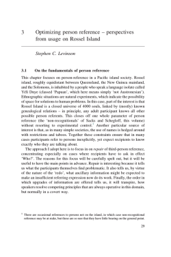
From www.bloodjournal.org by guest on February 6, 2015. For personal use only.
Scott Syndrome, Characterized by Impaired Transmembrane Migration of
Procoagulant Phosphatidylserine and Hemorrhagic Complications, Is an
Inherited Disorder
By Florence Toti, Nathalie Satta, Edith Fressinaud, Dominique Meyer, and Jean-Marie Freyssinet
An as yet single family with a bleeding historyis shown to
Epstein-Barrvirus-infectedBlymphocytes.Thedataare
present the characteristic lack of membrane expression of
compatible with homozygous status ofthe propositus and
procoagulantphospholipidsobserved in Scott syndrome.
heterozygousstatusofherchildren.
Scott syndrome apLow prothrombin consumption in the serum of the propospears to be transmitted as an autosomal recessive trait reitus, a 71-year-oldwoman, and two of her children was the
flecting the deletion or mutation of a putative outward phossole abnormal hemostasis parameter. The degree of expophatidylserine translocase. The detailed knowledge of this
sure of procoagulant phospholipids, chiefly phosphatidylser-transporter could have an impact in membrane physiology.
0 1996 by The American Society of Hematology.
he. was reduced in stimulated platelets, erythrocytes and
T
HE COMPLETION OF THE blood coagulation process
requires the surface assembly of the characteristic enzyme complexes involving several of the clotting factors.'
The exposure of negatively charged phospholipids, chiefly
phospatidylserine, sequestered in the inner leaflet of the
plasma membrane of resting cells, is a prerequisite for the
local concentration of actors playing a fundamental role in
the generation of thrombin. Stimulated platelets are thought
to be the major source of phosphatidylserine, previously referred to as platelet factor 3. When available at the surface
of activated cells or derived fragments, phosphatidylserine
acquires a procoagulant character due to its ability to complex vitamin K-dependent factors VII, IX, X, and 11, as well
as activated cofactors VI11 and V. The surface amplification
potential of such a system is linked to the feedback control
exerted by thrombin through retroactivation of cofactors VI11
and V and platelet ~timulation.'~~
Vitamin K antagonists enable efficient prevention of thromboembolic disorders by
interfering in the maturation process of vitamin K-dependent
clotting factors, resulting in an impaired capacity of the latter
to associate with procoagulant phospholipids.' The symmetrical approach consisting in the pharmacologic modulation of
the degree of exposure of phosphatidylserine by stimulated
platelets has been hampered by the lack of knowledge of the
mechanism governing its transmembrane migration.
The production of a procoagulant response is certainly not
the sole consequence of the loss of the asymmetric distribution of phospholipid species between the two leaflets of the
plasma membrane, but physiologic implications in cell types
other than blood cells remain poorly understood. Ubiquitous
cytodynamic events such as endocytosis or exocytosis are
accompanied by the loss of membrane a~ymmetry.~
Membrane fusion of enveloped viruses with target cells is facilitated in the presence of phosphatidyl~erine.~.~
Programmed
cell death, apoptosis, essential in many aspects of normal
development and occurring in major diseases such as cancer,
acquired immunodeficiency syndrome (AIDS), autoimmune
disorders, et^,^ results in the release of apoptotic fragments
bearing exposed pho~phatidylserine.~.~
Several protective
mechanisms could neutralize the pathogenic potential of circulating cell fragments. Phosphatidylserine is a determinant
of reticuloendothelial
and antiphospholipid antibodies could counteract its procoagulant character." Such
antibodies are precisely associated with an increased risk of
thrombosis."
The availability of procoagulant phospholipids is essential
Blood, Vol 87, No 4 (February 15). 1996: pp 1409-1415
for a normal hemostatic response, as clearly shown by the
severe to moderate bleeding episodes due to a deficiency of
platelet procoagulant activity.l3-I6Several isolated cases were
reported, but a unique one has been well documented as Scott
syndrome.17 The defect also results in decreased membrane
emission of procoagulant vesicles or microparticles.'s It is
not restricted to platelets but also affects red blood cells and
lymphocytes, suggesting that it reflects a mutation occurring
in multiple hematologic lineages.'' The possibility of an inherited disorder transmitted as an autosomal recessive trait
was discussed but not confirmed.I4 In a more recent report,
Epstein-Barr virus (EBV)-infected B cells from the first case
of Scott syndrome were fused with the myeloma cell line
UC-LUC, resulting in hybridomas in which the Scott abnormality was corrected. The investigators concluded to possible complementation of aberrant gene(s)." We report here
the first characterization of a familial Scott syndrome and
confirm that it is a genetic disorder affecting the outward
transmembrane migration of phosphatidylserine.
PATIENTS AND METHODS
Subject selection. The propositus [E51 (Fig l), a 71-year-old
woman born from first cousins, suffered from severe to moderate
hemorrhagic episodes for a long time, including epistaxis, traumarelated hematoma, bleeding after tooth extractions, and at childbirths.
She has been successfully treated using whole blood transfusions.
Her older sisters [11-1 and 11-31 died from hemorrhage in childbirth.
Her children [III-6 to 111-101, nephews [III-1 and 111-3 to 111-51, and
grandchildren [IV-I to IV-201 denied any bleeding tendency. No
From theInstitut d'He'matolgoie et Immunologie, Faculte' de Me'decine, Universite'Louis Pasteur,Strasbourg; the Laboratoire d'He'matologie, Institut de Biologie, CHU Hbtel-Dieu, Nantes; and the Unite'
I43 INSERM, Hbpital de Bicgtre, Le Kremlin-Bicgtre, France.
Submitted August 2, 1995; accepted September 28, 1995.
Supported by Grants from the Institut National de la Santd et de
la Recherche Me'dicale (CRE93-04-06), the Universite' Louis
Pasteur de Strasbourg (Equipe d'acceuil DREDno. 1313), the Fondation pour la Recherche Me'dicale, and the Fondation de France.
Address reprint requests to Jean-Marie Freyssinet, DSc, lnstitut
d'He'wtologieet
Imrnunologie, Faculte' de Mddecine, 4, rue
Kirschleger, F-67085 Strasbourg Cedex, France.
The publicationcosts of this article were defrayed in part by page
charge payment.This article must therefore be hereby marked
"advertisement" in accordance with I% U.S.C. section I734 solely to
indicate this fact.
0 1996 by The American Society of Hematology.
0006-4971/96/8704-0002$3.00/0
1409
From www.bloodjournal.org by guest on February 6, 2015. For personal use only.
TOTI ET AL
1410
I
II
J
111
IV
2F
1M
3M
ZF
M
ZF
other recurrent symptom was reported by any ofthe members of
this family. The nature of the study was explained to the patient,
one of her sons [ I I M ] , two of her daughters 1111-9 and 111-IO], two
ofher grandnieces [IV-l and !V-21 (nephew [Ill-11's daughters),
and I O control subjects who gave fully informed consent.
Hemosmsis pnrmneters. cell prepomtions. and culture. The hemostasis parameters were determined according to standard routine
procedures." For studies performed with cells, blood samples from
the propositus, her son [III-S], and hcr daughter [ I l l - I O ] were collected in National Institute of Health (NIH) formula A acid-citratedextrose ( I part anticoagulant:6 parts blood) and immediately processed for platelet"andredblood
cell isolation."' Lymphocytcs
were isolated under sterile conditions and stored in liquid nitrogen.
Infection of B lymphocytes by EBV (EBV B9S8, Marmouset) was
performed in RPM1 culture medium containing 20% fetal calf serum
(FCS; Life Technologies Inc, Paisley, U K ) in the presence of SO ngl
mL cyclosporine and was achieved within 3 weeks. EBV-infected B
cells were expanded in standard RPM1 culture medium supplemented
with S mmollL HEPES. S% FCS. S 8 Hyclone serum (Hyclone
Laboratories Inc. Logan, UT), and 50 nglmL gentamicin. In some
experiments, cells were cultured in the presence of recombinant
human interleukin-4 (!L-4; 1 nglmL; Promega Corp. Madison, W!)
to reduce mortality. Cell mortalitywas estimated by Trypan blue
staining (0.1% final concentration).
F~tnctioncrldetection of procorrgdnnt phospholipid.7. Procoagulant phospholipid exposure in stimulated cells and derived microparticles was detected using a human prothrombinase assay in which
phosphatidylserine promotes the activation of prothombin by factor
Xa in the presence of factor Va." Thrombin generated by assembled
prothombinase complex was measured using a chromogenic assay as
described el~ewhere.'~
Red blood cells, platelets, and EBV-infected
lymphocytes were studied at IO7 cellslmL, 7 X IO" cellslmL, and 2
X IO' cellslmL, respectively. The ability of each cell type to expose
procoagulant phosphatidylserine was examined after stimulation by
0 . 1 NIH UlmL human cy-thrombin'" + 12 pglmL equine tendon
collagen (Hormon-Chemie, Munich, Germany) or S pmollL calcium
ionophore A23187 for I O minutes at37°C for platelets, S pmol/L
A23 I X7 for 90 minutes at 37°C for red blood cells, S pmol/L A231 87
for I O minutes at 37°C for EBV-infected B cells. in the presence of
2 mmol/L external CaCI2 for each cell type. Red blood cells and
EBV-infected B lymphocytes were separated from derived microparticles by centrifugation at I2.000g for 30 seconds before the measurements. In each case. results were compared with the prothrombinase activities developed in samples from healthy volunteers. To
assess whether the phosphatidylserine content of any cell type was
normal, cells were disrupted by three successive freezinglthawingl
sonication steps, and the resulting cell fragment suspensions were
used in the prothrombinase assay as described above.
Flow c ~ t o m e t ~Analysis
~.
of the platelet membrane status was
,M
M
1F
1F
?F
,M
Fig 1. Pedigreeof the family with Scott syndrome. (Solid symbols1individualswith bleedingdisorders and elevated residualserum prothrombin
(measuredonly in the propositus111-51): (dotted symbok) individuals with elevated residualserumprothrombin (measured only insubjects 1111-81, 1111-91.
and 1111-101). Arrowsindicateindividualsexamined in
the whole study.
performed using annexin V as a protein probe of phosphatidylserine
Annexin V was purified from human placenta and
exp"ure,Z".'l.''.~h
conjugated to fluorcscein isothiocyanatc (FITC), as alrcady described." Ionophore-stimulated (see above) and nonstimulated cell
suspensions were incubated with S pglmL annexin V"'"' for IO
minutes at room temperature in the presence of 0.5 mmollL calcium
before data acquisition using a BectonDickinsonflow cytometer
(Becton Dickinson. San Jose, CA). Parameters weresetto assess
phosphatidylserine exposure evidenced by annexin V""' binding
andrecordedon
FLI Huorescencc scale, simultaneously for cells
and derived microparticles that were distinguished according to size
(forward light scatter). For each samplc. 10.000 events were recorded
and further analyzed in the appropriate gate delineated on dot plots.
as previously described."
Analysis of the ability of red blood cells to undergo shape change
and membrane vesiculation after stimulation by ionophore (see
above) was performed using forward-angle versus right-angle light
scatter dot plot representation" of I O . 0 0 0 events per sample.
Cytoske/eto/ protein onc11y.vis. Cytoskeletal proteins
of
ionophore-stimulated and unstimulated platelets were analyzed by polyacrylamide gel electrophoresis in thepresenceofsodiumdodecyl
sulphate, as described elsewhere.''
RESULTS
Table 1 is a representative example of the various assessments of the hemostasis parameters concerning the propositus [H-S], her daughter [Ill-IO], andherson [111-8], who
were investigated in this study (Fig l ) . The only abnormal
values are those of serum prothrombintimeand
consequently residual serum prothrombin. The patient had 68%
of residual prothrombin. whereas the levels of her children
was lower, but consistently higher than normal at 7.5% for
the daughter and 5.5% for the son. The propositus' other
daughter [W91could be examined only once and had 5.5%
residual serum prothrombin and no other anomaly. The two
grandnieces, nephew [Ill- 11's daughters, had normal hemostasis parameters.
In the absence of stimulation, platelets or erythrocytes
from the propositus exhibited a low prothrombinase activity.
This was also true for control samples, reflecting minimal
availability of negatively charged phospholipids for prothrombinase complex assembly in each case. Exposure of
procoagulant phospholipids remainedweak after various
stimulations of propositus' cells when compared with samples from healthy subjects (Figs 2 and 3). Prothrombinase
activities in stimulated propositus' red blood cells and corre-
From www.bloodjournal.org by guest on February 6, 2015. For personal use only.
INHERITED S C O T SYNDROME
U)
c
15
141 1
c] UnStd
T
Thr/Coll
Son
Controls
Propositus
Daughter
Fig 2. Procoagulant phospholipidexternalization in stimulated
platelets from the propositus
111-51 measured by prothrombinase
or
assay. Platelet suspensions wereeitherunstimulated(UnStd)
stimulated by the
association of thrombin andcollagen (Thr/Coll; 0.1
NIH UlmL and 0.12 pg/mL final respective concentrations1 for 10
minutes at 37°C or by the calcium ionophore A23187(A23187; 5
pmol/L final concentration) for10 minutes at37°C. Stimulations and
prothrombinase activitymeasurements were performed in the presence of 2 and 1 mmol/L external CaCI,, respectively. Data are expropospressed as the mean? SEM; n = 3 for the platelets from the
subjects.
itus andher offspring, and n = 10 for platelets from control
sponding supernatants were 7 to 10 times lower than those
measured with control cells.
Platelets from the patient’s son and daughter displayed a
35% to 45% reduction, respectively, of prothrombinase activity when compared with control cells after stimulation by
calcium ionophore (Fig 2). Such a reduction could have
been somewhat underestimated because measurements were
performed under drastic activation conditions.
Table 1. Assessment of theHemostasis Parameters in the
propositus [11-5], Her Son [111-81, and Her Daughter [111-101
Propositus
11-5
Son
Daughter
111-8
Ill-l0
Bleeding
(min)
time
3.30ND ND
Platelet count (giga/L) 150-500
237
223
395
Prothrombin
time
(S)
12-14
11.9
11.9
12.7
33-38
aPlT (S)35
34
38
Serum prothrombin
15.7 15.2 11.8
time ( S )
Residual serum
68 7.5
5.5
prothrombin (%)
Coagulation factors
105 72 102
FVlll (%)
>l20
ND
FV (%)
94
ND
FIX (%)
ND
>l20ND
FXll (%)
vWF (ristocetin cofactor)
(%)
118 50-20090
Fibrinogen
2-4 (g/L)
2.96
3
3.5
Abbreviation: ND, not determined.
The defect of phosphatidylserine exposure was more easily evidenced in red blood cells that elicit a delayed response
to calcium ionophore.’’ Erythrocytes from the propositus
were almost insensitive to ionophore treatment. Stimulated
red blood cells from the daughter exhibited about twice as
much prothrombinase activity as those of her mother,
whereas the son’s cells produced a response of about 60%
of normal (Fig 3). Diminished prothrombinase activity was
also observed in supernatants of corresponding stimulated
red blood cells from the three members of the family, testifying to a reduced ability to shed procoagulant microparticles. Again, the propositus’ cells showed the most pronounced defect.
Procoagulant phospholipid exposure occurred at comparable basal levels in nonstimulated control and patient’s EBVinfected B lymphocytes (Fig 4), probably as a result of cell
mortality, which was estimated between 10% and 15% in
both populations, depending on the culture stage. Mortality
could be reduced by about 30%, with concomitant reduction
of the same order of basal prothrombinase activity, when
cells were cultured in the presence of IL-4 (data not shown).
Surprisingly, the mortality of offspring’s nonstimulated
EBV-B cells was consistently lower, ranging between 3%
and 8%, resulting in lower basal prothrombinase activity.
Ionophore treatment of control EBV-B cells was responsible
for the development of a substantial phospholipid-dependent
procoagulant activity almost equally distributed between
cells and microparticles. The overall prothrombinase activity
(cell and microparticle-associated) of the stimulated pa-
l
0 UnStd RBC
SN from UnStd RBC
1 Std RBC
SN from Std RBC
0.5
Normal
Values
2.30-9.30
0
>25
<5
ND
ND
75
50-200
80-120
70-120
80-120
Controls
Propositus
Daughter
Son
Fig 3. Procoagulant phospholipid externalizationin calcium iono[11-51, her
phore-stimulatedredblood
cells fromthepropositus
daughter [111-101, and her son [Ill-81 measured b y prothrombinase
assay. Thrombin generated in the presence of unstimulated red blood
cells (Unstd RBC) and corresponding supernatant (SN from UnStd
RBC). Thrombin generated in the presence of stimulated cells (Std
RBC) and corresponding supernatant (SN from Std RBC). Cells were
stimulated by 5 pmollL calcium ionophore A23187 in the presence
of 2 mmol/L externalCaCI, for 90 minutes at37°C. The CaCI2concentration was 1 mrno1lL in the prothrombinase assay. Data are expressed as the mean 2 SEM; n = 4 for cells from the propositus and
her offspring, and n = 10 for cells from controlsubjects.
From www.bloodjournal.org by guest on February 6, 2015. For personal use only.
TOTI ET AL
1412
4-
0
0 UnStd
H A23187
h
v)
7-
her offspring’s cells elicited an intermediate response (Fig
6). The dot plot representation of forward-angle versus rightangle light scatter enabled us to clearly distinguish two populations. Cells showed the highest forward and side scatter
signals, whereas derived microparticles had lower ones.’’
Less than 1.5% of total events (cells + debris and/or machine
noise) was detected in the microparticle gate in any of unstimulated cell types. With stimulated red blood cells, the
proportion of microparticles was, respectively, 19.S%, 5%,
IO%, and 8% for the normal subject’s, propositus’, daughter’s, and son’s cells.
No difference was noticed in the electrophoretic patterns
of cytoskeletal proteins of patient’s and control platelets
(data not shown).
DISCUSSION
Cells MP
Cells MP
Cells MP
Cells MP
Controls
Propositus
Daughter
Son
Fig 4. Procoagulant phospholipidexternalization in calcium ionophore-stimulated EBV-infected B cellsfrom the propositus [ll-51, her
daughter [111-101, and herson[Ill-81measured
by prothrombinase
assay. Thrombin generated in the presence of unstimulated (UnStd)
or ionophore-stimulatedcells (A231871 and corresponding shed microparticles (MP). Stimulation was achieved in the presenceof5
pmol/L ionophore and 2 mmollL external CaCIz for 10 minutes at
37°C. The CaCI, concentrationwas 1 mmollL in the prothrombinase
assay. Data ere expressed as the mean 2 SEM; n = 4 for cells from
the propositus and her offspring, and n = 10 for control cells.
tient’s, daughter’s, and son’s EBV-B cells was, respectively,
2S%, 44%, and 53% that of control cells. These values would
be, respectively, 15%,’47%, and 56% if the basal corresponding activities were deduced. Microparticle-associated procoagulant activity was almost unaffected by ionophore treatment of propositus’ cells, whereas in her offspring’s cells it
remained lower than half of that of the cells. When any of
these EBV-B cells including control cells were disrupted,
the overall prothrombinase activity was about twice that induced by ionophore in control cells, showing that the absolute values were similar.
Disrupted platelets, red blood cells, and EBV-infected B
cells from the propositus and her children yielded the same
prothrombinase activity as thatof corresponding control cells
treated likewise, showing a normal content of procoagulant
phospholipids.
Flow cytometry analysis using annexin VFITCallowed us
to gather evidence regarding the defective exposure of phosphatidylserine and microparticle shedding bythe patient’s
platelets compared with control platelets exceedingly stimulated by calcium ionophore (Fig S). The latter underwent
massive phosphatidylserine exposure and membrane vesiculation. The patterns corresponding to the binding of annexin
Vmc to stimulated cells and derived microparticles were in
accordance with prothrombinase activity measurements (Fig
2).
Red blood cells from the patient appeared refractory to
shape change and showed reduced vesiculation after treatment with S pmol/L calcium ionophore for 90 minutes at
37°C in the presence of 2 mmoYL external Ca2’, whereas
The above results clearly establish close links between the
almost total absence of phosphatidylserine exposure byin
vitro stimulated platelets from the propositus and her hemorrhagic diathesis. The defect was observed not only with the
physiologically relevant agonist combination of thrombin
and collagen, but also under more drastic conditions using
calcium ionophore. The latter was not able to induce significant phosphatidylserine externalization in patient’s red blood
cells when compared with corresponding control cells. The
reduction of procoagulant phospholipid exposure was also
highly significant in her EBV-infected B cells despite interference of cell mortality. Two of her children could be explored for the same anomaly. The overall phospholipid-dependent procoagulant response did not exceed SO% to SS%
of normal with the daughter’s cells, whereas the son’s cells
showed a behavior closer to normal but consistently lower.
These observations are in agreement with measurements of
residual prothrombin in serum and have been confirmed by
flow cytometry. It has to be emphasized that residual serum
prothrombin is a complex function of the degree of availability of procoagulant lipids in whole blood. This could explain
the differences of serum prothrombin time and residual serum prothrombin between the propositus and her children.
The absence of bleeding in the offspring might precisely be
related to residual serum prothrombin close to normal. In
our routine practice, under no circumstance of unexplained
hemorrhagic disorder, these parameters were found to be
abnormal except in this family. The defect of phosphatidylserine exposure and consecutive reduced membrane vesiculation was further evidenced by the reduced binding of a
fluorescent derivative of annexin V used to detect available
phosphatidylserine. It has to be pointed out that none of
the above defects could be due to abnormal composition of
membrane phospholipids because patient’s cell fragments
bearing fully scrambled phospholipids produced a phosphatidylserine-dependent procoagulant response identical to that
of control cells when lysed likewise.
In a first instance, these observations appear compatible
with homozygous status of the propositus while her children
could be heterozygous with respect to Scott syndrome, which
wouldbe transmitted as an autosomal recessive trait. The
patient’s sisters can reasonably be considered as homozygous because they died from documented hemorrhage. The
From www.bloodjournal.org by guest on February 6, 2015. For personal use only.
1413
INHERITED SCOTT SYNDROME
3Normal + A23187
;
I
Propositus + A23187
Fig 5. Flow cytometry analysisofphosphatidylserine externalization and membrane microparticle
shedding in calcium ionophore-stimulated platelets
from the propositus[L51 and normal subject shown
by annexin VFmclabeling. Dot plot representations
of platelet suspensions showing size parameters on
forward light scatter scale and green fluorescence
associated to annexin VFmcbound to phosphatidylserine in cells and microparticles. MP, microparticle
gates with the proportion ofshedmicroparticles;
A23187,calcium ionophore-stimulated cells.Platelets were stimulated by 5 pmol/L ionophore in the
presence of 2 mmollL external CaCI, for 10 minutes
at 37°C. Thesepatterns correspond to a single analysis representative of results obtained in four independent experiments performed likewise.
b
Green fluorescence of bound annexin-VFITC
possibility of compound heterozygous status appears improbable because it should have been transmitted to the three
sisters. However, it has to be emphasized that such conclusions should be viewed as only partly qualified because the
genetic analysis could not be performed due to the absence
of candidate gene(s). In subjects born from first cousins,
' / , h of the genome has a homozygous character that implies
that at least six or seven subjects of this type have to be
examined for localization of mutated gene or regulatory element(s) by mapping homozygous loci. Unfortunately, such
a study could not be performed here for obvious reasons.
The first experimental evidence that a gene defect can give
rise to Scott syndrome was reported recently." Our familial
study confirms that it is indeed a genetic disorder. The identification of candidate gene(s) is now the next goal but remains
dependent on the feasibility of functional rescue methodology in Scott EBV-B cell lines.
The maintenance of the asymmetric distribution of phospholipid species between the two leaflets of the plasma membrane of a variety of cells involves a specific inward
transporter of aminophospholipids, chiefly phosphatidylethanolamine andphosphatidylserine:Although
the gene
coding for this transporter remains to be identified," it is
tempting to speculate that aminophospholipid translocase activity is increased in Scott syndrome. This would explain
the sustained sequestration of phosphatidylserine in the inner
membrane leaflet. However, experiments performed by other
investigators who had access to the red blood cells from the
above Scott subjects excluded this possibility as the inward
phosphatidylserine transport was found normal. In addition,
(arbitrary units)
once incorporated byred blood cells from the propositus,
labeled phosphatidylserine could not be re-externalized (A.
Zachowski and J.-C. Sulpice, personal communication, February 28, 1995). Identical preliminary observations were also
reported by another group who had access to the cells of the
first case of Scott syndrome."
The flip-flop or transmembrane migration of phospholipids remains mostly mysterious.'.'' Nevertheless, a phosphatidylcholine translocase has been recently characterized using
a gene disruption approach. It is the product of the mdr2
gene belonging to the family of P-glycoprotein multidrug
transporters. Its absence leads to liver disease due to the lack
of phosphatidylcholine in bile.'" Because aminophospholipid
translocase activity appears normal in Scott red blood cells,
it seems reasonable to hypothesize that the Scott defect could
be due to a mutation affecting a putative outward phosphatidylserine or aminophospholipid translocase. Another possibility would be that aminophospholipid translocase operates
as both an inward and outward transporter, because several
pumps do, depending on particular regulation. In this case,
only the outward function would be impaired. This hypothesis could be explored using cell mutants defective in inward
phosphatidylserine transport recently described.3n
A more general perspective of the identification of the gene(s)
and corresponding product(s) involved in phosphatidylserine or
minephospholipid externalization is to assess whether it is a
moreubiquitous feature involved in theprocessesregulating
communication between cells and their environment under
various physiologic or pathologiccircumstances. In this respect,
Scott syndrome would be an additional example of the prime
From www.bloodjournal.org by guest on February 6, 2015. For personal use only.
TOTI ET AL
1414
Control
A23187
Normal
Propositus
.
.
.*h;. .
'
I
.
'
SSC (Side light Scatter)
usefulness of genetic variants for the characterization of as yet
unknown physiologic mechanisms.
ACKNOWLEDGMENT
The authors dedicate this publication to Prof M.-J. Lameu and
are grateful to Family M... and Dr P. Roger for their kind cooperation. The expert technical assistance of M.-L. Temen is gratefully
acknowledged. We are indebted to Prof D. Hurez (Laboratoire d'lmmunologie, CHU d'Angers) for facilitated access to laboratory
equipment for experiments thathadto be performed on site. We
express our gratitude to Drs A. Zachowski and J.-C. Sulpice for
measurements of aminophospholipid translocase activity. We thank
Prof J.-L. Pasquali for helpful advice for EBV infection of B cells,
and Dr G.Lombard-Platet and Prof J.-L. Mandel for critical comments.
REFERENCES
1. Furie B, Furie BC: Molecular and cellular biology ofblood
coagulation. N Engl J Med 326:800, 1992
Fig 6. Flow cytometry analysis of phosphatidylserine externalization and membrane microparticle
shedding in calcium ionophore-stimulated erythroIll-51, her daughter [Ill-101,
cytes from the propositus
her son [111-81, and a normal subject. Dot plot representations of cell
suspensions showing size parameters on forward light scatter scale (arbitrary units)
versus right-angle scatter. Cells show the highest
forward and side scatter signals, whereas derived
microparticles have lower ones (see Results for proportions). Stimulation was
achieved in thepresence
of 5 pmol/L ionophore and2 mmol/L external CaCll
for 90 minutes at37°C. These patterns correspond t o
a single analysis representative of results obtainedin
six independent experiments performed likewise.
2. Zwaal RFA, Comfurius P, Bevers EM: Mechanisms and function of changes in membrane-phospholipid asymmetry in platelets
and erythrocytes. Biochem SOCTrans 2 I :248, 1993
3. Mann KG. Nesheim ME, Church WR, Haley P, Krishnaswamy
S : Surface-dependent reactions of the vitamin K-dependent enzyme
complexes. Blood 7 6 I , 1990
4. Devaux P F Static and dynamic lipid asymmetry in cell membranes. Biochemistry 30:1163, 1991
5. Hoekstra D, Kok JW: Entry mechanisms of enveloped viruses.
Implications for fusion of intracellular membranes. BiosciRep
9:273, I989
6. Yamada S , Ohnishi S : Vesicular stomatitis virus bindsand
fuses with phospholipid domain on target cell membrane. Biochemistry 25:3703, 1986
7. Thompson CB: Apoptosis in the pathogenesis andtreatment
of disease. Science 267:1456, 1995
8. Savill J, Fadok V, Henson P, Haslett C: Phagocyte recognition
of cells undergoing apoptosis. Immunol Today 14:131, 1993
9. Koopman G,Reutelingsperger CPM, Kuijten GAM, Keehnen
RMJ, Pals ST, van Oers MHJ: Annexin V for flow cytometric detec-
From www.bloodjournal.org by guest on February 6, 2015. For personal use only.
INHERITED S C O T SYNDROME
tion of phosphatidylserine expression on B cells undergoing
apoptosis. Blood 84:1415, 1994
10. Allen TM, Williamson P, Schlegel R: Phosphatidylserine as
a determinant of reticuloendothelial recognition of liposome models
of the erythrocyte surface. Proc Natl Acad Sci USA 85:8067, 1988
11. Galli M, Bevers EM, Comfurius P, Barbui T, Zwaal RFA:
Effect of antiphospholipid antibodies on procoagulant activity of
activated platelets and platelet-derived microvesicles. Br J Haematol
83:466, 1993
12. Santoro SA: Antiphospholipid antibodies and thrombotic predisposition: Underlying pathogenic mechanisms. Blood 83:2389,
1994
13. Weiss HJ, Vicic W, Lages B, Rogers J: Isolated deficiency
of platelet procoagulant activity. Am J Med 67:206, 1979
14. Miletich JP, Kane WH, Hofmann SL, Stanford N, Majerus
PW: Deficiency of factor Xa-factor Va binding sites on the platelets
of a patient with a bleeding disorder. Blood 54:1015, 1979
15. Rosing J, Bevers EM, Comfurius P, Hemker HC, van Dieijen
G, Weiss W,Zwaal RFA: Impaired factor X and prothrombin activation associated with decreased phospholipid exposure in platelets
from a patient with a bleeding disorder. Blood 65:1557, 1985
16. Ahmad S S , Rawala-Sheikh R, Ashby B, Walsh PN: Platelet
receptor-mediated factor X activation by factor IXa. High-affinity
factor IXa receptors induced by factor VI11 are deficient on Platelets
in Scott syndrome. J Clin Invest 84:824, 1989
17. Weiss HJ: Scott syndrome: A disorder of platelet procoagulant activity. Semin Hematol 31:312, 1994
18. Sims PJ, Wiedmer T, Esmon CT, Weiss HJ, Shattil SJ: Assembly of the prothrombinase complex is linked to the vesiculation
of the platelet plasma membrane. Studies in Scott sydrome: An
isolated defect in platelet procoagulant activity. J Biol Chem
264:17049, 1989
19. Bevers EM, Wiedmer T, Comfurius P, Shattil SJ, Weiss HJ,
Zwaal RFA, Sims PJ: Defective Caz+-inducedmicrovesiculation and
deficient expression of procoagulant activity in erythrocytes ofa
patient with a bleeding disorder: A study of the red blood cells of
Scott syndrome. Blood 79:380, 1992
20. Kojima H, Newton-Nash D, Weiss HJ, Zhao J, Sims PJ,
Wiedmer T Production and characterization of transformed B-lymphocytes expressing the membrane defect of Scott syndrome. J Clin
Invest 94:2237, 1994
1415
21. Dachary-Prigent J, Freyssinet JM, Pasquet JM, Canon JC,
Nurden A T Annexin V as a probe of aminophospholipid exposure
and platelet membrane vesiculation: A flow cytometric study showing a role for free sulfhydryl groups. Blood 81:2554, 1993
22. Connor J, Bucana C, Fidler IJ, Schroit AJ: Differentiationdependent expression of phosphatidylserine in mammalian plasma
membranes: Quantitative assessment of outer-leaflet lipid inprothrombinase complex formation. ptoc Natl Acad Sci USA 86:3184,
1989
23. Satta N, Toti F, Feugeas 0, Bohbot A, Dachary-Prigent J,
Eschwbge V, Hedman H, Freyssinet JM: Monocyte vesiculation: A
mechanism for dissemination of membrane-associated procoagulant
activities and adhesion molecules following stimulation by lipopolysaccharide. J Immunol 153:3245, 1994
24. Freyssinet J M , Gauchy J, Cazenave J P : The effect of phospholipids on the activation of protein C by the human thrombinthrombomodulin complex. Biochem J 238:151, 1986
25. Thiagarajan P, Tait JF: Binding of annexin V/placental anticoagulant protein I to platelets. Evidence for phosphatidylserine exposure in the procoagulant response of activated platelets. J Biol Chem
265:17420, 1990
26. Ravanat C, Archipoff G, Beretz A, Freund G, Cazenave JP,
Freyssinet JM: Use of annexin V todemonstrate the role of phosphatidylserine exposure in the maintenance of haemostatic balance by
endothelial cells. Biochem J 282:7, 1992
27. Auland ME, Roufogalis BD, Devaux PF, Zachowski A: Reconstitution of ATP-dependent aminophospholipid translocation in
proteoliposomes. Proc Natl Acad Sci USA 91:10938, 1994
28. Higgins CF: Flip-Flop: The transmembrane translocation of
lipids. Cell 79:393, 1994
29. Smit JJM, Schinkel AH, Oude Elferink RPJ, Groen AK, Wagenaar E, van Deemter L, Mol CAAM, Ottenhoff R, van der Lugt
NMT,van Roon, MA, van der ValkMA, Offerhaus GJA, Bems
MM, Borst P Homozygous disruption of the murine mdr2 P-glycoprotein gene leads to a complete absence of phospholipid from bile
and to liver disease. Cell 75:451, 1993
30. Hanada K, Pagano RE: A Chinese hamster ovary cell mutant
defective in the non-endocytic uptake of fluorescent analogues of
phosphatidylserine: Isolation using a cytosol acidification procedure.
J Cell Biol 199793, 1994
From www.bloodjournal.org by guest on February 6, 2015. For personal use only.
1996 87: 1409-1415
Scott syndrome, characterized by impaired transmembrane migration
of procoagulant phosphatidylserine and hemorrhagic complications,
is an inherited disorder
F Toti, N Satta, E Fressinaud, D Meyer and JM Freyssinet
Updated information and services can be found at:
http://www.bloodjournal.org/content/87/4/1409.full.html
Articles on similar topics can be found in the following Blood collections
Information about reproducing this article in parts or in its entirety may be found online at:
http://www.bloodjournal.org/site/misc/rights.xhtml#repub_requests
Information about ordering reprints may be found online at:
http://www.bloodjournal.org/site/misc/rights.xhtml#reprints
Information about subscriptions and ASH membership may be found online at:
http://www.bloodjournal.org/site/subscriptions/index.xhtml
Blood (print ISSN 0006-4971, online ISSN 1528-0020), is published weekly by the American
Society of Hematology, 2021 L St, NW, Suite 900, Washington DC 20036.
Copyright 2011 by The American Society of Hematology; all rights reserved.


© Copyright 2026