
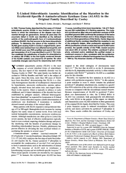
Multiple G6PD Mutations Are Associated With a Clinical and
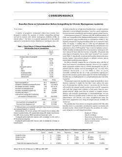
From www.bloodjournal.org by guest on February 6, 2015. For personal use only.
Multiple G6PD Mutations Are Associated With a Clinical and Biochemical
Phenotype Similar to That of G6PD Mediterranean
By Maria Domenica Cappellini, Franco Martinez di Montemuros, Gianluca De Bellis, Silvana Debernardi,
Chiara Dotti, and Gemino Fiorelli
Glucose-&phosphatedehydrogenase (GGPD) deficiency, one
of the most common red cell abnormalities. is characterized
by a wide clinical, biochemical, and molecularheterogeneity.
In this study we have determined the molecular basis of
GGPD deficiency in a sample of 70 male subjects, originating
from different parts of Italy, who all shared a clinical and
biochemical phenotype identical or very similar to that of
G6PD Mediiterranean, the most common variant in Italy. In
59 cases (84%) we found the mutation 563 C --t T, previously
known to be underlying the GGPD Mediterranean and the
two polymorphic variants GGPD Cagliari and GGPD Sassari.
From the remaining 11 we amplifiedthe entire coding region
of G6PD in 8 different fragments and subjected them to nonradioactive single-strand conformation analysis. Direct sequencing was then performed on abnormal fragments. By
this approach we found six cases (8.5%) with 1360 G --t A
mutation (GGPD Union) and two cases (2.8%) with 1376
G --* C (GGPD Cosenza). In the remaining three samples we
found two other mutations: 1342 A G (two cases, 2.8%)
and 1052 G --t T (one case, 1.4%). These two molecular defects have never been described before and were designated
by us as GGPD S. Antioco and G6PD Partenope, respectively.
Haplotype analysis suggested that all the non-Mediterranean mutations occurred independently on a normal GGPD
allele. This study shows that the G6PD Union mutation has
a high polymorphic frequency in the Italian population and
that the genetic heterogeneity of GGPD Mediterranean-like
variants is higher at the molecular level than expected from
biochemical characterization.
0 1996 by The American Society of Hematology.
G
phic variants also have similar clinical manifestations; although they are silent most of the time, they are responsible
for neonatal jaundice or favism. A previous study showed
that these three variants are due to the same single C -+ T
transition at nt 563 (exon 6), which results in a serine to
phenylalanine amino acid shift at position 188 together with
a C + T mutation at nt 1311 (exon ll),’ a polymorphic site
also present in approximately 20% of a normal Mediterranean p~pulation.~
Recently, Corcoran et allo described a 592‘ + mutation
responsible for a G6PD Mediterranean-like variant named
G6PD Coimbra, suggesting that the Mediterranean variant
may have a molecular heterogeneity higher than expected
from the biochemical and clinical features. Although the
563‘ + and 131 1‘
mutations have been detected in the
Mediterranean variant in other
the original
report described this molecular abnormality only in two
cases,’ and no extensive studies have been performed in a
larger population in Italy. Here we report the prevalence of
the nt 563 and 1311 mutations in Italian subjects having
LUCOSE-6-PHOSPHATE dehydrogenase (G6PD;
E.C. 1.1.1.49.) is the key enzyme of the pentose phosphate pathway and provides the NADPH essential for a number of biosynthetic and detoxifying reactions. G6PD deficiency is the most common enzymopathy of humans;
originally described as a single disorder, it soon became
evident that it occurs in many populations and also that its
clinical manifestations vary widely, suggesting a possible
biochemical heterogeneity.’ For these reasons various attempts have been made in the past to standardize the methods
for measuring the kinetic properties of this intriguing enzyme.’ According to the suggested World Health Organization (WHO) o rite ria,^ so far more than 400 different G6PD
variants have been described, grouped in five classes on
the basis of their activity and clinical manifestations. More
recently, the cloning and sequencing of the G6PD gene has
prompted a search for mutations responsible for such a wide
number of variants, but, to date, only approximately 80 different point mutations have been found. A relation between
a single point mutation and a single biochemical variant
could have been expected, but this turned out not to be
the case as some of the variants previously described as
biochemically distinct seem to be identical at the molecular
level and vice versa various biochemical variants share the
same m ~ t a t i o nStudies
.~
carried out on the Italian population
showed that G6PD Mediterranean is the most common variant in Italy, followed by two polymorphic variants named
G6PD Cagliari and G6PD Sassari. The activities of all three
variants is less than 10% of that of the normal enzyme; the
residual enzyme has peculiar kinetic properties such as an
increased use of substrates analogues and a low Michaelis
constant for the substrate glucose-6-phosphate (G6P).5
G6PD Mediterranean, Cagliari, and Sassari are distinguishable from each other mainly by the elution peak of purified
residual enzyme from a DEAE Sephadex A50 column by a
KCl gradient but also by slight differences in Km values
and substrates analogue utilization: The normal enzyme is
eluted at 230 (22) mmol/L KC1 and G6PD Mediterranean,
Cagliari and Sassari are eluted at 234 (+-3), 212 (t5), and
249 (22.5) mmol/L KCl, respectively.’ The three polymorBlood, Vol 87, No 9 (May 1). 1996 pp 3953-3958
-+
-
From Cenrro Anemie Congenite, Istituto di Medicina fnterna e
Fisiopatologia Medica, Universita di Milano, Ospedale Maggiore
di Milano, IRCCS; Istituto di Tecnologie Biomediche Avanzate, Consiglio Nazionale delle Ricerche, Milano, Italy.
Submitted March 20, 1995; accepted December 27, 1995.
Supported by a grant from Progetto Finalizzato CNR Ingegneria
Genetica Malattie Ereditarie to G.F. and by MURST 60%. F.M. di
M. is a scholar of CNR.
Presented in part at the 36th Annual Meeting of the American
Society of Hematology, Nashville, TN.
Address reprint requests to Maria Domenica Cappellini, MD,
Istituto di Medicina Intema e Fisiopatologia Medica, Pad. Litta,
Ospedale Maggiore Policlinico, IRCCS, via F. Sforza 35, 20122
Milano, Italy.
The publication costs of this article were defrayed in part by page
charge payment. This article must therefore be hereby marked
“advertisement” in accordance with 18 U.S.C. section 1734 solely to
indicate this fact.
0 1996 by The American Society of Hematology.
OOO6-4971/96/8709-O021$3.O0/0
3953
From www.bloodjournal.org by guest on February 6, 2015. For personal use only.
3954
CAPPELLINI ET AL
G6PD deficiency with G6PD Mediterranean-like variants, as
well as the presence of four other mutations also responsible
for variants with a clinical and biochemical phenotype similar to that of G6PD Mediterranean. Two of the latter have
never been described in the G6PD coding region.
MATERIALS AND METHODS
Subjects. From January 1986 to January 1994, we collected 324
cases of G6PD deficiency. All the subjects were of Italian ancestry
and most of them came to our attention because of a family history
of favism, geographical origin, and/or a previous hemolytic crisis.
Among them, 129 (39.8%) had a severe deficiency (activity <IO%
of normal, WHO class 1 and 2), 55 (17%) a moderate deficiency
(activity >15% of normal, WHO class 3), and 140 (43.2%) were
female heterozygotes. Seventy of the 129 men (16 from Northern
Italy, 17 from Southern Italy and 37 from Sardinia) with severe
G6PD deficiency were enrolled in this study for biochemical and
molecular characterization. After obtaining informed consent from
each patient, 15 mL of whole blood was collected in Acid-citratedextrose, for biochemical characterization of residual enzyme and
DNA analysis.
Enzyme puri5cation and biochemical characterization. G6PD
purification was performed as previously described, using a twostep method based on affinity chromatography on 2‘5’-ADP Sepharose 4B (Pharmacia, Uppsala, Sweden) followed by automated
anion-exchange chromatography on a DEAE 5PW column (Wat e r ~ ) .Characterization
’~
of G6PD was carried out on purified enzyme
preparations according to WHO and ICSH recommendations.’.’+‘The
following variables were determined: activity, electrophoretic mobility (performed in three different buffers), Michaelis constants for
substrates (KmG6P,K,NADP), inhibition constant (K,) for NADPH,
percentage utilization of substrate analogues (2dG6P, Gal6P,
dNADP), optimum pH, thermostability, KCI gradient elution peak.
A mixture of equal amounts of purified enzyme from three subjects
with G6PD Mediterranean, Cagliari, and Sassari was submitted to
the KCI gradient as previously described.’
mutation by polymerase chain reaction
Detection of 563‘
(PCR) and endonuclease cleavage. DNA was purified from leukocytes by standard methods. A 547 bp fragment encompassing exons
6 and 7 was amplified from genomic DNA by PCR with primers
described previously.” The synthetic oligonucleotides were assembled on a Cyclone DNA synthesizer (Biosearch, San Rafael, CA).
The amplification reaction was performed in I O mmoUL Tris-HCI
pH 8.3, 1.5 mmol/L MgCI2, 200 pmoVL of each dNTP, 20 pmol of
each primer in the presence of 0.5 p g of DNA, and 2.5 U of Taq
polymerase (Perkin Elmer-Cetus, Nonvalk, CT) in a final volume
of 50 pL. The reactions were carried out using a DNA Thermal
Cycler (Perkin Elmer-Cetus) for 32 cycles as follows: denaturation
at 92°C for 1 minute, annealing at 58°C for 1 minute, elongation at
72°C for 2 minutes, and a last elongation at 72°C for 10 minutes.
The amplified fragment was digested overnight at 37°C with I O U
of endonuclease Mboll (Amersham, UK) in order to detect the C +
T transition at nt 563. Restriction fragments were analyzed on a 3%
agarose gel containing ethidium bromide.
PCR-single-strand conformation polymorphism (SSCP) analysis.
In an attempt to detect mutations other than 563‘
in the G6PD
gene, the entire coding region was PCR-amplified in eight different
fragments using pairs of primers described previously.” All the
amplified exons were then submitted to nonradioisotopic SSCP analysis.“,” Briefly, 3 pL of the amplified product was added to 27 p L
of 92% formamide, 20 mmol/L EDTA, and 0.05% bromophenol
blue, denatured at 98°C for 10 minutes, and quenched on ice for 2
minutes, Ten to 15 fiL of this mixture was loaded on a 6% to 8%
polyacrylamide mini-gel (0.75 mm X 6 cm X 8 cm; acry1amide:bis
-
-
38:1.S), run at IS0 volts, and silver stained. The staining procedure
was performed according to Bassam et allHwith some personal modifications as follows: 4 minutes in 1 % nitric acid (fixing), 2 minutes
in deionized water, I O minutes in 0.2% wt/vol silver nitrate/0.037%
formaldehyde (staining), 5 minutes in 3% wt/vol sodium carbonate/
0.037% formaldehyde/0.002% sodium thiosulfate (developing), 2
minutes in 10% acetic acid (stopping). The gel was then dried for
further conservation.
Detection ojf31 I‘ ’ polymorphism. This silent mutation was
screened by PCR-SSCP analysis of exons I O and 11 as previously
described, using a positive sample determined by sequence analysis
and a normal sample as markers.”
DNA sequencing. A method based on the avidin-biotin system
was used to obtain single-strand DNA. PCR amplification was performed using the sense biotinylated oligonucleotide CTGAGAGAGCTGGTGCTA and the anti-sense oligonucleotide GGAGAGGCATGAGGTAGC spanning a region 733 bp long encompassing exons
I O to 12. The PCR product was incubated with pre-washed Dynabeads M-280 Streptavidin (Dynal, Oslo, Norway) for I O minutes at
room temperature, mixing occasionally to allow almost complete
binding of the biotinylated DNA fragment. The supernatant was then
removed using a magnetic device to retain the beads (MPC-E, Dynal), and the remaining DNA pellet was denatured with 0.1 mol/L
NaOH. After the supernatant removal, the Dynabeads-single-strand
DNA complex was washed once with sterile water and finally resuspended in I O p L of distilled water. The whole Dynabedds-singlestrand DNA complex was used as template for the sequencing
reaction performed with 5 pmol of the specific antisense internal
fluorescent primer CATAGCCCACAGGTATGC placed 4 I bp
downstream from the end of exon 11. The sequence reaction was
performed by the Sanger dideoxy termination method with the Auto
Read Sequencing Kit (Pharmacia LKB, Uppsala, Sweden) designed
for use with the Automated Laser Fluorescent DNA Sequencer
(Pharmacia LKB).”’.”
Haplotype analysis. Haplotype analysis was canied out on different amplified fragments encompassing exon 4, exon 5, IVS 5 , IVS
8, and exons I O and I1 using a combination of primers previously
described.” The following restriction fragment length polymorphisms were tested: NlaIII (exon 4), FokI (exon 5). PvuII (IVS 5 ) ,
BspHI (IVS 8). and PstI (exon 1 I).
RESULTS
Biochemical characterization. All 70 subjects enrolled
in this study had G6PD activity lower than 10% of normal
(0.3 +- 0.1 IU/gHb vs 6.05 t- 1 .O IU/gHb). On the basis of
the biochemical characteristics of the enzymes, subjects were
considered as carriers of the G6PD Mediterranean (27/70;
38.6%), G6PD Cagliari (29/70; 41.4%), and G6PD Sassari
(14/70; 20%) variants. The means of elution peaks of our specimens were 232 ? 12, 212 ? 2.8, and 248 t- 1.4 mom
KCl for the G6PD Mediterranean. Cagliari, and Sassari
variants, respectively. KmG6P and analogues utilization, although less significant, were useful in distinguishing the
three variants5
DNA analysis. The 563C’T mutation in exon 6, detected
by PCR and MboII restriction endonuclease cleavage, was
identified in 59 of 70 samples (84%). We found the expected
mutation in 26 of 27 (96%) G6PD Mediterranean samples,
in 21 of 29 (72%) G6PD Cagliari samples, and in 12 of 14
(86%)G6PD Sassari samples. To prove that we were dealing
with truly different enzymes due to a single molecular defect
and not with experimental errors, three G6PD-deficient sub-
From www.bloodjournal.org by guest on February 6, 2015. For personal use only.
MUTATIONS
OF GGPD MEDITERRANEAN-LIKE VARIANTS
246
3955
1 2 3 4
t
I-
-,
J
5
15
10
20
EFCLUENl (ml)
Fig 1. Chromatographicprofile of a mixture of G6PD Mediterranean, Cagliari, and Sassari from a DEAE-Sephadex A-50 column with
a KCI elution gradient. Fraction volume: 0.5 mL; flow rate: 0.5 mLI
min.
jects previously characterized as carriers of G6PD Mediterranean, G6PD Cagliari, and G6PD Sassari and having the
563"'
mutation have been selected for a new biochemical
characterization. The enzymes were purified as described
above and the biochemical data confirmed the previous analysis. Moreover, an additional experiment was performed by
mixing equal units of the three different enzymes and submitting the mixture to the KCI gradient on the DEAE-Sephadex
A-50 column. The results are shown in Fig 1: three peaks
were clearly distinguishable at 214. 236. and 246 mmol/L
KCI for G6PD Cagliari, G6PD Mediterranean, and G6PD
Sassari, respectively.
The 131 IT silent polymorphism, tested by SSCP analysis
(Fig 2). was found associated with the 563"'
mutation in
57 of the 59 cases (97%). To search for other molecular
defects in the 1 1 samples negative for the 563"'
mutation,
SSCP analysis was performed after amplification of the entire coding region in eight separate fragments encompassing
Fig 3. PCR-SSCP analysis of a 732 bp fragment surrounding exons
10, 11, and 12. Two normal samples (lanes 3 and 4) were run alongside two G6PD-deficient samples (lanes 1 and 21 showing a marked
mobility shift. A 6% polyacrylamide mini-gel (381.5 acry1amide:bis
ratio) was run at 150 V for 4 hours and 30 minutes and silver stained.
exons 2.4,s. 6 + 7.8.9, IO + 1 I , and IO + 12, respectively.
Nine samples showed three different abnormal patterns (A,
six samples: B, two samples; C, one sample) in the fragment
encompassing exons I O and 1 I (Fig 2). The six samples
with pattern A were submitted to sequence analysis, which
revealed a I36Oc"* (exon 1 I ) base substitution. No other
abnormalities were seen in other regions. This mutation has
previously been described as G6PD Union" and causes an
Arg to Cys amino acid shift at position 454. The G
A
mutation abolished a Fspl restriction endonuclease site, in
fact. the normal samples were cut in two fragments of 452
bp and 45 bp, whereas the mutant samples remained undigested. In the two samples showing pattern B, sequence
analysis revealed an A
G mutation at nt position 1342
(exon I I), which caused a Ser to Gly replacement at position
448. Also this mutation can be proved by restriction enzyme
digestion as it creates a new HnelII restriction site: one of
the normal fragments (144 bp) was replaced by two other
fragments of 1 15 bp and 29 bp in the presence of this mutation. In the sample with pattern C, a G T transition was
revealed at nt 1052, responsible for a Gly to Trp transition
at position 35 I . No restriction sites were created or abolished
in this case. The 1342 and 1052 mutations have not previously been described except in abstract form and were
designated by us as G6PD S. Antioco and G6PD Partenope,
respectively, according to the origin of the subjects." The
remaining two samples showed an abnormal pattern in the
amplified fragment encompassing exons IO, I I , and 12 (Fig
3). As a 1376 mutation (G6PD Cosenza) was previously
described in exon 12 by Calabrb et al in a subject of Italian
origin,'" we tested our samples for this molecular abnormality. A new Ddel restriction site was created by this molecular
defect, which was then tested by enzymatic digestion; the
normal fragment (733 bp) was undigested. whereas the mutant was cut into fragments of 548 and 185, respectively.
Both samples examined were positive for this mutation,
which causes an amino acid shift 459 Arg Pro. The biochemical data and the clinical manifestations of the I 1 samples negative for the 563"T mutation are reported in Table
-+
+
-+
1 2 3 4 5 6 7 8 910
I
Fig 2. PCR-SSCP analysis of a 497 bp fragment encompassing
exons 10 and 11. G6PD mutant samples show three different abnormal patterns: pattern A (lanes 6 to 10). pattern B (lanes 2 and 3).
pattern C (lane 4). Lane 1 is a sample with the pattern of 1311T
polymorphism.Lane 5 is a normal sample. Pattern A and B differ from
normal in the upper bands. An 8% polyacrylamide mini-gel (381.5
acry1amide:bi.sratio) was run at 150 V for 5 hours and silver stained.
-+
From www.bloodjournal.org by guest on February 6, 2015. For personal use only.
3956
CAPPELLINI ET AL
C
1. All mutations and digestion patterns are summarized in
Table 2.
Haplotype analysis. All the G6PD samples with the
Mediterranean mutation had the same haplotype (Type VII:
NZaIII-/FokI -/PvuII -/&pH1 -/PstI +/BcZI +) as that
commonly found in Europe and the Middle East.g," By contrast, G6PD Union, G6PD S. Antioco, G6PD Partenope, and
G6PD Cosenza showed a common different haplotype (type
I: NlaIII -/FokI -/PvuII -/BspHI -/Psi1 +/BcZI -) associated with the original G6PD B variant."
._
0
;F
m
.-C0
S
.P
.P
33t I
.g .P .P .P
%
C C C C C C u u t Q Q
T
T
t
T
t
t
T
T
T
T
t
v v v u u v a a u u u ~
#l
%%%%%SS2EE
r' %
c c c c c c c c ~ c c
I
C
m
- c
- m o
.m
-+
0 0 n
c c c c c c .o
" c 0
2.om
2 m~
C C C C C C
u 333333ujuj2::
E L. - . - . - . - . .-g 'E g . _ . _ . _i
.-.g 'f .-.% .E.E P .E .G ,E .E .m k .%
DISCUSSION
Identification of G6PD mutations. This paper reports the
molecular characterization of 70 Italian subjects with severe
G6PD deficiency, carriers of three Mediterranean-like polymorphic G6PD variants biochemically defined as Mediterranean, Cagliari, and Sassari. Although these variants are very
similar, the major discriminating feature among them is the
chromatographic pattern of a KC1 gradient of the purified
enzyme on a DEAE-Sephadex A50 column, which segregated together with G6PD deficiency in family pedigree
studies and proved to be indicative in the control experiment
carried out in this ~ o r k . ~ , ~The
, ~ "rationale
'~
of the present
study was to establish if the 563 C -+ T mutation, described
for all the three Mediterranean-like variants8 was really the
unique molecular defect and/or if other mutations could be
involved as a series of recent papers have shown that different G6PD biochemical variants share the same DNA mutation and vice versa that the same molecular abnormality may
be responsible for different biochemical phenotype^.'.^ In the
present extended study we identified five point mutations
associated with the Mediterrenean, Cagliari, and Sassari variants. Three of these base substitutions have thus far been
found in the Italian population: the 563 C + T referred to
as the "original" Mediterranean mutation,' the 1360 C + T
originally described for variants G6PD Union and G6PD
Maewo in Oriental populations,22*28
and the 1376 G
C
responsible for G6PD Cosenza in Southern Italy.24The 1052
G -+ T and 1342 A + G substitutions are new findings. The
first was observed in two subjects who had clinical history
of favism, originating from S. Antioco, a little island of
South Sardinia, carrying the G6PD Cagliari variant. The
second was identified in a subject born in Naples, who had
neonatal jaundice and several episodes of acute hemolytic
crisis triggered by different causes, also carrying the biochemical variant G6PD Cagliari. Since the name G6PD Napoli has already been used to define a biochemical sporadic
variant detected in this area,29to avoid misunderstandings
we designated our mutation G6PD Partenope from the ancient name of the city. Surprisingly, among the 11 samples
negative for the Mediterranean mutation, 8 had the G6PD
Cagliari biochemical phenotype, whereas only1 had the
G6PD Mediterranean and 2 the G6PD Sassari. The five DNA
defects described in this study, indicate a molecular heterogeneity higher than expected for the G6PD Mediterraneanlike variants in Italy, mainly for GBPD Cagliari. The fact
that different point mutations cause identical or very similar
enzymatic phenotypes raises intriguing questions. Interestingly, this phenomenon has been observed also for G6PD
0001
I
- m
m 0
c
P
U 'j
E
L
'-
miS
t
0
3
E
-m
'*0
e 855
U
I
S S ~ Z S S S S S ~ ~ ~
.gE .BE .gE .BE .EE .gE .FE
b c
.U e
c c
c
aaapaaaaal
€ E
.L? .L?
-m
;g
g
t#< g g g g g g g g g g g !
E
f z
.-c
4
I
.E
lp
E
P
A J J A J - l J - I J d A Z
'o
0
5
0
5
.o
'o
.o
'o 'o
'$
0 0 'I
0 0 '$
0 0
'$ 0 0
'I 0 0 -m
m m l U m m m t O m 6 m m E
r r r r c r r r r r r
gsggc?sggg.ggg
3
-e
gi e g g g g s c z $ g $ ;
O N U ) l r r N m ( D
B
1
Q
Q)
tzzzzzzzt;;g
0
ui
v)
4
iE o
8 .pgn~~c~$rQ$~rQ+l
S1 -S
r
(D"roz
-10
0
-
9
(D
m
0
P
8
E
E
c
c
B
f
n
'E
2s
'
y
~
o
N
*
m
o
r
N
?
r
~
~
~
~
~
x
z
m
i
kg
U 0
55
9"9Y'ot'9':+1
RRL:IEMRIzmo
r
?I!
f
-ui
g
w
-8
i
332 E ! 28c 8" r8c8r 8r r8c8c 6 8 8
3%
Y =
$2
N
g N= N pN ~
N
N
Pf
c
e W ~ " w 0 y q y q q
r N m m N r * * N r N
::
Eg ;
se
CS
S E
q NgN =N =
t l
N X~ ab ;e
;;;g
N
c
c
0 .-
.-:g
i
(
8 Eb
P- f
3:
-
2
Q
5 .E3
~
g ~ m a $ $ $ $ $ ~ i + l
ti
r N m * m " m m O r - l
"E
2$
g:
g .-::
.m p1
Q $ 5
* z
b
a*
L
?
From www.bloodjournal.org by guest on February 6, 2015. For personal use only.
MUTATIONS
3957
OF G6PD MEDITERRANEAN-LIKE VARIANTS
Table 2. Datection of Mutations by Endonuclease Cleavage
Fragment Size (bpl
No. of
GGPD Variant
Mutation
Exon
Cases
Enzyme
Uncut
Cut (normal)
Cut (mutant)
Mediterranean
Union
Cosenza
S. Antioco
563 C T
1360C-T
1376 G C
1342A-G
6
11
12
11
59
6
2
2
Mboll
FspI
Ddel
Haelll
547
497
733
497
377, 119, 26, 25
452,45
733
144, 130, 70, 67,
30, 25, 18, 12, 6
277, 119, 100,26,25
497
548, 185
130, 115, 70, 67, 30, 29,
25, 18, 12, 6
Partenope
1052 G
10
1
-
497
-
-
-
+
-
T
A-, which was generally regarded as a distinct homogeneous
mutation but then proved to be the result of the superimposition of several point substitutions on the background of
G6PD A376G.30
The original Mediterranean mutation (563T)
remains the most common among the Italian population and
has been found also in many other ethnic groups.'@'' The
original G6PD Union mutation (1360 C
T) also turned
out to be frequent in the Italian population among the Mediterranean-like variants.
Relationship between mutations and enzyme structure.
To date only one mutation placed at nt 592 (G6PD Coimbra)
other than the five reported in this paper has been identified
as responsible for a phenotype similar to that of G6PD Mediterranean." For this molecular defect, as the distance from
nt 563 is only 29 nucleotides, resulting in 10 amino acids
at the protein level, it could have been hypothesized that
alterations in this part of the gene could lead to similar
biochemical properties affecting the surrounding G6P binding site, which was supposed to be around nt 594-615.3'-32
The new finding that mutations in exons 10, 11, and 12 are
responsible for a similar phenotype is in contrast with this
hypothesis and suggests that more complex interactions exist
for this protein. In view of the recent crystallization of G6PD
from Leuconostoc mesenteroides and modelling of the tertiary structure of human G6PD we observed that four of five
amino-acidic substitutions detected in this work (188 Ser
Phe, 448 Ser -+ Gly, 454 Arg --* Cys, 459 Arg Pro) and the
198 Arg Cys (G6PD Coimbra) fall close to the substratebinding site identified to be a nonapeptide (aa 198-206) conserved in several G6PD specie^.^*-^^ This observation could
at least, in part, explain why different molecular defects
share similar biochemical phenotypes. However, it remains
unexplained why amino acid changes in different domains
of the tertiary structure should be responsible for similar
biochemical properties as in the case of the 351 Gly Trp.
Again, a similar behavior has been reported for G6PD A-,
common among Africans, which is determined by the
376* + G mutation associated with three different second mutations: 202A, 680T, and 968C. Recent studies demonstrated
that the enzyme activity is not affected unless the nt 376
mutation is pre~ent.'~
The three-dimensional data have suggested the interaction between amino acid residues 68 (nt
202) and 126 (nt 376), but cannot yet explain what happens
when other molecular abnormalities are involved.33These
results suggest that the biochemical properties of G6PD may
be conditioned by the three-dimensional structure of the protein but on the other hand strongly support the hypothesis
-+
+
-+
+
-+
that some genetically determined extragenic factor or post
translational modification could play a role in causing the
specific behavior of G6PD deficiency. This could explain
also the opposite situation, more frequently observed, in
which different biochemical variants have the same molecular defect; for example, in the case of G6PD Dallas and
G6PD Birmingham, they are biochemically distinct from
G6PD Mediterranean, Cagliari, and Sassari, but have the
same 563T mutation?
Haplotype and SSCP analysis. In order to obtain further
insights on the origin and spread of the different mutations
responsible for the G6PD Mediterranean-like variants, we
performed haplotype analysis according to Vulliamy et
al.25*35
Interestingly, in our samples we found a strong linkage
between mutation 563T and haplotype VII, previously reported in subjects of African origin also carrying the G6PD
Mediterranean mutation.' By contrast, mutations other than
563, in our hands, were linked to haplotype I, which is
peculiar to normal G6PD B. Since G6PD B is the starting
point of the evolutionary tree of the G6PD gene, as it is
found also in the ~himpanzee,~'
we may speculate that the
mutations at nt 1052, 1342, 1360, and 1376 occurred independently on a normal G6PD B allele. The sequential order
of these is difficult to assess without testing the new mutations and their linkage with different haplotypes in more
samples.
Finally, from the technical point of view, this study shows
the validity of the SSCP/silver staining method in detecting
G6PD DNA abnormalities. Four bands are detectable for
each single amplified denatured fragment, suggesting that
each single strand may present more than one conformation,
and therefore, that mutations can affect one or more of these
conformations causing different migration patterns. This
method allowed us to detect different patterns in all 11 samples with unknown molecular defect. Furthermore, with our
modifications, silver staining is fast and sensitive; only 1/
25th of the amplified product is necessary for satisfactory
staining, which could be performed in about 20 minutes.
REFERENCES
1. Luzzatto L, Metha A: Glucose-6-phosphate dehydrogenase deficiency, in Scriver CR, Beaudet AL, Sly WS, Valle D (eds): The
Metabolic Basis of Inherited Disease. London, McGraw-Hill, 1988,
p 2237
2. Beutler E: Study of glucose-6-phosphate dehydrogenase: History and molecular biology. Am J Hematol 4253, 1993
3. Betke K, Beutler E, Brewer GJ, Kirkman HN, Luzzatto L,
From www.bloodjournal.org by guest on February 6, 2015. For personal use only.
3958
Motulsky AG, Ramot B, Siniscalco M: Standardization of procedures for the study of glucose-6-phosphate dehydrogenase. Report
of a WHO scientific group. WHO Tech Rep Ser 366, 1967
4. Beutler E: G6PD deficiency. Blood 843613, 1994
5. Fiorelli G , Manoussakis C, Sampietro M, Pittalis S, Guglielmino CR, Cappellini MD: Different polymorphic variants of glucose-6-phosphate dehydrogenase (G6PD) in Italy. Ann Hum Genet
53:229, 1989
6. D’Urso M, Battistuzzi G, Luzzatto L: Genetic variants of human erythrocyte glucose-6-phosphate dehydrogenase: Automated
procedure for characterization by column chromatography. Anal
Biochem 108:146, 1980
7. Fenu MP, Finazzi G, Manoussakis C, Palomba V, Fiorelli G:
Glucose-6-phosphate dehydrogenase deficiency: Genetic heterogeneity in Sardinia. Ann Hum Genet 46:105, 1982
8. De Vita G. Alcalay M, Sampietro M, Cappellini MD, Fiorelli
G, Toniolo D: Two point mutations are responsible for G6PD polymorphism in Sardinia. Am J Hum Genet 44:233, 1989
9. Beutler E, Kuhl W: The NT 131 I polymorphism of G6PD:
G6PD Mediterranean mutation may have originated independently
in Europe and Asia. Am J Hum Genet 47:1008, 1990
10. Corcoran CM, Calabrb V, Tamagnini G, Town M, Haidar
B, Vulliamy TJ, Mason PJ, Luzzatto L: Molecular heterogeneity
underlying the G6PD Mediterranean phenotype. Hum Genet 88:688,
1992
1 1 . Kurdi-Haidar B, Mason PJ, Berrebi A, Ankra-Badu G, AlAli A, Oppenheim A, Luzzatto L: Origin and spread of the glucose6-phosphate dehydrogenase variant (G6PD-Mediterranean) in the
Middle East. Am J Hum Genet 47:1013, 1990
12. Oppenheim A, Jury CL, Rund D, Vulliamy TJ, Luzzatto L:
G6PD Mediterranean accounts for the high prevalence of G6PD
deficiency in Kurdish Jews. Hum Genet 91:293, 1993
13. Pittalis S, Martinez di Montemuros F, Tavazzi D, Fiorelli G:
Rapid isolation of glucose-6-phosphate dehydrogenase from human
erythrocytes by combined affinity and anion-exchange chromatography for biochemical characterization of variants. J Chromatogr
573:29, 1992
14. Beutler E, Blume KG, Kaplan PC, Lohr GN, Ramot B, Valentine WN: International Committee for Standardization in Haematology. Recommended methods for red cell enzyme analysis. Br J
Haematol 35:331, 1977
15. Poggi V. Town M, Foulkes NS, Luzzatto L: DNA sequence
abnormalities of human glucose-6-phosphate dehydrogenase gene
by PCR amplification of the entire coding region from genomic
DNA. Biochem J 271:157, 1990
16. Orita M, Suzuki Y, Sekiya T, Hayashi K: Rapid and sensitive
detection of point mutations and DNA polymorphisms using the
polymerase chain reaction. Genomics 5:874, 1989
17. Ainsworth PJ, Surch LC, Coulter-Mackie MB: Diagnostic
single-strand conformation polymorphism (SSCP): A simplified nonradioisotopic method as applied to a Tay-Sachs BI variant. Nucleic
Acids Res 19:405, 1991
18. Bassam BJ, Caetano-AnollCs G, Gresshoff PM: Fast and sensitive silver staining of DNA in polyacrylamide gels. Anal Biochem
196:80, 1991
19. Martinez di Montemuros F, Cappellini MD, Dotti C, Tavazzi
D, De Bellis G, Debernardi S, Fiorelli G: Molecular characterisation
CAPPELLINI ET AL
of an Italian G6PD variant responsible for chronic non-spherocytic
haemolytic anaemia. Clin Genet 46:357, 1994
20. Sanger F, Nicklen S, Coulson AR: DNA sequencing with
chain terminating inhibitors. Proc Natl Acad Sci USA 745463, 1977
21. Ansorge W, Sproat B, Stegemann J, Schwager C, Zenke M:
Automated DNA sequencing: Ultrasensitive detection of fluorescent
bands during electrophoresis. Nucleic Acids Res 15:4593, 1987
22. Rovira A, Vulliamy TJ, Pujades A, Luzzatto L, Corrons JL:
The glucose-6-phosphate dehydrogenase (G6PD) deficient variant
G6PD Union (454 Arg Cys) has a worldwide distribution possihly
due to recurrent mutation. Hum Mol Genet 3:833, 1994
23. Cappellini MD, Martinez di Montemuros F. Dotti C, Tavazzi
D, De Bellis G, Debemardi S, Fiorelli G: Molecular heterogeneity
of glucose-6-phosphate dehydrogenase (G6PD) Mediterranean type
in Italy. Blood 84:114a, 1994 (abstr, suppl I)
24. Calahro V, Mason PJ, Filosa S, Civitelli D, Cittadella R,
Tagarelli A, Martini G, Brancati C, Luzzatto L: Genetic heterogeneity of glucose-6-phosphate dehydrogenase deficiency revealed by
single-strand conformation and sequence analysis. Am J Hum Genet
52527, 1993
25. Vulliamy TJ, Othman A, Town M, Nathwani A, Falusi Y,
Luzzatto L: Linkage disequilibrium sites in the G6PD gene in African populations and the origin of G6PD A-. Gene Geography 5: 13.
1991
26. Luzzatto L, Allan NC: Different properties of glucose-6-phosphate dehydrogenase from human erythrocytes with normal and abnormal enzyme levels. Biochem Biophys Res Commun 21 547. I965
27. Testa U, Meloni T, Lania A, Battistuzzi G, Cutillo S, Luzzatto
L: Genetic heterogeneity of glucose-6-phosphate dehydrogenase deficiency in Sardinia. Hum Genet 56:99. 1980
28. Beutler E, Westwood B, Kuhl W, Hsia YE: Glucose-h-phosphate dehydrogenase variants in Hawaii. Hum Hered 42:327. 1992
29. De Flora A, Morelli A, Ben& U, Giuntini P, Ferraris AM,
Galiano S, Ravazzolo R. Gaetani GF: G6PD Napoli and Ferrara 11:
Two new glucose-6-phosphate dehydrogenase variants having similar characteristics but different intracellular lability and specific activity. Br J Haematol 48:417, 1981
30. Beutler E. Kuhl W, Vives-Corrons JL, Prchal JT: Molecular
heterogeneity of G6PD A-. Blood 742550, 1989
3 I . Camardella L, Caruso C, Rutigliano B, Romano M, Di Prisco
G, Descalzi-Cancedda F: Human glucose-6-phosphate dehydrogenase. Identification of a reactive lysil residue labelled with pyridoxal
5’-phosphate. Eur J Biochem 171 :485, 1988
32. Rowland P, Basak AK, Gover S, Levy HR, Adams MJ: The
three-dimensional structure of glucose-6-phosphate dehydrogenase
from Leuconostoc mesenteroides refined at 2.0 A resolution. Structure 2: 1073, 1994
33. Mason PJ, Vulliamy TJ, Bautista JM, Luzzatto L, Naylor C.
Adams M: The three dimensional structure of G6PD helps to explain
G6PD deficiency. Blood 84:14a, 1994 (abstr, suppl I )
34. Town M, Bautista JM, Mason PJ, Luzzatto L: Both mutations
in G6PD A- are necessary to produce the G6PD deficient phenotype.
Hum Mol Genet 3:171, 1992
35. Vulliamy TJ, Othman A, Town M, Nathwani A, Falusi AG,
Mason PJ, Luzzatto L: Polymorphic sites in the African population
detected by sequence analysis of the glucose-6-phosphate dehydrogenase gene outline the evolution of the variants A and A-. Proc
Natl Acad Sci USA 88:8568, 1991
+
From www.bloodjournal.org by guest on February 6, 2015. For personal use only.
1996 87: 3953-3958
Multiple G6PD mutations are associated with a clinical and
biochemical phenotype similar to that of G6PD Mediterranean
MD Cappellini, F Martinez di Montemuros, G De Bellis, S Debernardi, C Dotti and G Fiorelli
Updated information and services can be found at:
http://www.bloodjournal.org/content/87/9/3953.full.html
Articles on similar topics can be found in the following Blood collections
Information about reproducing this article in parts or in its entirety may be found online at:
http://www.bloodjournal.org/site/misc/rights.xhtml#repub_requests
Information about ordering reprints may be found online at:
http://www.bloodjournal.org/site/misc/rights.xhtml#reprints
Information about subscriptions and ASH membership may be found online at:
http://www.bloodjournal.org/site/subscriptions/index.xhtml
Blood (print ISSN 0006-4971, online ISSN 1528-0020), is published weekly by the American
Society of Hematology, 2021 L St, NW, Suite 900, Washington DC 20036.
Copyright 2011 by The American Society of Hematology; all rights reserved.

© Copyright 2026