
PDF (721 kB) - Lung Cancer
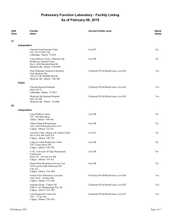
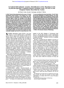
Lung Cancer 80 (2013) 249–255
Contents lists available at SciVerse ScienceDirect
Lung Cancer
journal homepage: www.elsevier.com/locate/lungcan
Review
Personalized therapy on the horizon for squamous cell carcinoma of the lung
Han Sang Kim a , Tetsuya Mitsudomi b , Ross A. Soo c , Byoung Chul Cho a,∗
a
Division of Medical Oncology, Yonsei Cancer Center, Yonsei University College of Medicine, Seoul, Republic of Korea
Department of Surgery, Kinki University Faculty of Medicine, Japan
c
Department of Haematology-Oncology, National University Cancer Institute, National University Health System, Singapore
b
a r t i c l e
i n f o
Article history:
Received 29 November 2012
Received in revised form 15 February 2013
Accepted 16 February 2013
Keywords:
Squamous cell carcinoma
Non-small cell lung cancer
Targeted therapy
Driver mutation
Biomarker
a b s t r a c t
Squamous cell carcinoma (SQCC) of the lung is the second-largest subtype of non-small cell lung cancer (NSCLC), causing an estimated 400,000 deaths per year worldwide. Recent developments in cancer
genome sequencing technology expanded our knowledge of driver mutations, which were identified as
novel candidates for targeted therapy in various cancers. Successful targeted treatments for lung adenocarcinoma, NSCLC’s primary subtype, with EGFR mutation or ALK fusion are clinically available, and a
clinical trial of personalized targeted therapy in patients with lung adenocarcinoma is underway by the
Lung Cancer Mutation Consortium. Although there are targeted treatments for lung adenocarcinoma, no
personalized therapies currently exist for SQCC. Recently, comprehensive genomic characterization of
lung SQCC using massively parallel sequencing has enabled us to identify several potential driver mutations/signaling pathways. These are FGFR1 amplifications, PI3KCA mutations, PTEN mutations/deletions,
PDGFRA amplifications/mutations, and DDR2 mutations. The march toward personalized therapy may
have taken a step forward with the discovery of these potential biomarkers for the treatment of SQCC of
the lung.
This article reviewed the current knowledge of genomic landscape of lung SQCC and summarized
ongoing clinical trials of targeted agents for lung SQCC. Also, we will suggest several other actionable
mutations with matching drugs that should be investigated in future clinical trials for the personalized
treatment of lung SQCC.
Crown Copyright © 2013 Published by Elsevier Ireland Ltd. All rights reserved.
1. Introduction
Lung cancer is the leading cause of cancer-related death worldwide, causing ∼1.5 million deaths per year [1]. Non-small cell lung
cancer (NSCLC) comprises about 85% of newly diagnosed lung cancers, which can be classified into two major histological subtypes:
adenocarcinoma (∼50%) and squamous cell carcinoma (∼30%).
Squamous cell carcinoma of the lung (SQCC) is closely associated
with tobacco smoking. The discovery of so-called ‘driver mutations’
such as the epidermal growth factor receptor (EGFR) and anaplastic
lymphoma kinase (ALK) has led to the remarkable improvement
in personalized therapy for lung adenocarcinoma [2–4]. However, no such available molecular target exists in squamous
histology. Patients with SQCC are treated with ‘one-size-fits-all’
platinum-based chemotherapy, and therapies developed for lung
adenocarcinoma such as bevacizumab, pemetrexed and EGFR
tyrosine kinase inhibitors are contraindicated or largely ineffective
∗ Corresponding author at: Division of Medical Oncology, Yonsei University College of Medicine, 50 Yonsei-ro, Seodaemun-gu, Seoul 120-752, Republic of Korea.
Tel.: +82 2 2228 8126; fax: +82 2 393 3652.
E-mail address: [email protected] (B.C. Cho).
in lung SQCC [5–7]. For this reason, the identification of novel
druggable molecular targets in SQCC is a top research priority.
Recent developments in genomics using next-generation
sequencing (NGS) technology allow us to capture information on
genetic alterations of the tumor including single nucleotide variation, copy number alterations, and chromosomal rearrangements
in whole-genome level [8]. Application of NGS in lung cancer
research has produced a comprehensive profile of genetic alteration and has led to the identification of potential targets for
personalized treatments [9–14].
In this review, we highlight the current knowledge of molecular
targets, ongoing clinical trials of targeted agents, and actionable
mutations with matching drugs in lung SQCCs.
2. Genomic landscape of lung SQCC using NGS technology
Whilst comprehensive genome-scale characterization has been
reported in lung adenocarcinoma [11,13,14], genetic alteration in
SQCC is less understood. Single platform studies have identified
several genetic changes related to SQCC, such as amplification
of p63, PI3KCA, PDGFRA, SOX2, or FGFR1 and mutation in p53,
EGFRvIII, PI3KCA, NRF2, PTEN, and DDR2 (Table 1) [15]. In 2012,
the cancer genome atlas (TCGA) researchers produced a comprehensive genomic landscape of SQCC using whole exome/genome
0169-5002/$ – see front matter. Crown Copyright © 2013 Published by Elsevier Ireland Ltd. All rights reserved.
http://dx.doi.org/10.1016/j.lungcan.2013.02.015
250
H.S. Kim et al. / Lung Cancer 80 (2013) 249–255
Table 1
Previously-identified genetic alterations in squamous cell carcinoma of the lung.
Genetic alteration
Chromosomal region
Year reported in SQCC
Reference
p53 Mutation
p63 Amplification
EGFRvIII mutation
PI3KCA mutation/amplification
NRF2 mutation
PDGFRA amplification
SOX2 amplification
PTEN mutation
FGFR1 amplification
DDR2 mutation
17p13.1
3q28
7p12
3q26.3
2q31
4q12
3q26.3–q27
10q23.3
8p12
1q23.3
1992
2000
2006
2007
2008
2009
2009
2010
2010
2011
[72]
[73,74]
[75]
[54]
[76]
[64]
[77]
[58]
[49]
[67]
Abbreviations: SQCC, squamous cell carcinoma of the lung.
sequencing, mRNA sequencing, mRNA expression and promoter
methylation for 178 lung SQCCs [12]. The major findings were
as follows: (i) lung SQCC is characterized by complex genomic
alterations, with a mean of 360 exonic mutations, 165 genomic
rearrangements, and 323 segments of copy number alterations
per tumor. They identified statistically recurrent mutations in 11
genes (TP53, CDKN2A, PTEN, PIK3CA, KEAP1, MLL2, HLA-A, NRF2,
NOTCH1, RB1 and HLA-A) and somatic copy number alterations
of chromosomal segments containing SOX2, PDGFRA and/or KIT,
EGFR, FGFR1 and/or WHSC1L1, CCND1, CDKN2A, NFE2L2, MYC,
CDK6, MDM2, BCL2L1, EYS, FOXP1, PTEN, and NF1. Of note, compared with lung adenocarcinoma, almost all lung SQCCs display
somatic mutation of TP53 (81%) and selective amplification of
chromosome 3q. (ii) There were frequent alterations in the cell
cycle control (CDKN2A/RB1, 72%), response to oxidative stress
(NFE2L2/KEAP1/CUL3, 34%), apoptotic signaling (PI3K/AKT, 69%)
and squamous differentiation (SOX2/TP63/NOTCH1, 44%). (iii)
Most importantly, they identified a potentially targetable gene or
pathway alteration in most lung SQCC samples studied (Fig. 1).
Although EGFR and KRAS mutations, the two most common
oncogenic aberrations in lung adenocarcinoma, are extremely
rare, there are frequent alterations in PI3K/AKT pathway and
receptor tyrosine kinase (RTK) signaling, including EGFR amplification, BRAF mutation or FGFR amplification or mutation, in 69%
of samples. Interestingly, loss-of-function mutations in HLA-A
class I major histocompatibility gene were found in 3% of the
tumors, suggesting a potential role of genotypic selection of
patients for immunotherapies, such as anti-programmed death
1 (PD1) and anti-cytotoxic T-lymphocyte antigen 4 (CTLA4)
antibodies.
In a study by Paik et al. [16], the incidence of known targetable driver mutations in SQCC was examined using multiplex
PCR sequencing. The driver mutations included PIK3CA mutations,
PTEN mutations/deletions, FGFR1 amplifications, and DDR2 mutations. Overall, 60% of patients were found to have druggable targets,
and the frequency of these mutations was similar to those in TCGA
genetic profiles (Fig. 1). Although functional dependence of lung
SQCC on these newly identified genetic mutations or pathway
alterations should be validated, these data can help to facilitate
effective personalized treatment for lung SQCC.
3. Currently ongoing clinical trials in lung SQCC
Several agents are expected to be effective for the treatment
of SQCC, and therefore currently are being tested in clinical trials
(Table 2). These are cytotoxic nab-paclitaxel, novel targeted agents,
and immunotherapies Table 3.
3.1. New cytotoxic agents
Next-generation taxane (nab-paclitaxel, abraxane) is currently
being evaluated in late phase trials. Nab-paclitaxel, composed
of paclitaxel nanoparticle and human albumin, utilizes albumin
receptor 60-kDa glycoprotein (gp60) and intracellular caveolin-1
and achieves higher intratumoral concentration compared with
previous taxanes [17]. In addition, SPARC (secreted protein, acidic
and rich in cysteine) increases nab-paclitaxel accumulation in the
tumor area [17]. In patients with stage IIIB/IV NSCLC, nab-paclitaxel
showed higher objective response rate (ORR) and less neuropathy and neutropenia than solvent-based paclitaxel (ORR, 33% vs.
25%; P = 0.005) [18]. In subgroup analysis, nab-paclitaxel had better efficacy in squamous histology (ORR, 41% vs. 24%, P < 0.001)
than non-squamous histology (ORR, 26% vs. 25%, P = 0.808). One
possible explanation for the better efficacy of nab-paclitaxel in
squamous histology may be aberrant caveolin-1 expression in SQCC
[19]. A study using SPARC, ceveolin-1, and miroRNAs as predictive
biomarkers in patients with advanced stage NSCLC treated with
carboplatin and nab-paclitaxel is ongoing (NCT00729612).
3.2. Targeted agents
3.2.1. Anti-EGFR therapy
The currently approved EGFR-targeted agents in lung SQCC
are small molecule tyrosine kinase inhibitors (TKIs), erlotinib and
gefitinib [20,21]. Although treatment with EGFR-TKIs has afforded
tremendous clinical benefits in lung adenocarcinomas with EGFR
Fig. 1. Frequency of potential actionable drug targets in squamous cell carcinoma of the lung.
H.S. Kim et al. / Lung Cancer 80 (2013) 249–255
251
Fig. 2. Alterations in actionable oncogenic signaling pathways in squamous cell carcinoma of the lung.
mutations, the same is not true for lung SQCCs, in which the ORR to
EGFR-TKIs is typically less than 10%. In a recent TCGA data, EGFR
mutations associated with the sensitivity to EGFR-TKIs was not
found in lung SQCCs [12]. This was also confirmed by Rekhtman
et al. [22], which reported that EGFR mutation does not occur in
pure lung SQCCs from which adenosquamous carcinoma and poorly
differentiated adenocarcinoma were excluded [22].
Cetuximab, the monoclonal antibody targeting EGFR protein,
in combination with cisplatin/vinorelbine had a minimal survival
benefit compared with chemotherapy alone in patients with stage
IIIB/IV NSCLC in the FLEX study (overall survival [OS], 11.3 vs. 10.1
months; P = 0.044) [23]. In a preplanned subset analysis, a survival
benefit was seen in squamous histology, albeit minimal (OS, 9.0 vs.
8.2 months). High tumor EGFR expression (immunohistochemistry
score ≥200), which was more common in SQCC than in non-SQCC
histology, may serve as a predictive biomarker for the selection of
patients who would be most likely to derive a clinical benefit from
treatment with chemotherapy plus cetuximab [24].
Other anti-EGFR agents, such as the second-generation EGFRTKI (afatinib) or the monoclonal antibody (necitumumab), are
currently being studied in lung SQCC patients (Table 2).
3.2.2. Anti-MET therapy
MET, a proto-oncogene located on 7q21-31, encodes a tyrosine
kinase receptor for hepatocyte growth factor (HGF). MET protein
expression, positivity defined as more than grade 1+ staining by
immunohistochemistry, was observed in about 40% of lung SQCCs,
and was associated with poor prognosis [25]. MET gene amplification, albeit not reported in TCGA data, was reported to be more
frequent in patients with lung SQCC than in those with adenocarcinoma [26]. Furthermore, MET gene amplification has been shown
to play a role in primary or acquired resistance to EGFR-TKI [27,28].
Onartuzumab, the monoclonal antibody against MET, in combination with erlotinib showed improvements in overall survival (OS)
and progression-free survival (PFS) compared with erlotinib alone
in a MET-overexpressed (Met Dx+) subgroup of advanced NSCLC
patients in a randomized phase II study [29]. A randomized phase
II study to test activity of onartuzumab in combination with a platinum doublet in lung SQCC is currently ongoing (NCT01519804).
3.2.3. Anti-insulin-like growth factor 1 (IGF-1) therapy
Insulin-like growth factor-1 receptor (IGF-1R, located in
15q26.3) is a cell surface receptor with tyrosine kinase activity
Table 2
Ongoing clinical trials exclusively for squamous cell carcinoma of the lung.
Regimen
Newer cytotoxic agents
Albumin-bound paclitaxel/carboplatin vs. Gemcitabine/carboplatin
Targeted agents
Afatinib (EGFR) vs. Erlotinib
Necitumumab (EGFR)/gemcitabine/cisplatin vs. Gemcitabine/cisplatin
Onartuzumab (MET)/paclitaxel/platinum vs. Paclitaxel/platinum
AXL1717 (IGF-1R) vs. docetaxel
Gemcitabine/carboplatin/Iniparib (PARP-1) vs. Gemcitabine/carboplatin
Immunotherapeutic agents
Reolysin/paclitaxel/carboplatin
Ipilimumab/paclitaxel/carboplatin vs. Paclitaxel/carboplatin
BMS-936558 vs. docetaxel
Phase
Line of therapy
II
1st
III
III
II
II
III
2nd
1st
1st
2nd
1st
800
1097
110
140
780
II
III
III
1st
1st
2nd
55
920
264
Abbreviations: ORR, objective response rate; PFS, progression-free survival; OS, overall survival.
Planned N
120
Primary endpoint
ClinicalTrials.gov identifier
ORR
NCT01236716
PFS
OS
PFS
PFS
OS
NCT01523587
NCT00981058
NCT01519804
NCT01561456
NCT01082549
ORR
OS
ORR
NCT00998192
NCT01285609
NCT01642004
252
H.S. Kim et al. / Lung Cancer 80 (2013) 249–255
Table 3
Selected potential actionable genetic alterations with matched targeted agents in squamous cell carcinoma of the lung.
Molecular alteration/drug
Target
FGFR amplification
FGFR1-3
AZD4547
BGJ398
FGFR1-3
LY2874455
FGFR1-4
FGFR1-2, VEGFR2
Brivanib
FGFR, VEGFR, PDGFR, FLT3, c-KIT
Dovitinib
PI3KCA mutation
PI3K, mTOR
BEG235
BKM120
PI3KC-A,B,D,G
GDC0941
PI3KC-A,B,D,G
PX-866
PI3KC-A,D,G
PTEN mutation/deletion
PI3KC-B
GSK2636771
PDGFRA amplification/mutation
PDGFRA
Crenolanib
PDFGR, VEGFR, FGFR
BIBF1120
PDFGR, VEGFR, FGFR, c-KIT
Pazopanib
DDR2 mutation
DDR2, BCR/ABL, SRC, c-KIT
Dasatinib
BRAF mutation
MEK1, MEK2
Trametinib
MEK1, MEK2
MEK162
ERBB2 amplification
EGFR, ERBB2, ERBB4
Afatinib
EGFR, ERBB2
Lapatinib
T-DM1
ERBB2, cytotoxic
Sponsor
Study phase (cancer type)
ClinicalTrials.gov identifier
AstraZeneca
Novartis
Eli Lilly
Bristol Myers Squibb
Novartis
II (breast, stomach)
I (solid tumors)
I (solid tumors)
III (liver, colon)
III (kidney), II (lung)
NCT01202591, NCT01457846
NCT01004224
NCT01212107
NCT00858871, NCT01367275
NCT01223027, NCT01676714
Novartis
Novartis
Genentech
Oncothyreon
II (breast)
II (lung), I (breast)
II (lung), I (breast)
II (prostate) I (solid tumors)
NCT00620594
NCT01297491, NCT01339442
NCT01493843, NCT00960960
NCT01331083, NCT01204099
Glaxo Smith Kline
I (solid tumors)
NCT01458067
Arog Pharmaceuticals
Boehringer Ingelheim
Glaxo Smith Kline
II (glioma, GIST)
III (lung, ovary)
III (kidney, sarcoma) II (lung)
NCT01229644, NCT01243346
NCT00805194, NCT01015118
NCT00720941, NCT00753688 NCT01027598
Bristol-Myers Squibb
II (lung) II (breast)
NCT01491633 NCT00371345
Glaxo Smith Kline
Norvatis
III (melanoma) II (lung)
II (melanoma)
NCT01245062 NCT01362296
NCT01320085
Boehringer Ingelheim
Glaxo Smith Kline
Roche
III (lung, breast)
III (breast), I (lung)
III (breast, stomach)
NCT01523587, NCT01125566
NCT00078572, NCT00528281
NCT00829166, NCT01641939
that activates the PI3K/Akt/mTOR and RAS/RAF/MAPK signaling
pathways by binding IGF-1 or IGF-2 ligand [30]. The protein or
mRNA expression of IGF-1R, but not IGF-1R gene copy number,
was reported to be higher in lung SQCC than in other histologic
subtypes [31]. In a phase II study with NSCLC, CP-751871 (figitumumab) with paclitaxel and carboplatin showed a response rate
of 78% in patients with advanced SQCC [32]. However, two subsequent phase III studies of figitumumab with paclitaxel/carboplatin
or erlotinib were discontinued due to non-significant survival benefit and increased toxicity [33,34]. A phase II study is ongoing to
compare anti-tumor activity of AXL1717, a small-molecule IGF-1R
inhibitor, with docetaxel as a second-line treatment of advanced
lung SQCC (NCT01561456).
3.2.4. PARP-1 inhibitor
Poly (ADP-ribose) polymerase-1 (PARP-1) is an important DNA
repair enzyme, which is implicated in base excision repair and
single-strand break (SSB) repair. When PARP-1 is inhibited, accumulated unrepaired SSBs are converted to double-strand breaks
(DSB), inducing cell death [35]. PARP-1 is significantly upregulated
at the mRNA level in multiple cancer types, including lung SQCC
[36]. Iniparib is a novel PARP-1 inhibitor and has demonstrated
synergistic activity with gemcitabine and carboplatin in preclinical
studies [37]. A phase III trial of iniparib in combination with gemcitabine/carboplatin versus chemotherapy alone in SQCC is currently
ongoing (NCT01082549) with the primary endpoint being OS [38].
3.3. Immunotherapeutic agents
Immunotherapy utilizes antibodies, vaccines, or viruses to
modulate T-cell activity. Tumor infiltration of CD4+ /CD8+ T-cell
was associated with better prognosis while higher expression of
immunosuppressive regulatory T-cell was related to disease recurrence in operable NSCLC patients [39]. Two immunologic targets
of current interest are cytotoxic T-lymphocyte antigen-4 (CTLA-4)
and programmed death-1 (PD-1), which are negative regulators of
T-cell activation [40,41]
The preliminary efficacy of ipilimumab, the fully human monoclonal antibody against CTLA-4, has recently been reported in
NSCLC [40]. In a randomized phase II study, a phased ipilimumab
regimen (two doses of placebo plus paclitaxel and carboplatin
followed by four doses of ipilimumab plus paclitaxel and carboplatin) showed better immune-related PFS (irPFS) compared to
paclitaxel/carboplatin alone in patients with stage IIIB/IV NSCLC
(5.7 vs. 4.6 months; P = 0.05), though statistically not significant
[40]. The improvement in irPFS with the addition of ipilimumab to
chemotherapy was greater in SQCC (HR, 0.55 [95% CI, 0.27–1.12])
than in non-squamous histology (HR, 0.82 [95% CI, 0.52–1.28]).
Although caution is warranted in interpreting these subset data,
it is notable that tumor-infiltrating T cells are more abundant in
lung SQCC [42].
The aberrant increase in the expression of ligands for PD-1 was
observed in SQCC including B7-H1, also known as PD-L1, and B7H3 [43]. Blockade of PD-1, a co-inhibitory receptor expressed by
activated T cells, can overcome immune resistance and mediate
tumor regression [44]. In a phase I study in patients with advanced
solid tumors including NSCLC patients (n = 76), BMS-936558, the
fully human monoclocal antibody against PD-1, showed a response
rate of 18% and a PFS rate at 24 weeks of 26% [41]. The expression
of PD-1 ligand on tumor was suggested as a potential biomarker for
response.
Oncolytic viruses have been investigated for use in cancer treatment. The Reoviruses are double-stranded RNA viruses targeting
RAS-activated cells [45]. Preliminary results from a single-arm
phase II trial of Reolysin, an isolated Reovirus, in combination with
carboplatin and paclitaxel, showed a response rate of 26% and a
disease control rate of 83% in lung SQCC (NCT00998192).
4. Promising druggable targets in SQCC
Based on the recent findings on comprehensive genomic characterization of lung SQCC [12,16], promising druggable mutations
on the horizon for targeted therapy will be reviewed (Fig. 2).
4.1. FGFR1 amplification
The fibroblast growth factor receptor (FGFR) tyrosine kinase
family is comprised of four kinases, FGFR 1–4, and plays a crucial
H.S. Kim et al. / Lung Cancer 80 (2013) 249–255
role in cancer cell growth, survival, and resistance to chemotherapy. Furthermore, an elevated concentration of FGF in serum is
an independent adverse prognostic factor in NSCLC [46,47]. FGFR1
has been shown to be a target for deregulation by amplification,
point mutation, or translocation [48]. Weiss et al. [49] examined
high-resolution genomic profiles of 77 lung adenocarcinomas and
155 SQCCs and identified amplifications of FGFR1 exclusively in
lung SQCC. Examination of an independent series by fluorescent
in situ hybridization (FISH) revealed FGFR1 amplification of 22%
in SQCC samples. FGFR1 amplification drives downstream activation of mitogen-activated protein kinase (MAPK) signaling in cancer
cells. Treatment with FGFR inhibitor showed downstream inhibition and induction of apoptosis in FGFR1-amplified cell lines as
well as strong antitumor activity in xenograft model, strongly supporting the utility of FGFR1 amplification as a relevant therapeutic
target in lung SQCC.
Recently, Cho et al. [50] observed FGFR1 amplification in 13%
and found that FGFR1 amplification is a negative prognostic factor
in patients with resected SQCC (disease-free survival, 26.9 vs. 94.6
months, P < 0.0001). The incidence of FGFR1 amplification was also
associated with smoking status (current smoker, 28.9% vs. former
smoker, 2.5% vs. non-smoker, 0%; P < 0.0001). This finding suggests that FGFR1 amplification is an oncogenic aberration caused
by cigarette smoking in SQCC, while Heist et al. reported that there
was no significant difference in overall survival stratified by FGFR
amplification status [51].
4.2. Phosphoinositide 3-kinase (PI3K) pathway
The PI3K pathway is a major oncogenic signaling pathway which
functions in cell survival and proliferation [52]. Somatic mutation
or amplification of PIK3CA, encoding the p110␣ catalytic subunit, is
known to be more prevalent in lung SQCCs than lung adenocarcinomas [53–55]. The incidence of PIK3CA mutation and amplification
in lung SQCCs was reported to be 8–16% and 33–43%, respectively
[12,16,53]. In phase I trials, response rate was significantly higher
for patients with PIK3CA mutations treated with PI3K/AKT/mTOR
pathway inhibitors than for those without documented mutations
(35% vs. 5%, P < 0.001) [56]. Because lung SQCC patients were not
included in these trials, further studies are needed to determine
whether PIK3CA is a predictive marker to PIK3CA inhibition or not,
in lung SQCC.
The tumor suppressor PTEN confers phosphatase activity,
which inhibits the PI3K/AKT/mTOR signaling pathway. Loss of
PTEN expression has been reported in 74% of NSCLC [57]. Several mechanisms are associated with loss of PTEN expression,
including PTEN point mutation/deletion/hypermethylation. The
incidence of PTEN mutation/deletion was reported to be 15–28%
in lung SQCC [12,16,58]. PTEN loss is associated with a poorer
response to erlotinib in EGFR-mutant NSCLC [59]. Drugs targeting the PI3K/AKT/mTOR signaling pathway may be effective in
PTEN-deficient tumors. In particular, a crucial role of PIK3CB has
been reported in PTEN-deficient tumors [60,61]. In a prostate
cancer model, the inhibition of PIK3CB reduced Akt phosphorylation and prevented tumorigenesis [60]. While PIK3CA depletion
did not affect PI3K signaling in colorectal cancer cells, downregulation of PIK3CB is required for PI3K pathway inhibition in
PTEN-deficient tumors [61]. These data supports the selective inhibition of PIK3CB may be effective in the treatment of PTEN-deficient
tumors.
4.3. PDGFRA amplification/mutation
Platelet-derived growth factor receptor (PDGFR) tyrosine
kinase, which is classified as PDGFRA and PDGFRB, plays a
crucial role in cell proliferation and angiogenesis [62,63]. The
253
amplification of the chromosomal segment of 4q12 harboring
PDGFR-␣ has been reported in ∼9% of lung SQCCs [12,64]. Several multi-targeted TKIs targeting PDGFRA, such as sorafenib,
sunitinib, and imatinib, are currently available. Despite initial enthusiasm for the anti-PDGFRA therapy, the addition
of sorafenib to platinum-based chemotherapy disappointingly
failed to improve survival in a randomized phase III study
[65]. Notably, the addition of sorafenib increased mortality
in a subset of lung SQCCs. A selective anti-PDGFRA agent,
crenolanib, is now tested in a phase II study in patients
with glioma and gastrointestinal stromal tumor (NCT01229644,
NCT01243346).
4.4. Anti-DDR2 therapy
Discoidin domain receptor 2 (DDR2) tyrosine kinase has been
known to promote cell proliferation and survival when activated by its collagen ligand [66]. Mutation in the kinase domain
of DDR2 was identified in 3.8% of lung SQCC samples (11/290)
[67]. DDR2 mutation promotes malignant transformation and
is associated with sensitivity to shRNA-mediated depletion of
DDR2 or dasatinib, which was approved for the treatment of
Bcr-Abl-positive chronic myelogenous leukemia. It also promotes
malignant transformation, suggesting that gain-of-function mutations in DDR2 may be a promising therapeutic target in lung
SQCCs [67]. A phase II trial to confirm the efficacy of dasatinib in lung SQCC with DDR2 mutation is currently underway
(NCT01491633).
4.5. BRAF mutation
BRAF mutation leads to the development of cancer through constitutional serine/threonine kinase activity [68]. BRAF mutation is
present in ∼4% of SQCC, all of which are not canonical V600E mutation [69]. As mitogen-activated protein kinase (MEK) dependency
was observed in BRAF mutant tumor, MEK inhibitor may benefit
from non-V600E BRAF mutant SQCC [70].
4.6. ERBB2 amplification
Amplification of ERBB2, an EGFR family receptor tyrosine kinase,
was found to be present in ∼4% of lung SQCC [12,71]. Further
research needs to be done to confirm the efficacy of ERBB2-directed
therapy in ERBB2-amplified lung SQCC.
5. Perspectives
The recent expansion of our knowledge on the genomic landscape of lung SQCC leads to the identification of potential driver
mutations, including FGFR1 amplification, PIK3CA mutation, PTEN
mutation/deletion, PDGFRA amplification/mutation, DDR2 mutation, and BRAF mutation. The impact of these mutations on the
response to the matching targeted therapy should be validated in
future clinical trials.
Similar to routine EGFR mutation testing in lung adenocarcinoma at diagnosis, better understanding of driver mutations and
molecular pathways may lead to the development of mutation
panels for the selection of patients with lung SQCC for a specific
targeted therapy at the time of diagnosis or even disease progression. Furthermore, as the acquired resistance to targeted therapy is
common and inevitable, identification of driver oncogene leading
to drug resistance should be a research priority to achieve future
personalized therapies for lung SQCC.
254
H.S. Kim et al. / Lung Cancer 80 (2013) 249–255
Conflict of interest statement
The authors have no conflicts of interest to declare.
[24]
Acknowledgements
[25]
This work was supported by the National Research Foundation
of Korea (NRF) grants funded by the Korea government (MEST)
(2012R1A2A2A01046927).
[26]
[27]
References
[28]
[1] Jemal A, Bray F, Center MM, Ferlay J, Ward E, Forman D. Global cancer statistics.
CA Cancer J Clin 2011;61:69–90.
[2] Sandler AB, Johnson DH, Herbst RS. Anti-vascular endothelial growth factor
monoclonals in non-small cell lung cancer. Clin Cancer Res 2004;10:4258s–62s.
[3] Giaccone G. Epidermal growth factor receptor inhibitors in the treatment of
non-small-cell lung cancer. J Clin Oncol 2005;23:3235–42.
[4] Kwak EL, Bang YJ, Camidge DR, Shaw AT, Solomon B, Maki RG, et al. Anaplastic lymphoma kinase inhibition in non-small-cell lung cancer. N Engl J Med
2010;363:1693–703.
[5] Johnson DH, Fehrenbacher L, Novotny WF, Herbst RS, Nemunaitis JJ, Jablons
DM, et al. Randomized phase II trial comparing bevacizumab plus carboplatin and paclitaxel with carboplatin and paclitaxel alone in previously
untreated locally advanced or metastatic non-small-cell lung cancer. J Clin
Oncol 2004;22:2184–91.
[6] Hanna N, Shepherd FA, Fossella FV, Pereira JR, De Marinis F, von Pawel J, et al.
Randomized phase III trial of pemetrexed versus docetaxel in patients with
non-small-cell lung cancer previously treated with chemotherapy. J Clin Oncol
2004;22:1589–97.
[7] Scagliotti GV, Parikh P, von Pawel J, Biesma B, Vansteenkiste J, Manegold C,
et al. Phase III study comparing cisplatin plus gemcitabine with cisplatin plus
pemetrexed in chemotherapy-naive patients with advanced-stage non-smallcell lung cancer. J Clin Oncol 2008;26:3543–51.
[8] Meyerson M, Gabriel S, Getz G. Advances in understanding cancer genomes
through second-generation sequencing. Nat Rev Genet 2010;11:685–96.
[9] Campbell PJ, Stephens PJ, Pleasance ED, O’Meara S, Li H, Santarius T, et al. Identification of somatically acquired rearrangements in cancer using genome-wide
massively parallel paired-end sequencing. Nat Genet 2008;40:722–9.
[10] Pleasance ED, Stephens PJ, O’Meara S, McBride DJ, Meynert A, Jones D, et al. A
small-cell lung cancer genome with complex signatures of tobacco exposure.
Nature 2010;463:184–90.
[11] Lee W, Jiang Z, Liu J, Haverty PM, Guan Y, Stinson J, et al. The mutation spectrum revealed by paired genome sequences from a lung cancer patient. Nature
2010;465:473–7.
[12] The Cancer Genome Atlas Research N, Genome sequencing centres: Broad I,
Hammerman PS, Lawrence MS, Voet D, Jing R, et al. Comprehensive genomic
characterization of squamous cell lung cancers. Nature 2012;489:519–25.
[13] Imielinski M, Berger AH, Hammerman PS, Hernandez B, Pugh TJ, Hodis E,
et al. Mapping the hallmarks of lung adenocarcinoma with massively parallel
sequencing. Cell 2012;150:1107–20.
[14] Govindan R, Ding L, Griffith M, Subramanian J, Dees ND, Kanchi KL, et al.
Genomic landscape of non-small cell lung cancer in smokers and neversmokers. Cell 2012;150:1121–34.
[15] Weir BA, Woo MS, Getz G, Perner S, Ding L, Beroukhim R, et al. Characterizing
the cancer genome in lung adenocarcinoma. Nature 2007;450:893–8.
[16] Paul Paik AH, Lu Wang, Natasha Rekhtman, Marc Ladanyi, Kris Mark G. Multiplex testing for driver mutations in squamous cell carcinomas of the lung. J
Clin Oncol 2012;30 (suppl; abstr 7505).
[17] Gradishar WJ. Albumin-bound paclitaxel: a next-generation taxane. Expert
Opin Pharmacother 2006;7:1041–53.
[18] Socinski MA, Bondarenko I, Karaseva NA, Makhson AM, Vynnychenko I,
Okamoto I, et al. Weekly nab-paclitaxel in combination with carboplatin versus
solvent-based paclitaxel plus carboplatin as first-line therapy in patients with
advanced non-small-cell lung cancer: final results of a phase III trial. J Clin Oncol
2012;30:2055–62.
[19] Yoo SH, Park YS, Kim HR, Sung SW, Kim JH, Shim YS, et al. Expression of caveolin1 is associated with poor prognosis of patients with squamous cell carcinoma
of the lung. Lung Cancer 2003;42:195–202.
[20] Shepherd FA, Rodrigues Pereira J, Ciuleanu T, Tan EH, Hirsh V, Thongprasert S,
et al. Erlotinib in previously treated non-small-cell lung cancer. N Engl J Med
2005;353:123–32.
[21] Kim ES, Hirsh V, Mok T, Socinski MA, Gervais R, Wu YL, et al. Gefitinib versus
docetaxel in previously treated non-small-cell lung cancer (INTEREST): a randomised phase III trial. Lancet 2008;372:1809–18.
[22] Rekhtman N, Paik PK, Arcila ME, Tafe LJ, Oxnard GR, Moreira AL, et al. Clarifying
the spectrum of driver oncogene mutations in biomarker-verified squamous
carcinoma of lung: lack of EGFR/KRAS and presence of PIK3CA/AKT1 mutations.
Clin Cancer Res 2012;18:1167–76.
[23] Pirker R, Pereira JR, Szczesna A, von Pawel J, Krzakowski M, Ramlau R, et al. Cetuximab plus chemotherapy in patients with advanced
[29]
[30]
[31]
[32]
[33]
[34]
[35]
[36]
[37]
[38]
[39]
[40]
[41]
[42]
[43]
[44]
[45]
[46]
[47]
non-small-cell lung cancer (FLEX): an open-label randomised phase III trial.
Lancet 2009;373:1525–31.
Pirker R, Pereira JR, von Pawel J, Krzakowski M, Ramlau R, Park K, et al. EGFR
expression as a predictor of survival for first-line chemotherapy plus cetuximab
in patients with advanced non-small-cell lung cancer: analysis of data from the
phase 3 FLEX study. Lancet Oncol 2012;13:33–42.
Masuya D, Huang C, Liu D, Nakashima T, Kameyama K, Haba R, et al. The tumourstromal interaction between intratumoral c-Met and stromal hepatocyte
growth factor associated with tumour growth and prognosis in non-small-cell
lung cancer patients. Br J Cancer 2004;90:1555–62.
Go H, Jeon YK, Park HJ, Sung SW, Seo JW, Chung DH. High MET gene copy
number leads to shorter survival in patients with non-small cell lung cancer. J
Thorac Oncol 2010;5:305–13.
Engelman JA, Zejnullahu K, Mitsudomi T, Song Y, Hyland C, Park JO, et al. MET
amplification leads to gefitinib resistance in lung cancer by activating ERBB3
signaling. Science 2007;316:1039–43.
Tanizaki J, Okamoto I, Sakai K, Nakagawa K. Differential roles of transphosphorylated EGFR, HER2, HER3, and RET as heterodimerisation partners
of MET in lung cancer with MET amplification. Br J Cancer 2011;105:
807–13.
Spigel DRTJE, Ramlau R, Daniel DB, Goldschmidt JH, Blumenschein GR, Krzakowski MJ, et al. Final efficacy results from OAM4558g, a randomized phase II
study evaluating MetMAb or placebo in combination with erlotinib in advanced
NSCLC. J Clin Oncol 2011;29 (suppl; abstr 7505).
Pollak MN, Schernhammer ES, Hankinson SE. Insulin-like growth factors and
neoplasia. Nat Rev Cancer 2004;4:505–18.
Dziadziuszko R, Merrick DT, Witta SE, Mendoza AD, Szostakiewicz B, Szymanowska A, et al. Insulin-like growth factor receptor 1 (IGF1R) gene copy
number is associated with survival in operable non-small-cell lung cancer: a
comparison between IGF1R fluorescent in situ hybridization, protein expression, and mRNA expression. J Clin Oncol 2010;28:2174–80.
Karp DD, Paz-Ares LG, Novello S, Haluska P, Garland L, Cardenal F, et al.
Phase II study of the anti-insulin-like growth factor type 1 receptor antibody CP-751,871 in combination with paclitaxel and carboplatin in previously
untreated, locally advanced, or metastatic non-small-cell lung cancer. J Clin
Oncol 2009;27:2516–22.
Jassem JCJL, Karp DD, Mok T, Benner RJ, Green SJ, Park K, et al. Randomized,
open label, phase III trial of figitumumab in combination with paclitaxel and
carboplatin versus paclitaxel and carboplatin in patients with non-small cell
lung cancer (NSCLC). J Clin Oncol 2010;28:15s (suppl; abstr 7500) 2010.
Yee D. Insulin-like growth factor receptor inhibitors: baby or the bathwater? J
Natl Cancer Inst 2012;104:975–81.
Krishnakumar R, Kraus WL. The PARP side of the nucleus: molecular actions,
physiological outcomes, and clinical targets. Mol Cell 2010;39:8–24.
Ossovskaya V, Koo IC, Kaldjian EP, Alvares C, Sherman BM. Upregulation of poly
(ADP-ribose) polymerase-1 (PARP1) in triple-negative breast cancer and other
primary human tumor types. Genes Cancer 2010;1:812–21.
Davar D, Beumer JH, Hamieh L, Tawbi H. Role of PARP inhibitors in cancer
biology and therapy. Curr Med Chem 2012;19:3907–21.
Spigel DRPGH, Hainsworth JD, De Marinis F, Kabbinavar FF, Kim ES, Lynch TJ,
et al. Sarah Cannon Research Institute and Tennessee Oncology, Nashville, TN.
Randomized phase III trial of gemcitabine/carboplatin with or without iniparib
(BSI-201) in patients with previously untreated stage IV squamous non-small
cell lung cancer (NSCLC). J Clin Oncol 2011;29 (suppl; abstr TPS220).
Petersen RP, Campa MJ, Sperlazza J, Conlon D, Joshi MB, Harpole Jr DH, et al.
Tumor infiltrating Foxp3+ regulatory T-cells are associated with recurrence in
pathologic stage I NSCLC patients. Cancer 2006;107:2866–72.
Lynch TJ, Bondarenko I, Luft A, Serwatowski P, Barlesi F, Chacko R, et al. Ipilimumab in combination with paclitaxel and carboplatin as first-line treatment
in stage IIIB/IV non-small-cell lung cancer: results from a randomized, doubleblind, multicenter phase II study. J Clin Oncol 2012;30:2046–54.
Suzanne Louise Topalian JRB, Stephen Hodi F, David F McDermott, David C
Smith, Scott N Gettinger, Janis M Taube, et al. Anti-PD-1 (BMS-936558, MDX1106) in patients with advanced solid tumors: Clinical activity, safety, and a
potential biomarker for response. 2012 ASCO Annual Meeting. J Clin Oncol
2012;30 (suppl; abstr CRA2509).
Hiraoka K, Miyamoto M, Cho Y, Suzuoki M, Oshikiri T, Nakakubo Y, et al.
Concurrent infiltration by CD8+ T cells and CD4+ T cells is a favourable
prognostic factor in non-small-cell lung carcinoma. Br J Cancer 2006;94:
275–80.
Boland JM, Kwon ED, Harrington SM, Wampfler JA, Tang H, Yang P, et al. Tumor
B7-H1 and B7-H3 expression in squamous cell carcinoma of the lung. Clin Lung
Cancer 2012;14:157–63.
Pardoll DM. The blockade of immune checkpoints in cancer immunotherapy.
Nat Rev Cancer 2012;12:252–64.
Strong JE, Coffey MC, Tang D, Sabinin P, Lee PW. The molecular basis of
viral oncolysis: usurpation of the Ras signaling pathway by reovirus. EMBO
J 1998;17:3351–62.
Joensuu H, Anttonen A, Eriksson M, Makitaro R, Alfthan H, Kinnula V, et al. Soluble syndecan-1 and serum basic fibroblast growth factor are new prognostic
factors in lung cancer. Cancer Res 2002;62:5210–7.
Fischer H, Taylor N, Allerstorfer S, Grusch M, Sonvilla G, Holzmann K, et al.
Fibroblast growth factor receptor-mediated signals contribute to the malignant
phenotype of non-small cell lung cancer cells: therapeutic implications and
synergism with epidermal growth factor receptor inhibition. Mol Cancer Ther
2008;7:3408–19.
H.S. Kim et al. / Lung Cancer 80 (2013) 249–255
[48] Turner N, Grose R. Fibroblast growth factor signalling: from development to
cancer. Nat Rev Cancer 2010;10:116–29.
[49] Weiss J, Sos ML, Seidel D, Peifer M, Zander T, Heuckmann JM, et al. Frequent
and focal FGFR1 amplification associates with therapeutically tractable FGFR1
dependency in squamous cell lung cancer. Sci Transl Med 2010;2, 62ra93.
[50] Hye Ryun Kim DJK, Dae Ryong Kang, Jin Gu Lee, Sun Min Lim, Chang Young
Lee, Sun Young Rha, et al. Fibroblast growth factor receptor 1 gene amplification is associated with poor survival and cigarette smoking dosage in resected
squamous cell lung cancer patients. J Clin Oncol 2012;31:731–7.
[51] Heist RS, Mino-Kenudson M, Sequist LV, Tammireddy S, Morrissey L, Christiani
DC, et al. FGFR1 amplification in squamous cell carcinoma of the lung. J Thorac
Oncol 2012;7:1775–80.
[52] Liu P, Cheng H, Roberts TM, Zhao JJ. Targeting the phosphoinositide 3-kinase
pathway in cancer. Nat Rev Drug Discov 2009;8:627–44.
[53] Kawano O, Sasaki H, Endo K, Suzuki E, Haneda H, Yukiue H, et al. PIK3CA mutation status in Japanese lung cancer patients. Lung Cancer 2006;54:209–15.
[54] Okudela K, Suzuki M, Kageyama S, Bunai T, Nagura K, Igarashi H, et al.
PIK3CA mutation and amplification in human lung cancer. Pathol Int 2007;57:
664–71.
[55] Yamamoto H, Shigematsu H, Nomura M, Lockwood WW, Sato M, Okumura N,
et al. PIK3CA mutations and copy number gains in human lung cancers. Cancer
Res 2008;68:6913–21.
[56] Janku F, Tsimberidou AM, Garrido-Laguna I, Wang X, Luthra R, Hong DS,
et al. PIK3CA mutations in patients with advanced cancers treated with
PI3K/AKT/mTOR axis inhibitors. Mol Cancer Ther 2011;10:558–65.
[57] Marsit CJ, Zheng S, Aldape K, Hinds PW, Nelson HH, Wiencke JK, et al. PTEN
expression in non-small-cell lung cancer: evaluating its relation to tumor
characteristics, allelic loss, and epigenetic alteration. Hum Pathol 2005;36:
768–76.
[58] Jin G, Kim MJ, Jeon HS, Choi JE, Kim DS, Lee EB, et al. PTEN mutations and
relationship to EGFR, ERBB2, KRAS, and TP53 mutations in non-small cell lung
cancers. Lung Cancer 2010;69:279–83.
[59] Sos ML, Koker M, Weir BA, Heynck S, Rabinovsky R, Zander T, et al. PTEN loss
contributes to erlotinib resistance in EGFR-mutant lung cancer by activation of
Akt and EGFR. Cancer Res 2009;69:3256–61.
[60] Jia S, Liu Z, Zhang S, Liu P, Zhang L, Lee SH, et al. Essential roles of
PI(3)K-p110beta in cell growth, metabolism and tumorigenesis. Nature
2008;454:776–9.
[61] Wee S, Wiederschain D, Maira SM, Loo A, Miller C, deBeaumont R,
et al. PTEN-deficient cancers depend on PIK3CB. Proc Natl Acad Sci USA
2008;105:13057–62.
[62] Zhang H, Bajraszewski N, Wu E, Wang H, Moseman AP, Dabora SL, et al. PDGFRs
are critical for PI3K/Akt activation and negatively regulated by mTOR. J Clin
Invest 2007;117:730–8.
255
[63] Williams LT. Signal transduction by the platelet-derived growth factor receptor.
Science 1989;243:1564–70.
[64] Ramos AH, Dutt A, Mermel C, Perner S, Cho J, Lafargue CJ, et al. Amplification
of chromosomal segment 4q12 in non-small cell lung cancer. Cancer Biol Ther
2009;8:2042–50.
[65] Scagliotti G, Novello S, von Pawel J, Reck M, Pereira JR, Thomas M, et al. Phase
III study of carboplatin and paclitaxel alone or with sorafenib in advanced nonsmall-cell lung cancer. J Clin Oncol 2010;28:1835–42.
[66] Day E, Waters B, Spiegel K, Alnadaf T, Manley PW, Buchdunger E, et al. Inhibition
of collagen-induced discoidin domain receptor 1 and 2 activation by imatinib,
nilotinib and dasatinib. Eur J Pharmacol 2008;599:44–53.
[67] Hammerman PS, Sos ML, Ramos AH, Xu C, Dutt A, Zhou W, et al. Mutations in
the DDR2 kinase gene identify a novel therapeutic target in squamous cell lung
cancer. Cancer Discov 2011;1:78–89.
[68] Wan PT, Garnett MJ, Roe SM, Lee S, Niculescu-Duvaz D, Good VM, et al. Mechanism of activation of the RAF-ERK signaling pathway by oncogenic mutations
of B-RAF. Cell 2004;116:855–67.
[69] Paik PK, Arcila ME, Fara M, Sima CS, Miller VA, Kris MG, et al. Clinical characteristics of patients with lung adenocarcinomas harboring BRAF mutations. J Clin
Oncol 2011;29:2046–51.
[70] Solit DB, Garraway LA, Pratilas CA, Sawai A, Getz G, Basso A, et al. BRAF mutation
predicts sensitivity to MEK inhibition. Nature 2006;439:358–62.
[71] Grob TJ, Kannengiesser I, Tsourlakis MC, Atanackovic D, Koenig AM, Vashist YK,
et al. Heterogeneity of ERBB2 amplification in adenocarcinoma, squamous cell
carcinoma and large cell undifferentiated carcinoma of the lung. Mod Pathol
2012;25:1566–73.
[72] Kishimoto Y, Murakami Y, Shiraishi M, Hayashi K, Sekiya T. Aberrations of the
p53 tumor suppressor gene in human non-small cell carcinomas of the lung.
Cancer Res 1992;52:4799–804.
[73] Yamaguchi K, Wu L, Caballero OL, Hibi K, Trink B, Resto V, et al. Frequent
gain of the p40/p51/p63 gene locus in primary head and neck squamous cell
carcinoma. Int J Cancer 2000;86:684–9.
[74] Hibi K, Trink B, Patturajan M, Westra WH, Caballero OL, Hill DE, et al. AIS is
an oncogene amplified in squamous cell carcinoma. Proc Natl Acad Sci USA
2000;97:5462–7.
[75] Ji H, Zhao X, Yuza Y, Shimamura T, Li D, Protopopov A, et al. Epidermal growth
factor receptor variant III mutations in lung tumorigenesis and sensitivity to
tyrosine kinase inhibitors. Proc Natl Acad Sci USA 2006;103:7817–22.
[76] Shibata T, Ohta T, Tong KI, Kokubu A, Odogawa R, Tsuta K, et al. Cancer related
mutations in NRF2 impair its recognition by Keap1-Cul3 E3 ligase and promote
malignancy. Proc Natl Acad Sci USA 2008;105:13568–73.
[77] Bass AJ, Watanabe H, Mermel CH, Yu S, Perner S, Verhaak RG, et al. SOX2 is
an amplified lineage-survival oncogene in lung and esophageal squamous cell
carcinomas. Nat Genet 2009;41:1238–42.

© Copyright 2026